Choroid Plexuses: Location, Structure, and CSF Function
By Dr. Zubair Khalid, DVM, MS, PhD ·
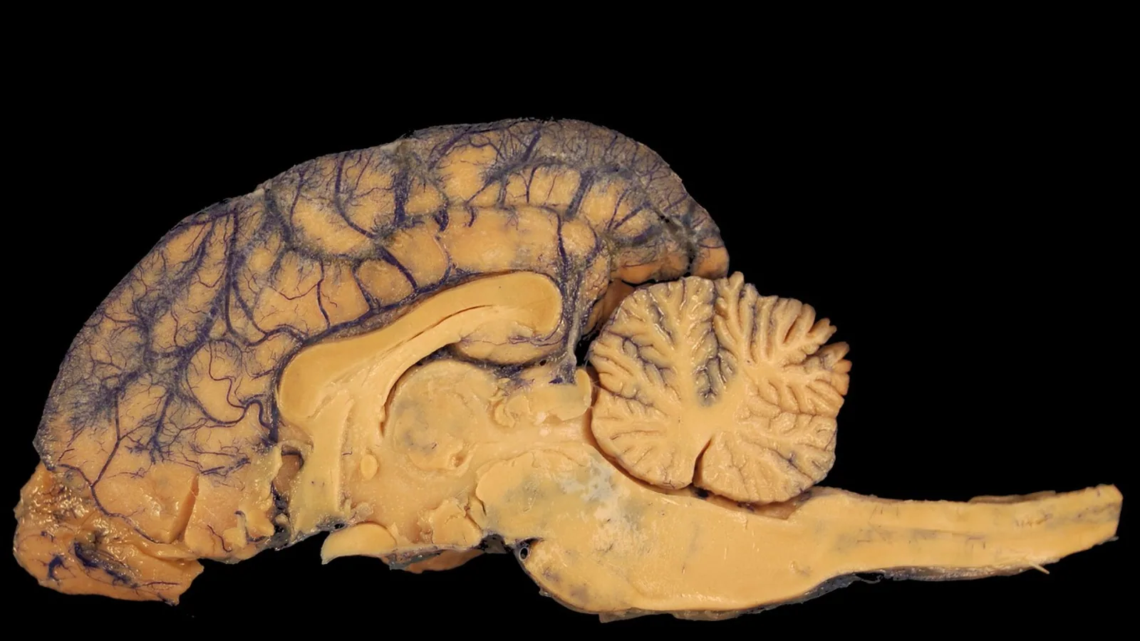
The choroid plexuses are vascularized, frond-like tufts of pia mater and epithelium that sit inside all four brain ventricles and produce most of the cerebrospinal fluid (CSF). They are found in the bodies and inferior horns of the lateral ventricles, in the roof of the third ventricle, and in the roof of the fourth ventricle, where the fluid they make exits through the lateral apertures of Luschka and the median aperture of Magendie.
This article covers the gross and microscopic anatomy of the choroid plexuses, the transport machinery inside their epithelial cells, the blood-CSF barrier, and the flow of CSF from ventricle to subarachnoid space. Comparative notes for dogs, cats, and other domestic mammals are included where the anatomy differs in a way that matters clinically.
What the Choroid Plexuses Are and Where They Sit


A choroid plexus is a specialized secretory tissue. Each plexus has a core of pia mater carrying blood vessels, covered by a single layer of cuboidal to columnar epithelial cells that face the ventricular lumen. The epithelium is continuous with the ependyma that lines the rest of the ventricular system, but it is structurally and functionally distinct. Choroid plexus epithelial cells are joined by tight junctions and adherens junctions, and they express a characteristic set of transporters and ion channels that ependymal cells do not [1].
The name comes from the Greek for "membrane" and the Latin for "braid," which describes the folded, ribbon-like appearance of the tissue. In the fresh brain, the plexuses look like reddish, granular clusters because of the dense capillary network inside them.
Lateral Ventricles
Each lateral ventricle contains choroid plexus in two regions. The larger portion runs along the floor of the central body of the ventricle, continuing from the interventricular foramen (of Monro) caudally. A second, narrower portion extends into the inferior (temporal) horn. This inferior horn segment is the one surgeons and imaging specialists watch most closely, because its size and position change with ventricular dilation.
Third Ventricle
In the third ventricle, the choroid plexus hangs from the roof, suspended in the tela choroidea. It forms two longitudinal fringes that run the length of the ventricular roof. The third ventricle plexus is smaller in volume than the lateral ventricle plexus but is a consistent source of CSF.
Fourth Ventricle
The fourth ventricle plexus occupies the roof of the ventricle, in the caudal part of the rhombencephalon. It has a distinctive T or inverted-L shape in many species, with paired lateral extensions. This is the segment closest to the apertures that drain CSF out of the ventricular system.
The Ventricular Apertures
CSF leaves the fourth ventricle through three openings. The lateral apertures of Luschka sit at the far lateral recesses of the fourth ventricle, one on each side. The median aperture of Magendie sits in the midline of the caudal roof. From these apertures, fluid enters the subarachnoid space, including the cerebellomedullary cistern (the cisterna magna), which is the site veterinarians most often sample for CSF collection in dogs and cats.
| Ventricle | Plexus location | Main CSF drainage route |
|---|---|---|
| Lateral ventricle | Floor of the body and inferior horn | Interventricular foramen into third ventricle |
| Third ventricle | Roof, suspended in tela choroidea | Cerebral aqueduct into fourth ventricle |
| Fourth ventricle | Roof, with paired lateral extensions | Lateral apertures of Luschka and median aperture of Magendie into the subarachnoid space |
| Subarachnoid space | No plexus tissue | Absorption at arachnoid granulations and along cranial and spinal nerve roots |
How Much CSF Is Produced and How Fast It Turns Over
The choroid plexuses produce roughly 60 to 70 percent of CSF in most mammals. The remaining 30 to 40 percent comes from interstitial fluid that drains from brain parenchyma into the ventricular system. This split matters because it explains why removing or cauterizing the plexuses does not stop CSF production entirely.
Total CSF volume in dogs is about 1 to 2 mL per kilogram of body weight. A 20 kg dog therefore carries roughly 20 to 40 mL of CSF at any moment. Turnover is several times daily, meaning the entire volume is replaced multiple times in 24 hours. This high turnover is why CSF composition reflects current plasma and brain conditions rather than a long historical average.
The rate of secretion is not constant. The choroid plexus epithelium contains a robust circadian clock, and expression of key transport proteins fluctuates across the day. In rats, aquaporin-1 (AQP1) in the fourth ventricle plexus and claudin-2 (CLDN2) in the lateral and fourth ventricle plexuses show clear diurnal variation at the protein level, and the timing of peak expression for AQP1, NKCC1, and CLDN2 is coordinated [2]. The authors of that work concluded that AQP1 and CLDN2 levels are likely to determine how CSF secretion volume changes across a day [2]. This is a useful concept for anyone interpreting serial CSF sampling or serial ventricular measurements.
Structure of the Choroid Plexus Epithelium
The Blood-CSF Barrier
The blood-CSF barrier is the functional gate between blood and ventricular fluid. It has three anatomical components:
- Fenestrated capillaries in the stromal core. These vessels are leaky by design, so plasma proteins and small solutes move freely out of the blood into the surrounding pia mater.
- A layer of pia mater and connective tissue around the vessels.
- Tight junctions between adjacent choroid plexus epithelial cells.
The tight junctions are the actual barrier. Because the capillaries are fenestrated, the selectivity of the blood-CSF barrier comes entirely from the epithelial cell layer and its junctional complexes [1]. This is the opposite of the blood-brain barrier, where tight junctions sit in the capillary endothelium itself.
Zonula occludens-1 (ZO1) is a key tight junction protein in this epithelium, and it is used as a marker of barrier integrity in cultured choroid plexus cells [3]. Claudins, including CLDN2, contribute to both barrier formation and fluid secretion [1]. Junctional proteins are not passive seals. They participate in the regulation of CSF production, and changes in their expression are part of how the plexus responds to injury and inflammation [1].
Transport Machinery That Drives Secretion
CSF is not a filtrate. It is an actively secreted fluid, and the epithelium spends energy to make it. The key players are:
- Na+/K+-ATPase on the apical membrane. In most secretory epithelia, the sodium pump sits on the basolateral side. In choroid plexus epithelium, it sits on the luminal (apical) surface facing the ventricle [4]. This unusual placement is one of the defining features of the tissue and is central to the ion gradients that drive water movement.
- Aquaporin-1 (AQP1). A water channel concentrated on the apical membrane. It allows rapid osmotic water flow into the ventricle. AQP1 expression varies with the time of day, which links water permeability to the circadian regulation of CSF output [2]. A subset of human choroid plexus epithelial cells also express aquaporin-4 (AQP4), the main water channel of brain parenchyma [3].
- NKCC1. A sodium-potassium-chloride cotransporter that moves ions into the cell and contributes to the osmotic gradient for secretion [5].
- Kir7.1. An inwardly rectifying potassium channel at the apical membrane. It sets the membrane potential of the epithelial cell and regulates the potassium concentration of CSF. Its conductance is unusually independent of extracellular potassium, which suits it to a buffering role [5].
- Glut1. A glucose transporter that supplies the metabolic fuel for this high-rate secretory tissue. Glut1 mRNA shows diurnal variation in the lateral and fourth ventricle plexuses [2].
The net effect is directional. Ions move from blood into the epithelial cell and then into the ventricle, water follows osmotically through AQP1, and the result is a continuous flow of fluid with a composition that the plexus actively sets.
Cilia and Mechanical Sensing
Choroid plexus epithelial cells are multiciliated. The cilia are exposed to CSF flow and to contact with neighboring cells, and they are maintained by mechanotransduction signaling. Piezo1, a mechanically activated ion channel, is expressed on the apical and lateral membranes of mature choroid plexus epithelial cells. It mediates calcium influx in response to fluid flow and to the pharmacological activator Yoda1, and it maintains calcium homeostasis in a cell-contact-dependent manner [6].
The consequence of losing Piezo1 is severe. Either knockout or overexpression of Piezo1 in these cells causes hydrocephalus in mice, reduces CSF flow, disrupts ciliary maintenance, and alters the expression of cilia-related genes [6]. Piezo1-mediated calcium signaling also regulates ciliogenesis in cultured choroid plexus epithelial cells [6]. This work establishes the plexus as a mechanically sensitive tissue, not just a passive filter.
Other mechanosensitive channels contribute as well. TRPV4 is a cation channel on choroid plexus epithelial cells, and its activity is phosphorylation-dependent. Activating TRPV4 alters the phosphorylation of tight junction proteins including ZO-1 and claudin-7, and it increases inhibitory phosphorylation of AMP-activated protein kinase (AMPK) [7]. The signaling axis between TRPV4 and AMPK is an active area of research in hydrocephalus because it offers a potential target for modifying CSF production [7].
The flow diagram below summarizes the path from blood to ventricular fluid and then out of the ventricular system.
flowchart TD
    A[Fenestrated capillary in plexus core] --> B[Stromal pia mater]
    B --> C[Choroid plexus epithelial cell]
    C --> D[Apical sodium potassium pump]
    C --> E[Aquaporin 1 water channel]
    D --> F[Ion gradient established]
    E --> G[Water enters ventricle]
    F --> G
    G --> H[Lateral ventricle]
    H --> I[Third ventricle]
    I --> J[Fourth ventricle]
    J --> K[Luschka and Magendie apertures]
    K --> L[Subarachnoid space]
CSF Circulation After It Leaves the Plexus
Fluid made in the lateral ventricles flows through the interventricular foramen into the third ventricle. It then passes down the cerebral aqueduct into the fourth ventricle. From the fourth ventricle, it exits through the lateral apertures of Luschka and the median aperture of Magendie into the subarachnoid space.
Once in the subarachnoid space, CSF bathes the brain and spinal cord, enters the perivascular spaces around penetrating vessels, and is absorbed at arachnoid granulations and along the sheaths of cranial and spinal nerve roots. A portion also drains along lymphatic pathways associated with the meninges. The movement is driven by a combination of bulk flow from continued secretion, arterial pulsation, respiratory pressure changes, and posture.
CSF motion is not uniform. Shear forces and pulsatile pressure vary along the production-to-outflow axis, and these mechanical cues are sensed by specialized modules in the choroid plexus epithelium, the ventricular ependyma, perivascular astrocytic endfeet, and meningeal outflow pathways [8]. Abnormal CSF motion can be converted into barrier dysfunction, which is one mechanism linking altered fluid dynamics to disease [8].
The plexus also does more than move water. It transports proteins into CSF and participates in immune surveillance at the brain-periphery interface [9]. In the immature brain, choroid plexus epithelial cells package insulin-like growth factor 1 (IGF-1) into extracellular vesicles and transport it directionally from the basolateral to the apical side, where it can reach the hippocampus [10]. This shows that the blood-CSF barrier is a selective delivery system, not a wall.
Comparative Notes for Domestic Species
The basic plan is conserved across mammals. The zebrafish choroid plexus expresses transporters involved in CSF secretion and is conserved with mammals, and ablating its epithelial cells reduces ventricular size without disrupting the blood-CSF barrier [11]. That finding supports the idea that the plexus controls ventricular volume through secretion rather than through barrier function alone.
In dogs and cats, the practical differences are in size and accessibility rather than in fundamental structure. The cisterna magna is large and reachable in most dogs, which is why it is the standard site for CSF collection. The lateral apertures of Luschka open into the lateral recesses of the fourth ventricle, and their position relative to the cerebellomedullary cistern explains why CSF collected there reflects fourth ventricle fluid.
Species differences in plexus volume and in the relative contribution of the fourth ventricle plexus have been described in rodents, where the lateral and fourth ventricle plexuses can be sampled separately and show different patterns of transporter expression [2]. Similar regional specialization is likely in domestic mammals, though the specific data are less complete.
Clinical Relevance, Limitations and Common Mistakes
The choroid plexus is the target of surgical and pharmacological interventions in veterinary neurology. Endoscopic choroid plexus cauterization is used in some hydrocephalus cases to reduce CSF production, and it is combined with endoscopic third ventriculostomy in certain patients [12]. The rationale is straightforward: if the plexus makes most of the fluid, reducing plexus tissue reduces fluid output. The limitation is equally straightforward: 30 to 40 percent of CSF comes from interstitial fluid, so cauterization is rarely a complete solution.
Choroid plexus enlargement is a recognized imaging finding. In a community-based cohort, larger plexus volume was associated with brain fluid dysregulation, indexed by elevated extracellular free water and reduced diffusion along perivascular spaces, and with progression of cerebral small vessel disease and white matter microstructural deterioration [9]. In multiple sclerosis, larger plexus volume has been associated with poorer information-processing speed and visuospatial memory in several studies, though longitudinal predictive value has not been consistent [13]. These are human imaging findings, but the underlying biology of plexus enlargement and fluid dysregulation is relevant to veterinary patients with similar imaging changes.
Common mistakes in interpreting plexus anatomy and function:
- Assuming the plexus is the only source of CSF. Interstitial fluid contributes a substantial fraction, and this fraction increases in some disease states.
- Treating the blood-CSF barrier as identical to the blood-brain barrier. The barriers differ in where the tight junctions sit and in what they exclude.
- Expecting CSF production to be constant. Circadian variation in AQP1 and CLDN2 expression changes secretion volume across the day [2].
- Overlooking the role of junctional proteins in secretion. Tight junctions are part of the secretory machinery, not just a seal [1].
- Assuming plexus enlargement always means pathology. Plexus volume changes with age, inflammation, and fluid status, and the clinical meaning depends on context.
What remains uncertain is how much of the circadian and mechanotransduction regulation seen in rodent and cell culture models translates quantitatively to dogs, cats, and horses. The specific contribution of individual transporters to CSF production rate in each species has not been fully mapped. The relationship between plexus volume on imaging and actual CSF production rate is also not established, which limits how much a single measurement can tell a clinician.
This article is educational and is not a substitute for veterinary diagnosis or treatment.
Frequently Asked Questions
What are the choroid plexuses?
They are vascularized, frond-like structures inside all four brain ventricles that produce most of the cerebrospinal fluid.
Where are the choroid plexuses located?
They sit in the bodies and inferior horns of the lateral ventricles, in the roof of the third ventricle, and in the roof of the fourth ventricle.
How much CSF do the choroid plexuses produce?
They produce roughly 60 to 70 percent of CSF in most mammals, with the rest coming from interstitial fluid.
What is the blood-CSF barrier?
It is the selective interface formed by fenestrated capillaries, pia mater, and tight junctions between choroid plexus epithelial cells.
What is the normal CSF volume in a dog?
Total CSF volume in dogs is about 1 to 2 mL per kilogram of body weight, and it turns over several times per day.
How does CSF leave the ventricular system?
It exits the fourth ventricle through the lateral apertures of Luschka and the median aperture of Magendie into the subarachnoid space.
What pumps drive CSF secretion?
Apical Na+/K+-ATPase and aquaporin-1 are the main drivers, supported by NKCC1, Kir7.1, and other transporters.
Can the choroid plexus be removed to treat hydrocephalus?
Parts of it can be cauterized surgically to reduce CSF production, but interstitial fluid still contributes a significant fraction, so it is not a complete cure.
Related Articles
- Prokaryotic DNA: Location, Structure, and Function
- RNA Location: Types, Functions, and Cellular Distribution
- Z-DNA Found in Biology: Location, Function, and Detection
- G-Quadruplex Telomere Structure and Function
- Equine Hoof Anatomy: Structures and Function
- Plasmid in Bacteria: Structure, Types, and Functions
- Pacinian Corpuscle: Structure, Function and Location
- Fibrous Cartilage: Structure, Types, and Locations
- Ependymal Cells: Function, Location, and CSF Production
Sources
- Transporters, Ion Channels, and Junctional Proteins in Choroid Plexus Epithelial Cells.
- Indication of diurnal variations in the rat choroid plexus for cerebrospinal fluid secretion.
- Primary cell and tissue cultures of human choroid plexus epithelial cells.
- Mechanisms of cerebrospinal fluid secretion by the choroid plexus epithelium: Application to various intracranial pathologies.
- Pathological mutation L241P in Kir7.1 channel inhibits its activity in the choroid plexus epithelium and decreases cerebrospinal fluid K(+) concentration in the mouse.
- Piezo1-mediated mechanotransduction in choroid plexus epithelial cells governs ciliogenesis and cerebrospinal fluid homeostasis.
- Phosphoproteomic Insights into TRPV4-AMPK Signaling Axis in the Choroid Plexus Epithelium: Implications for Therapeutic Targeting in Hydrocephalus.
- Cerebrospinal fluid mechanics across CNS barriers: from production, circulation, and clearance to mechanomedicine.
- Choroid plexus enlargement contributes to CSVD progression and white matter microstructural deterioration via brain-fluid dysregulation.
- Choroid plexus extracellular vesicle transport of blood-borne insulin-like growth factor 1 to the hippocampus of the immature brain.
- The evolutionarily conserved choroid plexus contributes to the homeostasis of brain ventricles in zebrafish.
- Imaging of Cerebrospinal Fluid Shunts and Their Complications.
- Choroid Plexus MRI Features and Cognitive Outcomes in Multiple Sclerosis: A Scoping Review.