Bones of the Face: Facial Skeleton Anatomy
By Dr. Zubair Khalid, DVM, MS, PhD ·
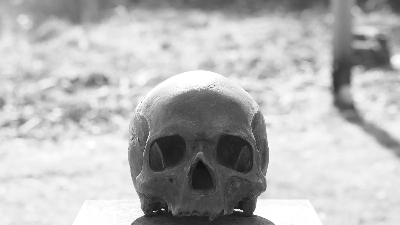
The facial skeleton is the set of paired and unpaired bones that form the muzzle, orbit, hard palate, and jaw, and that house the oral cavity, nasal cavity, and much of the dentition. In domestic mammals these bones are the maxilla, zygomatic, nasal, lacrimal, palatine, vomer, pterygoid, mandible, and the nasal conchae, together with the incisive bone in species that have one.
These bones matter to every clinician who works on the head. They define where local anesthetic blocks can be placed, where fractures occur after trauma, where dental roots sit, and where surgical approaches to the nasal cavity and sinuses are safe. A mental nerve block in a dog, a maxillary nerve block in a horse, or a dehorning procedure in a calf all depend on knowing the facial skeleton and the foramina that perforate it.
This article is educational and is not a substitute for veterinary diagnosis or treatment.
What Counts as a Facial Bone


The skull is divided into the neurocranium, which surrounds the brain, and the viscerocranium or splanchnocranium, which forms the face. This article covers the viscerocranium only. The cranial vault bones (frontal, parietal, occipital, interparietal, temporal, sphenoid, ethmoid) are covered separately.
Facial bones share four jobs:
- Support the soft tissues of the muzzle and cheeks.
- Form the bony walls of the nasal cavity, oral cavity, and orbit.
- Anchor the upper and lower teeth.
- Provide conduits (foramina and canals) for nerves and vessels.
Species differences in head shape drive most of the clinically important variation. Dolichocephalic breeds such as the greyhound have a long, narrow facial skeleton. Brachycephalic breeds such as the pug have a short, wide one. The same bones are present, but their proportions and the size of the paranasal sinuses change dramatically.
Paired Versus Unpaired Bones
| Bone | Paired or unpaired | Core location | Signature feature |
|---|---|---|---|
| Maxilla | Paired | Rostral upper jaw and lateral nasal wall | Alveoli of upper cheek teeth, infraorbital foramen |
| Zygomatic | Paired | Lateral orbit and cheek | Zygomatic arch, facial crest in the horse |
| Nasal | Paired | Dorsal muzzle | Nasal bone length sets muzzle profile |
| Lacrimal | Paired | Rostral orbit | Lacrimal fossa and nasolacrimal canal |
| Palatine | Paired | Caudal hard palate | Greater and lesser palatine foramina |
| Vomer | Unpaired | Midline nasal septum base | Keel-shaped bone on the nasal floor |
| Pterygoid | Paired | Caudal nasal cavity wall | Pterygoid hamulus |
| Mandible | Unpaired in adults | Lower jaw | Mental foramina, mandibular canal |
| Nasal conchae | Paired (multiple) | Inside nasal cavity | Scrolls of thin bone covered in mucosa |
The Maxilla
The maxilla is the largest facial bone and the workhorse of the upper jaw. It forms the rostral part of the hard palate, the lateral wall of the nasal cavity, and the medial wall of the orbit. Its alveolar process holds the upper canine and cheek teeth, and its palatine process meets its partner at the midline palatine fissure in many species.
The maxilla is perforated by the infraorbital canal, which opens rostrally as the infraorbital foramen. The infraorbital nerve and vessels pass through this foramen. The infraorbital nerve is the terminal branch of the maxillary nerve and supplies sensation to the upper lip, nose, and adjacent skin. This is the nerve blocked during dental procedures on the upper jaw and during surgery of the upper lip.
Topographic studies of the maxilla show that the distance from the infraorbital foramen to the zygomaticomaxillary suture is about 24 to 25 mm in humans, with a small but statistically significant difference between right and left sides [1]. Veterinary anatomy follows the same principle: the foramen sits at a predictable point above the upper fourth premolar in the dog, and this is the landmark used to place an infraorbital block.
The maxilla also contains the maxillary recess in the dog, a large paranasal sinus cavity that extends into the bone. In the horse, the maxilla is expanded by the caudal maxillary sinus and the ventral conchal sinus, which are clinically important because they can become infected or fill with blood after trauma.
The Zygomatic Bone
The zygomatic bone forms the lateral wall of the orbit and the rostral part of the zygomatic arch. It articulates with the maxilla, frontal, temporal, and lacrimal bones. In the horse, the zygomatic bone carries the facial crest, a prominent ridge on the lateral surface of the skull that is a key surgical landmark for the maxillary sinus and for the transverse facial artery.
The zygomaticofacial nerve exits through the zygomaticofacial foramen on the lateral surface of the bone. This nerve can be injured during facial surgery, particularly during zygomatic implant procedures and orbital approaches [1]. In veterinary patients, the zygomatic arch is a common site of fracture after blunt trauma, and the bone is thin enough that it can be penetrated by bite wounds from larger animals.
The Nasal Bone
The nasal bone is a paired, elongated bone on the dorsum of the muzzle. It forms the roof of the nasal cavity and articulates caudally with the frontal bone. The length of the nasal bone is the main determinant of the muzzle profile in dogs, and breed-specific shortening in brachycephalic dogs is largely a nasal bone and maxillary change.
The nasal bone is thin and easily fractured. In the horse, the nasal bone is thick and forms part of the nasal diverticulum, which is a blind pouch of skin and mucosa that opens into the nostril. This diverticulum is why a horse can be nasally intubated without passing the tube into the trachea if the tube enters the diverticulum instead.
The Lacrimal Bone
The lacrimal bone is a small paired bone at the rostral edge of the orbit. It contains the lacrimal fossa, which holds the lacrimal sac, and the opening of the nasolacrimal canal. Tears drain from the eye through the lacrimal puncta, into the lacrimal sac, and then through the nasolacrimal duct to the nasal cavity.
The nasolacrimal canal is a clinically important structure in all domestic species. Obstruction of the duct causes epiphora (overflow of tears onto the face). In the horse, the lacrimal bone is large and the nasolacrimal duct is long, which makes catheterization of the duct a routine procedure for treating nasolacrimal obstruction.
The Palatine Bone
The palatine bone is a paired bone that forms the caudal part of the hard palate and part of the lateral nasal wall. It contains the greater palatine foramen, through which the greater palatine nerve and vessels pass to supply the caudal palate and soft tissues. The lesser palatine foramina transmit the lesser palatine nerves.
The palatine bone also contributes to the pterygopalatine fossa, a small space behind the maxilla that contains the maxillary nerve, the pterygopalatine ganglion, and branches of the maxillary artery. This fossa is a target for maxillary nerve blocks in horses and cattle, and it is a route for surgical access to the caudal nasal cavity.
The Vomer
The vomer is an unpaired, midline bone that forms the caudal part of the nasal septum and sits on the floor of the nasal cavity. It is keel-shaped in cross section and articulates with the sphenoid, ethmoid, palatine, and maxilla. The vomer is thin and rarely fractured in isolation, but it can be involved in nasal cavity tumors and in congenital abnormalities of the nasal septum.
The Pterygoid Bone
The pterygoid bone is a paired bone that forms the caudal wall of the nasal cavity and part of the pterygopalatine fossa. Its ventral end forms the pterygoid hamulus, a small hook-like process that is a landmark for the pterygoid canal. The pterygoid canal transmits the nerve of the pterygoid canal and vessels.
In the Persian squirrel, a fossa has been identified between the pterygoid bone and the pterygoid process of the basisphenoid, and the pterygoid canal lies between the presphenoid, palatine, and pterygoid bones [2]. This level of detail matters in comparative anatomy because the pterygoid region is a common site of surgical approach to the caudal nasal cavity and the basisphenoid.
The Mandible
The mandible is the largest facial bone and the only movable bone of the skull. It is unpaired in adult domestic mammals because the two halves fuse at the mandibular symphysis. The mandible consists of a body, which holds the lower teeth, and a ramus, which articulates with the temporal bone at the temporomandibular joint.
The mandible contains the mandibular canal, which runs through the body of the bone and transmits the inferior alveolar nerve and vessels. The canal opens on the lateral surface as the mental foramen, through which the mental nerve and vessels exit. The mental nerve supplies sensation to the lower lip and chin. The mental foramen is the target for mental nerve blocks in dogs, cats, and other species.
Studies of mandibular morphology show that the shape of the mandible varies significantly with sagittal skeletal pattern and sex, with the first principal component of variation being sagittal length [3]. In veterinary patients, this translates to breed and species differences in mandibular length and angle. The Beetal goat has an elongated, narrow, dolichocephalic skull, while the Kajali sheep has a shorter, broader, mesaticephalic to brachycephalic skull [4]. Accessory mental foramina are common in sheep (87.5%) and less common in goats (12.5%) [4].
The mandible is also a site of pathological change. In a mouse model of sclerosteosis, prominent mandibles highlight hyperostosis in the jaw [5]. This is relevant to veterinary dentistry because mandibular enlargement can change occlusion and make dental procedures more difficult.
The Nasal Conchae
The nasal conchae are thin, scroll-like bones inside the nasal cavity. They are covered by respiratory mucosa and increase the surface area for warming, humidifying, and filtering inspired air. Domestic mammals have several conchae, including the dorsal, middle, and ventral nasal conchae, and the ethmoidal conchae.
The conchae are delicate and can be damaged by nasal trauma, foreign bodies, or overzealous nasal flushing. In the horse, the ventral conchal sinus is part of the paranasal sinus system and can become infected, leading to sinusitis. In the dog, the conchae are complex and fill most of the nasal cavity, which is why nasal tumors in dogs often present with epistaxis and obstruction.
Foramina of the Facial Skeleton
Foramina are the openings that allow nerves and vessels to pass through bone. Knowing them is essential for local anesthesia, surgery, and interpretation of radiographs and CT scans.
| Foramen or canal | Bone | Structures transmitted | Clinical use |
|---|---|---|---|
| Infraorbital foramen | Maxilla | Infraorbital nerve and vessels | Infraorbital nerve block for upper jaw dental work |
| Mental foramen | Mandible | Mental nerve and vessels | Mental nerve block for lower lip and chin surgery |
| Mandibular canal | Mandible | Inferior alveolar nerve and vessels | Mandibular nerve block for lower jaw dental work |
| Optic canal | Sphenoid (presphenoid) | Optic nerve and ophthalmic artery | Not a facial bone foramen, but relevant to orbital surgery |
| Zygomaticofacial foramen | Zygomatic | Zygomaticofacial nerve | Landmark for orbital and zygomatic surgery |
| Greater palatine foramen | Palatine | Greater palatine nerve and vessels | Palatal surgery and anesthesia |
| Lesser palatine foramina | Palatine | Lesser palatine nerves | Palatal anesthesia |
| Pterygoid canal | Pterygoid and sphenoid | Nerve of pterygoid canal and vessels | Surgical landmark for caudal nasal cavity |
| Nasolacrimal canal | Lacrimal and maxilla | Nasolacrimal duct | Tear drainage and catheterization |
| Accessory mental foramina | Mandible | Branches of mental nerve | Variation in sheep and goats [4] |
The optic canal is not part of the facial skeleton, but it is included here because it is frequently confused with facial foramina. The optic nerve passes through the optic canal, which is located in the presphenoid bone. Damage to the optic nerve during orbital surgery or trauma can cause blindness.
Species Differences in the Facial Skeleton
Dog
The dog has a long snout in most breeds, with a large maxillary recess that is part of the paranasal sinus system. The infraorbital foramen is located above the upper fourth premolar. The mandible is relatively straight and the mental foramen is located below the lower third premolar. Brachycephalic breeds have a shortened maxilla and nasal bone, which changes the position of the infraorbital foramen and makes intubation and dental radiography more challenging.
Horse
The horse has a prominent facial crest on the zygomatic bone, which is a key landmark for the maxillary sinus and for the transverse facial artery. The paranasal sinuses are large and complex, including the caudal maxillary, rostral maxillary, dorsal conchal, ventral conchal, and sphenopalatine sinuses. The infraorbital foramen is located above the upper third premolar. The mandible is large and the mental foramen is located on the lateral surface of the body, below the lower second premolar.
Cow
The cow has a cornual process, which is the bony core of the horn. It is an extension of the frontal bone, not a facial bone, but it is included here because dehorning is a common procedure that involves the facial skeleton. The cornual process is hollow in young animals and becomes pneumatized as the frontal sinus extends into it. The maxilla and mandible are large and the dental formula includes a diastema between the incisors and cheek teeth. The mental foramen is located on the lateral surface of the mandible, below the lower second premolar.
Sheep and Goat
Sheep and goats have similar facial skeletons, but there are differences. Beetal goat skulls are elongated and narrow, while Kajali sheep skulls are shorter and broader [4]. Accessory mental foramina are more common in sheep than in goats [4]. The facial tuberosity, a bony prominence on the maxilla, is positioned posteriorly in goats and at the premolar level in sheep [4]. These differences matter for regional anesthesia and for dehorning.
How the Facial Skeleton Is Examined in Practice
Physical examination of the facial skeleton starts with inspection and palpation. Swelling, asymmetry, crepitus, and pain on palpation suggest fracture or infection. The oral cavity is examined for dental disease, and the nasal cavity is examined for discharge, obstruction, or masses.
Radiography is the first-line imaging modality for the facial skeleton. Standard views include lateral, dorsoventral, and oblique projections. Intraoral radiography is used for dental assessment. Computed tomography (CT) provides detailed cross-sectional images and is superior for evaluating complex fractures, sinus disease, and nasal tumors. In the Persian squirrel, CT and radiographic features of the skull have been described in detail, including the absence of the alar canal and the presence of a distinct spinous foramen [2].
Advanced imaging is also used in research. Three-dimensional geometric morphometrics has been used to analyze the mandible and midfacial skeleton across different sagittal skeletal patterns and sexes, showing that shape variation is dominated by sagittal length [3]. This technique is not yet routine in veterinary practice, but it illustrates how detailed the study of facial bones has become.
Clinical Relevance, Limitations and Common Mistakes
The facial skeleton is involved in a wide range of clinical conditions. Fractures of the maxilla, zygomatic arch, and mandible are common after road traffic accidents, falls, and bite wounds. Nasal cavity tumors, sinusitis, and dental abscesses all involve facial bones. Dehorning in cattle and disbudding in goats and sheep require knowledge of the cornual process and the frontal sinus.
Local anesthesia of the face depends on accurate knowledge of foramina. The infraorbital nerve block is used for upper jaw dental procedures and is placed at the infraorbital foramen. The mental nerve block is used for lower lip and chin surgery and is placed at the mental foramen. The mandibular nerve block is used for lower jaw dental procedures and is placed near the mandibular foramen on the medial surface of the ramus.
Common mistakes in facial skeleton anatomy include:
- Confusing the infraorbital foramen with the zygomaticofacial foramen. The infraorbital foramen is on the maxilla and transmits the infraorbital nerve. The zygomaticofacial foramen is on the zygomatic bone and transmits the zygomaticofacial nerve [1].
- Assuming the mandible is paired in adults. In domestic mammals, the two halves fuse at the mandibular symphysis, so the mandible is unpaired.
- Forgetting that the optic canal is not a facial bone foramen. It is in the presphenoid and transmits the optic nerve.
- Missing accessory mental foramina. These are common in sheep and can be a source of incomplete anesthesia if only the main mental foramen is blocked [4].
- Overlooking species differences in the facial crest and paranasal sinuses. The horse has a prominent facial crest and large sinuses, while the dog has a large maxillary recess and a long snout.
- Assuming that facial bone anatomy is the same across breeds. Brachycephalic dogs have shortened maxillae and nasal bones, which changes the position of foramina and the shape of the nasal cavity.
Individual cases require a veterinarian for diagnosis and treatment. The information here is a foundation for study, not a substitute for clinical judgment.
Quick Review
- The facial skeleton includes the maxilla, zygomatic, nasal, lacrimal, palatine, vomer, pterygoid, mandible, and nasal conchae.
- The infraorbital foramen transmits the infraorbital nerve and vessels and is the target for infraorbital nerve blocks.
- The mental foramen transmits the mental nerve and vessels and is the target for mental nerve blocks.
- The optic canal transmits the optic nerve and is located in the presphenoid bone, not in a facial bone.
- The dog has a long snout and a large maxillary recess. The horse has a prominent facial crest and large paranasal sinuses. The cow has a cornual process.
- The mandible is unpaired in adult domestic mammals because the two halves fuse at the mandibular symphysis.
- Accessory mental foramina are common in sheep and less common in goats.
Frequently Asked Questions
What are the bones of the facial skeleton?
The bones of the facial skeleton are the maxilla, zygomatic, nasal, lacrimal, palatine, vomer, pterygoid, mandible, and nasal conchae. The incisive bone is also present in species that have one.
What passes through the infraorbital foramen?
The infraorbital nerve and vessels pass through the infraorbital foramen. The infraorbital nerve supplies sensation to the upper lip, nose, and adjacent skin.
What passes through the mental foramen?
The mental nerve and vessels pass through the mental foramen. The mental nerve supplies sensation to the lower lip and chin.
What passes through the optic canal?
The optic nerve and ophthalmic artery pass through the optic canal. The optic canal is located in the presphenoid bone, not in a facial bone.
How does the dog facial skeleton differ from the horse?
The dog has a long snout with a large maxillary recess. The horse has a prominent facial crest and large paranasal sinuses. Both have the same facial bones, but the proportions and sinus development differ.
What is the cornual process?
The cornual process is the bony core of the horn in cattle. It is an extension of the frontal bone and becomes pneumatized as the frontal sinus extends into it.
Related Articles
- Bovine Musculoskeletal Anatomy: Axial Skeleton and Joints
- Comparative Anatomy of the Avian Skeleton: Flight and Locomotion
- Equine Hindlimb Anatomy: Bones, Joints, and Ligaments
- Do Frogs Have Bones? Skeletal Anatomy and Pet Care
- Dog Itching Ears And Face
- How To Treat Ringworm At Home On Face
- Face Muscles: Anatomy, Functions, and Comparative Notes
- Cat Skeleton: Anatomy, Bones, and Function
Further Reading
- [[Application of free tissue flaps using the facial artery and vein/retromandibular vein as recipient vessels in the repair of postoperative nasopharyngeal skull base necrosis].](https://pubmed.ncbi.nlm.nih.gov/42706185/)
Sources
- Topographic and Morphometric Assessment of Facial Foramina in Maxilla, Mandible, and Zygomatic Bones: Anatomical and Clinical Relevance.
- Anatomic, Radiographic, and Computed Tomographic Study of the Skull in the Persian Squirrel (Sciurus anomalus).
- Morphological variations and differences of the mandible and midfacial skeleton across different sagittal skeletal patterns and sexes: a three-dimensional geometric morphometric analysis.
- Diagnostic anatomy of the head region of Beetal goat and Kajali sheep with special reference to its importance in regional anaesthesia.
- Characterising skull pathologies in the Sost-deficient mouse for preclinical evaluation of sclerosteosis treatments.