Areolar Connective Tissue: Histology and Function
By Dr. Zubair Khalid, DVM, MS, PhD ·
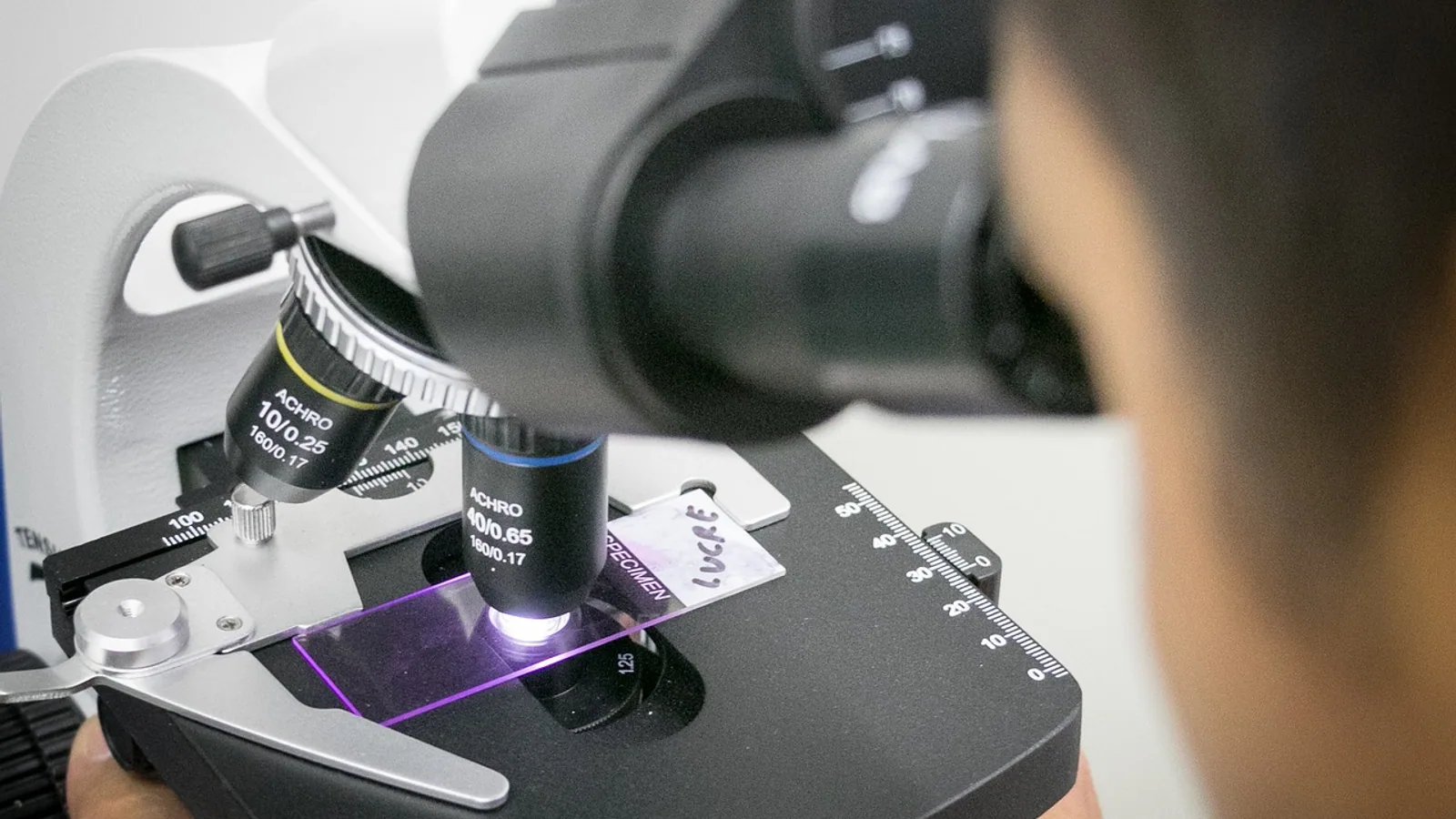
Areolar connective tissue is a loose connective tissue composed of scattered fibroblasts, mast cells, macrophages and other cell types suspended in a hydrated ground substance that carries loosely woven collagen and elastic fibers. It is the most widely distributed connective tissue in the body, forming the packing material beneath epithelia, around blood vessels and nerves, and within the mesentery.
Areolar tissue is the tissue students see first in almost every histology slide set, yet it does more physiological work than its unremarkable appearance suggests. It anchors epithelium to deeper layers, permits the diffusion of nutrients and oxygen from small vessels to cells that have no direct blood supply, and provides the loose cellular terrain through which immune cells travel to sites of injury. In veterinary practice, areolar tissue appears in surgical anatomy (the loose plane a surgeon follows to separate skin from fascia), in wound healing, in edema (where it swells with fluid), and in comparative anatomy, where its thickness and distribution vary markedly among dogs, cats, horses, cattle and pigs.
This article is educational and is not a substitute for veterinary diagnosis or treatment.
What Areolar Connective Tissue Is, and Why It Matters
Connective tissue proper falls into two broad families: loose connective tissue and dense connective tissue. Areolar tissue is the archetypal loose connective tissue. The word "areolar" comes from the small clear spaces (areolae) that appear between the fibers in a stained preparation, giving the tissue a lacy, open, almost sponge-like look under the microscope [1]. Those apparent empty spaces are not truly empty. In the living animal they hold ground substance, tissue fluid, and the cells that maintain and patrol the tissue.
Three features define areolar connective tissue:
- Abundant ground substance relative to fibers. The amorphous, gel-like matrix dominates the volume, which is why the tissue is soft, pliable and permeable.
- A loose, irregular weave of fibers. Collagen bundles run in multiple directions with no consistent orientation, and fine elastic fibers interlace among them. This is the key structural difference from dense regular connective tissue, where collagen is packed into parallel bundles.
- A mixed, mobile cell population. Fibroblasts build the matrix, but mast cells, macrophages, plasma cells, adipocytes, lymphocytes and occasional eosinophils and neutrophils are normal residents.
Why does this matter to a veterinary student or clinician? Because areolar tissue is the default connective tissue of the body's interstitial spaces. It is the tissue that swells in edema, the tissue that surgeons bluntly dissect, the tissue that delivers oxygen and glucose to avascular epithelium, and the tissue that hosts the first wave of the inflammatory response. Understanding its histology explains a large share of general pathology, wound healing and surgical anatomy.
Histology: How Areolar Tissue Looks Under the Microscope
The ground substance
Ground substance is the amorphous, colorless material that fills the space between cells and fibers. It is composed chiefly of glycosaminoglycans (GAGs, long unbranched polysaccharides with repeating disaccharide units) and proteoglycans, with glycoproteins such as fibronectin. Hyaluronic acid, a large GAG, gives the ground substance its viscous, water-binding character. Because ground substance is highly hydrated, it acts as a molecular sieve: water, ions, oxygen, nutrients, metabolites and small signaling molecules diffuse through it readily, while large proteins and most bacteria move slowly. This sieve behavior is the physical basis for the tissue's role in nutrient diffusion and in limiting the early spread of infection.
Ground substance is nearly invisible in routine hematoxylin and eosin (H&E) preparations because it does not stain strongly. It appears as pale, faintly basophilic or clear material between fibers. Special stains such as Alcian blue or periodic acid-Schiff highlight GAGs and glycoproteins when a laboratory needs to demonstrate matrix composition.
The fibers
Collagen fibers are the dominant fibrous component. In areolar tissue they are type I collagen chiefly, arranged as wavy, branching bundles of varying thickness. They stain eosinophilic (pink) on H&E and appear as irregular, nonparallel strands. Their tensile strength resists stretching, but because they are loosely arranged and not aligned, they do not confer the high unidirectional strength seen in tendons and ligaments. A histological study of human lateral ankle ligaments illustrates the contrast well: the calcaneofibular ligament shows dense, well-organized parallel collagen bundles with low cellularity and high mechanical stability, whereas the anterior talofibular ligament shows loosely arranged collagen fibers, lower density and higher cellularity and vascularity, indicating active remodeling but reduced structural strength [2]. Areolar tissue sits at the loose end of that spectrum.
Elastic fibers are thin, branching, refractile fibers that allow the tissue to stretch and recoil. They are less abundant than collagen in areolar tissue but are functionally important in sites subject to repeated deformation, such as the loose tissue around blood vessels and in the lungs. Elastin is the core protein, surrounded by microfibrils. On H&E, elastic fibers are difficult to distinguish from collagen, so laboratories use special stains such as orcein, resorcin-fuchsin, Verhoeff's or Weigert's stain to demonstrate them.
Reticular fibers are fine, branching networks of type III collagen that form supporting scaffolds in lymphoid organs, bone marrow and endocrine glands. They are a distinct fiber type associated with reticular tissue, not the defining feature of areolar tissue. Students frequently conflate the two (see the misconceptions section below).
The cells
Areolar tissue contains a resident population of cells that maintain the matrix and a transient population that arrives from the blood during inflammation. The table below summarizes the main cell types and their functions.
| Cell type | Origin | Main function in areolar tissue |
|---|---|---|
| Fibroblast | Mesenchymal (resident) | Synthesizes collagen, elastin, GAGs and proteoglycans. The dominant cell in most preparations. Maintains and remodels the matrix. |
| Mast cell | Bone marrow (resident) | Stores histamine, heparin and proteases in cytoplasmic granules. Releases mediators that dilate vessels, increase permeability and recruit immune cells during inflammation and allergy. |
| Macrophage | Bone marrow (monocyte-derived, resident or recruited) | Phagocytoses debris, microbes and dead cells. Presents antigen. Secretes cytokines that orchestrate repair and inflammation. |
| Plasma cell | Bone marrow (B-lymphocyte-derived) | Produces antibodies directed against local antigens. Common in tissue subject to chronic antigen exposure. |
| Adipocyte | Mesenchymal (resident) | Stores lipid. Present singly or in small clusters in areolar tissue, becoming the dominant cell in adipose tissue. |
| Lymphocyte | Bone marrow or thymus | Provides immune surveillance. Passes through areolar tissue en route between blood, lymph and lymphoid organs. |
| Neutrophil | Bone marrow (transient) | First responder to acute bacterial injury. Short-lived, phagocytic, recruited from blood within minutes to hours. |
| Eosinophil | Bone marrow (transient) | Defends against parasites and participates in allergic responses. Contains granules with major basic protein. |
| Mesenchymal stem cell (pericyte or adventitial cell) | Resident | Provides a reservoir for regeneration and repair. Differentiates into fibroblasts, adipocytes, chondrocytes or osteoblasts under appropriate signals. |
The relative proportions of these cells vary with site and with the state of the tissue. A resting subcutaneous sample is dominated by fibroblasts and adipocytes. A sample from inflamed tissue is dominated by neutrophils, macrophages and plasma cells.
Recognizing areolar tissue on a slide
A student can identify areolar connective tissue by four features:
- Open, lacy appearance with pale ground substance between fibers.
- Irregular, nonparallel collagen bundles of mixed thickness.
- Scattered, spindle-shaped fibroblasts with elongated nuclei, usually the most numerous cell type.
- A mixed cell population, often including round, granule-filled mast cells and large, irregular macrophages.
A useful comparison is a tendon or ligament section placed beside an areolar section. The tendon shows dense, parallel, wavy collagen with sparse, flattened fibroblasts squeezed between bundles. Areolar tissue shows the opposite: sparse, disorganized fibers with abundant space and cells.
Locations and Roles in the Body
Areolar tissue appears wherever the body needs a soft, pliable, well-vascularized packing material. The table below lists the major locations and the functional role at each site.
| Location | Role of areolar tissue |
|---|---|
| Beneath epithelia (lamina propria of mucous membranes, papillary dermis of skin) | Anchors epithelium to underlying tissue. Supplies nutrients to avascular epithelium by diffusion. Hosts immune cells that intercept antigens crossing the epithelial barrier. |
| Around blood vessels, nerves and lymphatics | Forms the adventitial layer that allows vessels and nerves to move without kinking. Provides a loose plane for surgical dissection. |
| Subcutaneous tissue (hypodermis) | Cushions the skin against mechanical trauma. Insulates. Stores energy as fat. Provides a mobile plane between skin and deep fascia. |
| Mesentery and omentum | Suspends and supports abdominal organs. Carries blood vessels, lymphatics and nerves. Provides a mobile, permeable sheet for fluid exchange and immune surveillance. |
| Between muscle groups and around fascia | Allows muscles to slide against one another during contraction. Provides a loose cleavage plane for surgical approaches. |
| Within the orbit, around the eye | Cushions the globe. Permits eye movement within the orbit. |
| Around joints and within tendon sheaths | Lubricates and cushions. Allows smooth gliding of tendons and joint structures. |
| In the marrow cavity and around bone trabeculae | Fills the spaces between trabeculae. Contains a rich vascular bed and a population of mesenchymal cells and adipocytes. |
| Around the internal carotid artery within the carotid canal | Provides a loose, adherent layer between the artery and surrounding dura mater. A cadaveric study of 16 sides found the membrane surrounding the petrous internal carotid artery was consistently dura mater, with a dural border cell layer loosely applied to the adventitial layer of the artery [3]. |
The functions of areolar tissue can be summarized in four categories:
- Cushioning and mechanical support. The hydrated ground substance and loose fiber network absorb compressive and shear forces, protecting delicate structures such as blood vessels and nerves.
- Diffusion of nutrients and metabolites. Because the matrix is highly hydrated and permeable, small molecules move freely between capillaries and cells. This is essential for avascular tissues such as epithelium and cartilage, which depend on adjacent areolar tissue for their supply.
- Immune surveillance and response. The resident macrophage and mast cell populations, plus the continuous traffic of lymphocytes and neutrophils, make areolar tissue a frontline immune compartment. Mast cell activation, for example, is a recognized mechanism in connective tissue disorders that affect vascular regulation [4].
- Facilitation of tissue movement. The loose, deformable matrix allows skin to slide over fascia, muscles to glide against one another, and vessels and nerves to accommodate movement without tearing.
Comparative Notes: Species Differences in Subcutaneous Areolar Tissue
The thickness, fat content and mechanical behavior of subcutaneous areolar tissue vary considerably among domestic species. These differences matter for surgery, for drug administration (subcutaneous injections), for thermoregulation and for wound healing.
Dogs. Subcutaneous areolar tissue is typically thin and loosely attached to the underlying fascia, particularly over the trunk and limbs. This loose attachment is why skin on the dorsum can be lifted and moved freely, and why blunt dissection planes are easy to develop in many surgical approaches. The subcutaneous layer contains variable amounts of fat, more in well-conditioned animals and less in thin or cachectic patients.
Cats. The subcutaneous areolar layer is thin and the skin is notably mobile, especially over the trunk. The loose attachment allows cats to twist and right themselves during falls and contributes to the characteristic "tenting" of the skin when it is lifted. Subcutaneous fat is present but generally less abundant than in dogs, and the layer is easily separated from the underlying musculature.
Horses. The subcutaneous areolar tissue is thin over most of the body but is specialized in certain regions. The loose areolar tissue beneath the skin of the neck and trunk allows the skin to move independently of the underlying musculature, which is important for the horse's ability to twitch the skin to dislodge insects. In the limbs, the subcutaneous tissue is tightly bound to the underlying fascia and bone, which is why wounds and swelling in the distal limb behave differently from those on the trunk.
Cattle. The subcutaneous areolar layer is well developed over the trunk and contributes to the thick, loose skin characteristic of the species. The panniculus muscle (cutaneous trunci) lies within this layer and is responsible for the skin twitch reflex commonly used in physical examination. Subcutaneous fat deposition is pronounced in feedlot cattle and is a major determinant of carcass grade.
Pigs. Subcutaneous areolar tissue is thick and contains abundant fat, forming the layer that produces bacon and lard. The loose, well-vascularized nature of this layer in pigs is one reason subcutaneous injections are technically straightforward in this species.
Birds. Subcutaneous areolar tissue is present but generally thinner than in mammals, and in many regions the skin is closely applied to underlying structures. The subcutaneous layer is more prominent in areas such as the crop region and the abdominal wall.
These differences have practical implications. A subcutaneous injection technique that works well in a dog (lifting a tent of skin over the dorsal trunk) may need modification in a horse or a pig because of differences in skin thickness and subcutaneous fat. Similarly, surgical approaches that rely on developing a loose areolar plane in a dog may encounter a tighter, more adherent layer in the distal limb of a horse or a cow.
How Areolar Tissue Is Studied and Observed
Areolar connective tissue is examined in the laboratory by standard histological methods. The steps are:
- Fixation. Tissue is fixed in neutral buffered formalin (typically 10%) to preserve structure.
- Processing and embedding. The sample is dehydrated through graded alcohols, cleared in xylene or a substitute, and embedded in paraffin wax.
- Sectioning. Sections are cut at 4 to 6 micrometers on a microtome and mounted on glass slides.
- Staining. Routine H&E staining shows nuclei in blue-purple and cytoplasm and collagen in pink. Special stains are used to demonstrate specific components: Masson's trichrome for collagen (blue or green), Verhoeff's or orcein for elastic fibers (black or brown), Alcian blue for GAGs (blue), and silver stains for reticular fibers (black).
- Microscopy. The slide is examined under a light microscope at magnifications from 4x (overview) to 100x (oil immersion, for cell detail).
In a clinical or research setting, areolar tissue may also be evaluated by immunohistochemistry, which uses antibodies against specific cell markers to identify cell types. A study of bone tissue in aseptic necrosis of the femoral head, for example, used histopathological and immunohistochemical methods to show that damaged trabeculae were replaced by large areolar cavities filled with connective tissue rich in adipocytes, with a low reaction of bone reparatory processes [5]. This illustrates how areolar tissue can expand to fill spaces left by tissue loss.
Experimental models also use areolar-like substrates. A study of cancer cell dynamics on silica fibers used a three-dimensional nonwoven scaffold to simulate loose connective tissue containing collagen fibers adjacent to a primary tumor, and found that cancer cells initially attached loosely to the fibers before extending stable, elongated membrane protrusions [6]. This kind of model helps researchers study how cells behave in a loose connective tissue environment.
Clinical Relevance, Limitations and Common Mistakes
Clinical relevance
Areolar tissue is involved in a wide range of clinical conditions.
Edema. Because the ground substance is highly hydrated and the fiber network is loose, areolar tissue is the first compartment to accumulate excess fluid when capillary filtration exceeds lymphatic drainage. Pitting edema, the classic sign in which a finger pressed into the skin leaves an indentation, reflects fluid movement within the subcutaneous areolar layer. The distribution of edema varies with species and with the underlying cause (cardiac, renal, hepatic, inflammatory or lymphatic).
Inflammation and immune response. Mast cells in areolar tissue release histamine and other mediators that increase vascular permeability and recruit neutrophils and macrophages. This is the cellular basis of the cardinal signs of inflammation: redness, heat, swelling, pain and loss of function. Connective tissue laxity and mast cell activation are also recognized mechanisms in hypermobility disorders, where altered connective tissue behavior contributes to vascular and autonomic dysfunction [4].
Wound healing. After injury, fibroblasts in areolar tissue proliferate and synthesize new collagen and ground substance to fill the defect. The loose, well-vascularized nature of areolar tissue supports the early inflammatory and proliferative phases of healing. In surgical wounds, the areolar plane is the layer that surgeons approximate when closing dead space.
Surgical anatomy. Surgeons use areolar tissue planes to separate structures with minimal trauma. The loose areolar tissue around blood vessels and nerves allows these structures to be mobilized without damaging them. In skull base surgery, for example, knowledge of the tissue surrounding the petrous internal carotid artery is important for safe exposure and mobilization of the vessel [3].
Tumor biology. Some soft tissue tumors arise from or resemble areolar connective tissue. Malignant fibrous histiocytoma, a tumor of fibroblasts with similarities to loose areolar connective tissue, illustrates the diagnostic challenge of distinguishing tumors that mimic normal loose connective tissue from other sarcomas [7]. Adaptive remodeling of loose connective tissue can also occur in response to mechanical displacement, as seen in the temporomandibular joint, where dense connective tissue and cartilaginous masses replace loose connective tissue after disc displacement [8].
Limitations
Areolar tissue is not a homogeneous entity. Its composition varies by site, by species, by age and by physiological state. A sample from the mesentery looks different from a sample from the dermis, and a sample from a young, well-nourished animal looks different from one from an aged or cachectic animal. Histological interpretation therefore requires knowledge of the site and the clinical context. Individual cases require veterinary assessment, and this article is not a substitute for diagnosis or treatment.
Common mistakes
Mistake 1: Confusing areolar tissue with reticular tissue. Reticular tissue is a distinct type of loose connective tissue characterized by a network of reticular fibers (type III collagen) that forms the structural framework of lymphoid organs, bone marrow and endocrine glands. Areolar tissue has a mixed fiber population dominated by collagen and elastin, with no specialized reticular framework. The two are sometimes grouped together as "loose connective tissue," but they are histologically and functionally distinct.
Mistake 2: Confusing areolar tissue with adipose tissue. Adipose tissue is a specialized connective tissue in which adipocytes are the dominant cell type, organized into lobules by thin septa of areolar tissue. Areolar tissue contains adipocytes but is not dominated by them. The distinction matters because adipose tissue has specialized metabolic and endocrine functions that areolar tissue does not share.
Mistake 3: Confusing areolar tissue with dense regular connective tissue. Dense regular connective tissue (tendon, ligament) has collagen fibers packed in parallel bundles with sparse cells and little ground substance. Areolar tissue has loosely arranged, nonparallel fibers with abundant ground substance and a mixed cell population. The functional difference is that dense regular tissue resists high unidirectional tensile forces, while areolar tissue accommodates multidirectional movement and diffusion.
Mistake 4: Assuming areolar tissue is inert packing material. Areolar tissue is metabolically active and immunologically important. It is a site of fluid exchange, immune surveillance, wound repair and cell signaling. Treating it as passive filler misses most of its physiology.
Mistake 5: Assuming all loose connective tissue is the same. Loose connective tissue is a category that includes areolar tissue, adipose tissue and reticular tissue. Each has a different composition and different functions. The term "loose connective tissue" is useful as a category, but it is not a synonym for areolar tissue.
Quick Review
- Areolar connective tissue is a loose connective tissue with abundant ground substance, a loose irregular weave of collagen and elastic fibers, and a mixed cell population dominated by fibroblasts.
- Its resident cells include fibroblasts, mast cells, macrophages, plasma cells and adipocytes, with transient neutrophils, eosinophils and lymphocytes arriving during inflammation.
- It is the most widely distributed connective tissue, found beneath epithelia, around blood vessels and nerves, in the mesentery, in subcutaneous tissue and in the spaces between muscle groups.
- Its main functions are cushioning, mechanical support, diffusion of nutrients and metabolites, immune surveillance and facilitation of tissue movement.
- It is distinct from dense regular connective tissue (parallel collagen, high tensile strength), adipose tissue (adipocyte-dominated) and reticular tissue (reticular fiber framework of lymphoid organs).
- Subcutaneous areolar tissue varies among species: thin and mobile in dogs and cats, tightly bound in the distal limbs of horses, thick and fat-rich in pigs and cattle.
- Clinical relevance includes edema, inflammation, wound healing, surgical dissection planes and soft tissue tumor biology.
Frequently Asked Questions
What is areolar connective tissue?
Areolar connective tissue is a loose connective tissue made of fibroblasts, mast cells, macrophages and other cells in a hydrated ground substance with loosely woven collagen and elastic fibers. It is the most widely distributed connective tissue in the body.
Where is areolar connective tissue found?
It is found beneath epithelia, around blood vessels and nerves, in the mesentery and omentum, in subcutaneous tissue, between muscle groups and around joints. Any site that needs soft, pliable packing material with good diffusion and immune access is likely to contain areolar tissue.
What is the difference between areolar tissue and adipose tissue?
Areolar tissue is a loose connective tissue with a mixed cell population and abundant ground substance. Adipose tissue is a specialized connective tissue dominated by adipocytes. Areolar tissue may contain scattered adipocytes, but it is not primarily a fat-storage tissue.
What is the difference between areolar tissue and reticular tissue?
Areolar tissue has a mixed fiber population dominated by collagen and elastin, with no specialized framework. Reticular tissue has a network of reticular fibers (type III collagen) that forms the structural scaffold of lymphoid organs, bone marrow and endocrine glands.
Why is areolar tissue important in wound healing?
After injury, fibroblasts in areolar tissue proliferate and produce new collagen and ground substance to fill the wound. The tissue's rich vascular supply and immune cell population support the inflammatory and proliferative phases of repair.
Does areolar tissue differ among animal species?
Yes. Subcutaneous areolar tissue varies in thickness, fat content and attachment to underlying fascia among dogs, cats, horses, cattle, pigs and birds. These differences affect surgical approaches, subcutaneous injection technique and the distribution of edema.
Related Articles
- Tissues Biology Definition
- Histology Technician Career Path: From Training to Advancement
- Histology Technician Salary Guide: How Certification and Experience Impact Pay
- Histology Technician Career Guide: Duties, Training, and Certification Paths
- Cytotechnologist vs. Histology Technician: Key Differences and Career Paths
- Histology Technician Jobs: Training, Certification, and Work Settings
- Dense Irregular Connective Tissue: Histology Guide
Sources
- Connective Tissue - Histology Guide
- A Histological Evaluation of Lateral Ankle Ligaments in Human Cadavers: Implications for Connective Tissue Integrity.
- Anatomy and Histology of the Petrous Carotid Membrane: Application to Skull Base Surgery.
- Too high and too loose: dysautonomia and the hypertensive paradox in hypermobility disorders.
- Histopathological and immunohistochemical aspects of bone tissue in aseptic necrosis of the femoral head.
- Cancer cell dynamics on silica fibers.
- Malignant fibrous histiocytoma. History, histology, histogenesis.
- Distinct mural cells and fibroblasts drive fibrochondrogenesis in retrodiscal tissue following temporomandibular joint disc displacement.