Afferent vs Efferent Nerves: Which Carry Impulses to CNS
By Dr. Zubair Khalid, DVM, MS, PhD ·
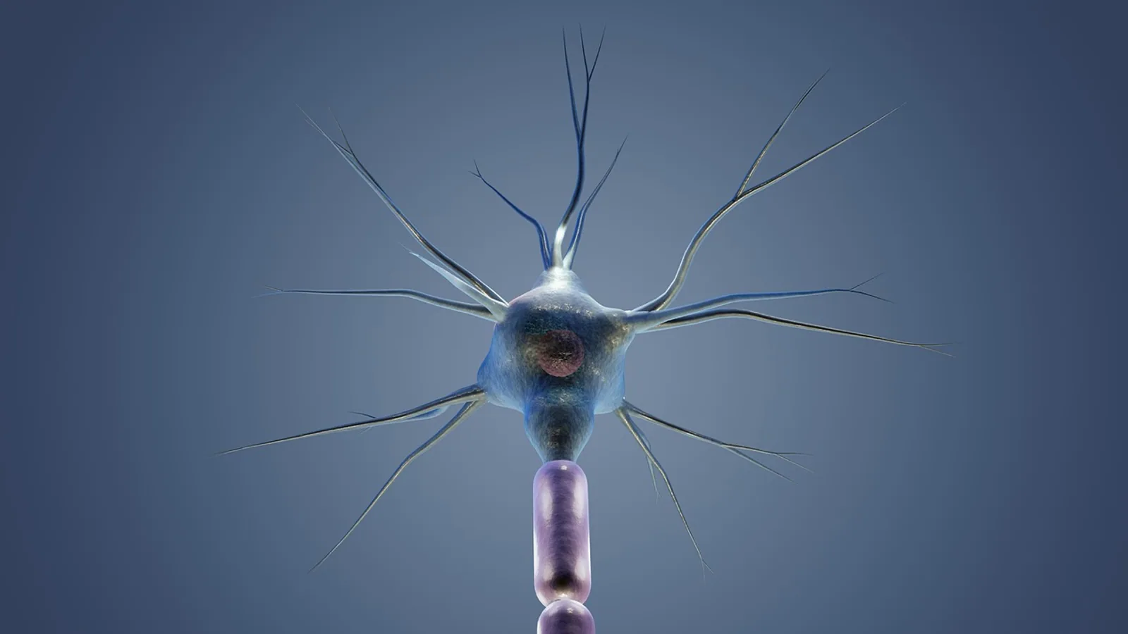
Afferent nerves carry nerve impulses toward the central nervous system, and efferent nerves carry nerve impulses away from it. The central nervous system (CNS) is the brain and spinal cord, and every peripheral nerve fiber in the body can be sorted by which side of that boundary it points toward.
That single rule decides how you read every nerve in a dissection lab, every reflex arc on an exam, and every mixed nerve you will ever trace in a dog, horse, or cat. Get the direction right and the rest of neuroanatomy falls into place. Get it backwards and you will mislabel roots, misread lesions, and miss why vagus nerve stimulation does two different things depending on which fibers you activate.
The Direction Rule, Stated Plainly
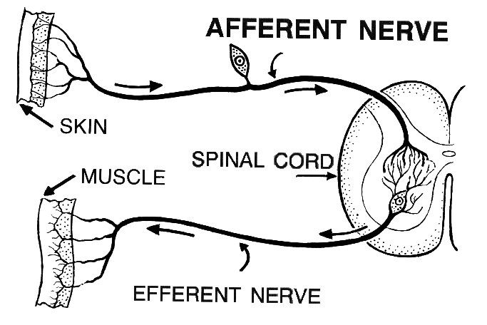
.jpg)
Afferent means conducting toward a center. Efferent means conducting away from a center. In neuroanatomy the center is the CNS, so afferent fibers travel toward the brain or spinal cord and efferent fibers travel away from it.
This is a direction rule, not a sensory versus motor rule. That distinction matters more than most students expect. Most afferent fibers are sensory, but not all. Most efferent fibers are motor, but not all. The autonomic nervous system contains afferent fibers that report on the state of internal organs, and it contains efferent fibers that are autonomic rather than somatic. A fiber that carries information about blood pressure from the carotid sinus toward the brainstem is afferent even though nobody would call it a classic "sensory" pathway in the way they describe touch or pain. A fiber that carries a command to a sweat gland or to the smooth muscle of the gut is efferent even though it is not a somatic motor neuron.
The mnemonic that keeps this straight is simple. A for Arriving. E for Exiting. Afferent arrives at the CNS. Efferent exits the CNS.
Why This Matters in Veterinary Practice
Every neurologic examination you perform on an animal is, at its core, a test of afferent and efferent function. When you tap the patellar tendon and watch the quadriceps contract, you are testing an afferent limb (the stretch receptors and their fibers carrying the signal to the spinal cord) and an efferent limb (the motor fibers carrying the command back to the muscle). When you check a pupillary light reflex, you are testing an afferent limb through the optic nerve and an efferent limb through the oculomotor nerve. When you assess whether a horse can swallow, you are testing a reflex that depends on afferent input from the pharynx and efferent output through the glossopharyngeal and vagus nerves.
Localizing a lesion depends on knowing which side of the reflex is broken. A dog that cannot feel a pinprick but can still move the limb has an afferent problem. A dog that can feel the pinprick but cannot move the limb has an efferent problem. The same logic extends into the autonomic nervous system, where afferent and efferent vagal fibers run in the same nerve trunk but do entirely different jobs [1][2].
The Spinal Cord: Dorsal Root Versus Ventral Root
The clearest place to see the afferent-efferent split is the spinal cord, because the two fiber populations are physically separated at the level of the roots.
Dorsal Root
The dorsal root is the afferent root. It enters the dorsal (upper) surface of the spinal cord. Its cell bodies sit in the dorsal root ganglion, a swelling on the dorsal root just outside the cord. These are pseudounipolar neurons: one process leaves the cell body and splits into a peripheral branch that reaches the receptor and a central branch that enters the spinal cord. The dorsal root carries afferent fibers only.
Ventral Root
The ventral root is the efferent root. It leaves the ventral (lower) surface of the spinal cord. Its fibers arise from motor neurons whose cell bodies sit in the ventral horn of the gray matter. The ventral root carries efferent fibers, both somatic motor fibers to skeletal muscle and autonomic efferent fibers that will synapse in a peripheral ganglion before reaching their target. The ventral root carries efferent fibers only.
Why the Roots Are Separated
This separation exists because of how the spinal cord develops. The dorsal portion of the neural tube becomes specialized for receiving input, and the ventral portion becomes specialized for sending output. That developmental logic is preserved in the adult animal, and it is why a lesion that destroys the dorsal root produces loss of sensation in a specific area while a lesion that destroys the ventral root produces weakness or paralysis in a specific muscle group.
The brainstem does not preserve this clean separation. According to a 2026 review of brainstem anatomy, the motor and sensory nuclei of cranial nerves III through XII are arranged in a mediolateral direction, and unlike the spinal cord, their afferent and efferent fibers are not macroscopically separated [3]. That is one reason cranial nerve anatomy feels harder than spinal cord anatomy. The physical landmark that makes the dorsal and ventral roots so easy to tell apart simply is not there.
Summary Table: Afferent Versus Efferent
| Term | Direction | Root | Typical fiber type | Example |
|---|---|---|---|---|
| Afferent | Toward the CNS | Dorsal root | Somatic sensory, visceral sensory | Stretch receptor fiber from the quadriceps reporting muscle length to the spinal cord |
| Efferent | Away from the CNS | Ventral root | Somatic motor, autonomic motor | Motor fiber from the ventral horn telling the quadriceps to contract |
| Afferent (autonomic) | Toward the CNS | Dorsal root or cranial nerve | Visceral sensory | Vagal afferent reporting gut distension to the brainstem [4] |
| Efferent (autonomic) | Away from the CNS | Ventral root or cranial nerve | Preganglionic and postganglionic autonomic | Vagal efferent carrying parasympathetic output to the heart or gut [1][2] |
The Flow of Information, Step by Step
The simplest way to understand afferent and efferent function is to follow one signal through a reflex arc.
- A receptor detects a change. A stretch receptor in a muscle, a thermoreceptor in the skin, or a mechanoreceptor in the gut wall fires.
- An afferent fiber carries the signal toward the CNS. The fiber travels through a peripheral nerve, enters the dorsal root, and reaches the spinal cord or brainstem.
- The CNS processes the signal. This may happen in the spinal cord gray matter for a simple reflex, or it may involve the brainstem, thalamus, and cortex for a conscious sensation.
- An efferent fiber carries the response away from the CNS. The fiber leaves through the ventral root or a cranial nerve and travels to a muscle, gland, or organ.
- The target responds. A skeletal muscle contracts, a gland secretes, or smooth muscle changes its tone.
That five-step sequence is the skeleton of every reflex in the body, from the withdrawal reflex in a dog's paw to the vago-vagal reflexes that control heart rate and digestion. In a vago-vagal reflex, both the afferent and efferent limbs travel in the vagus nerve, which is why the vagus is such a useful teaching example [2].
A Note on the Word "Effect"
Students sometimes confuse efferent with the everyday word effect. The two share a Latin root, but they are not the same idea. Efferent describes a direction of travel, not a consequence. An efferent fiber is not "the fiber that causes an effect." It is the fiber that exits the CNS. An afferent fiber can produce effects too, because the signals it delivers to the CNS trigger responses. The direction is what defines the term.
A related trap is the word "sensory." Sensory is a functional category, and afferent is a directional category. They overlap heavily but they are not interchangeable. The vagus nerve in most species of animals predominantly comprises afferent fibers [1], and many of those afferent fibers are visceral sensory fibers that never produce a conscious sensation. They still count as afferent because they carry impulses toward the CNS.
Cranial Nerves: Mixed Nerves and the Vagus
Cranial nerves complicate the clean dorsal root and ventral root picture because several of them carry both afferent and efferent fibers in the same trunk. These are called mixed nerves.
The vagus nerve (cranial nerve X) is the classic example. It carries afferent fibers from the pharynx, larynx, heart, lungs, and abdominal organs toward the brainstem. It carries efferent fibers from the brainstem to the same organs, providing parasympathetic control of heart rate, gut motility, and secretion. A 2026 review of vagus nerve stimulation notes that afferent and efferent vagal fibers modulate immune responses, mood regulation, and neurotransmitter systems [5]. The same nerve trunk does both jobs.
The auricular branch of the vagus is a useful contrast. According to a 2026 study comparing cervical and auricular vagus nerve stimulation, the cervical vagus nerve contains both afferent and efferent fibers, whereas the auricular branch is a purely afferent nerve [6]. That anatomical difference explains why stimulating the two branches produces different physiological responses. The cervical vagus can drive efferent parasympathetic output directly. The auricular branch can only send afferent signals that the brainstem must then process.
Other mixed cranial nerves include the trigeminal (V), facial (VII), glossopharyngeal (IX), and the spinal accessory (XI) has a motor component. The optic nerve (II) is purely afferent. The oculomotor (III), trochlear (IV), abducens (VI), and hypoglossal (XII) are primarily efferent. Knowing which cranial nerves are mixed and which are pure is a standard part of veterinary neuroanatomy, and it matters for lesion localization.
How Afferent and Efferent Fibers Are Studied
Researchers distinguish afferent from efferent fibers using several methods, and understanding these methods helps you read the primary literature.
Tract Tracing
Anterograde and retrograde tracers are injected into a brain region or nerve. A retrograde tracer is taken up by axon terminals and carried back to the cell body, labeling the neurons that project to the injection site. An anterograde tracer is taken up by cell bodies and carried forward along axons, labeling the terminals. By combining both, researchers can map which regions send afferents to a structure and which regions receive its efferents. This approach has been used extensively to map olfactory, visual, and auditory pathways in birds such as the pigeon [7][8][9][10][11][12].
Nerve Stimulation
Electrical or optogenetic stimulation can activate afferent or efferent fibers selectively. A 2026 study used channelrhodopsin-assisted circuit mapping to confirm that some vagal afferent fibers synapse directly onto dorsal motor nucleus of the vagus motor neurons, bypassing the nucleus of the solitary tract [2]. That finding challenges the textbook model of vago-vagal reflexes, which assumed the nucleus of the solitary tract was always the relay point.
Selective Denervation
Surgical transection of a specific nerve branch can isolate the contribution of afferent versus efferent fibers. A 2025 study in mice transected specific vagal branches to the gastrointestinal tract and then tested whether capsaicin could still activate afferent responses [4]. When the branches were cut, the afferent responses disappeared, confirming that those branches carried the relevant afferent fibers.
Pharmacological Dissection
Capsaicin is a standard tool for activating vagal afferents selectively. A 2025 study used capsaicin injections into the stomach, duodenum, and portal vein to identify which vagal branches carry afferent fibers from each organ [4]. This kind of experiment lets researchers map afferent pathways without cutting anything.
Comparative Notes Across Species
The afferent-efferent principle is universal across vertebrates, but the anatomy varies in ways that matter for veterinary practice.
In mammals, the dorsal root ganglion is a discrete swelling on the dorsal root, and the ventral root is a clean bundle of efferent fibers. This basic plan holds in dogs, cats, horses, cattle, pigs, and sheep. The pig has been used as a model for studying primary sensory neurons supplying the vas deferens, and those neurons were found mainly in the lumbar L2 and L3 and sacral S2 and S3 dorsal root ganglia, with a clear ipsilateral projection pattern [13]. That study also showed that most of those afferent neurons contained substance P or calcitonin gene-related peptide, which are markers of specific sensory fiber types.
In birds, the same afferent-efferent logic applies, but the brainstem and forebrain organization differs. Pigeon studies have mapped afferent and efferent connections of the olfactory bulb, the mesopallium, the nucleus rotundus, and other regions [7][8][9][10][11][12]. These studies use the same terminology because the direction rule is conserved. An afferent projection to the olfactory bulb is still an afferent projection, even though the bird brain is organized differently from the mammalian brain.
The vagus nerve varies in its afferent-to-efferent ratio across species. The vagus nerve in most species of animals predominantly comprises afferent fibers [1]. That ratio matters for interpreting vagus nerve stimulation studies, because stimulating the nerve will activate far more afferent fibers than efferent fibers in most species. The therapeutic effects of vagus nerve stimulation therefore depend heavily on which fiber population is being recruited, and a 2026 review notes that afferent and efferent vagal fibers modulate immune responses, mood, and neurotransmitter systems through distinct mechanisms [5].
Clinical Relevance, Limitations and Common Mistakes
Clinical Relevance
Lesion localization depends on the afferent-efferent distinction. A spinal cord lesion that destroys the dorsal columns will impair proprioception and fine touch while sparing motor function. A lesion that destroys the ventral horn or ventral root will cause weakness or paralysis while sparing sensation. A lesion that destroys both will cause mixed deficits.
Autonomic reflexes depend on both limbs. The baroreceptor reflex, which controls blood pressure, uses afferent fibers from the carotid sinus and aortic arch and efferent fibers through the vagus and sympathetic nerves. A 2026 study on electroacupuncture at PC6 in mice with myocardial infarction found that the cardioprotective effect was mediated through a vagal afferent to sympathetic efferent pathway, not through the classical vagal efferent anti-inflammatory pathway [14]. That finding shows how the afferent and efferent limbs of an autonomic reflex can use different nerves and different neurotransmitters.
Vagus nerve stimulation is a clinical tool that depends on understanding which fibers are being activated. A 2021 study showed that inhibition of inflammation by efferent vagal nerve stimulation requires T-cell derived acetylcholine, whereas inhibition by afferent vagal nerve stimulation does not [1]. The two stimulation modes work through different neuroimmune circuits. A 2026 review of vagus nerve stimulation notes that despite progress, clinical results remain mixed, and next-generation closed-loop devices and circuit-specific targeting may improve efficacy [5].
Limitations
Individual animals vary in their response to nerve injury, and the same lesion can produce different deficits depending on the species, the age of the animal, and the presence of concurrent disease. The studies cited here were performed in mice, rats, pigs, pigeons, chicks, and humans, and the findings may not translate directly to every domestic species. A veterinarian who examines your animal can assess the specific situation.
Common Mistakes
Mistake 1: Treating afferent as a synonym for sensory. Afferent is a direction. Sensory is a function. Autonomic afferents are afferent but not consciously sensory.
Mistake 2: Treating efferent as a synonym for motor. Efferent is a direction. Motor is a function. Autonomic efferents are efferent but not somatic motor.
Mistake 3: Confusing efferent with effect. The words share a root but mean different things. Efferent describes direction of travel.
Mistake 4: Assuming the dorsal root carries all afferents and the ventral root carries all efferents in every part of the nervous system. That is true in the spinal cord, but cranial nerves mix afferent and efferent fibers in the same trunk. The brainstem does not separate them macroscopically [3].
Mistake 5: Forgetting that the vagus carries both. The vagus is not a sensory nerve or a motor nerve. It is a mixed nerve with both afferent and efferent fibers, and the afferent fibers predominate in most species [1].
Mistake 6: Assuming all vagal reflexes route through the nucleus of the solitary tract. A 2026 study found that some vagal afferents synapse directly onto dorsal motor nucleus of the vagus motor neurons, bypassing the nucleus of the solitary tract [2].
Quick Review
- Afferent carries impulses toward the CNS. Efferent carries impulses away from the CNS.
- A for Arriving. E for Exiting.
- The dorsal root is afferent. The ventral root is efferent.
- Afferent and efferent are direction terms, not sensory and motor terms.
- Autonomic afferents exist, and autonomic efferents exist.
- Cranial nerves can be mixed. The vagus carries both afferent and efferent fibers.
- Efferent is not the same word as effect.
Frequently Asked Questions
Which nerves carry impulses toward the CNS only?
The dorsal root of the spinal cord carries afferent fibers only, and the optic nerve is a purely afferent cranial nerve. The auricular branch of the vagus is also purely afferent [6].
Is afferent sensory or motor?
Afferent is neither. Afferent is a direction term meaning toward the CNS. Most afferent fibers are sensory, but autonomic afferents exist and are not consciously sensory.
What is the difference between afferent and efferent neurons?
Afferent neurons carry signals toward the CNS, and efferent neurons carry signals away from it. The direction of travel is the defining difference.
Which root is efferent?
The ventral root is efferent. Its fibers arise from motor neurons in the ventral horn and carry commands away from the spinal cord.
Is the vagus nerve afferent or efferent?
Both. The vagus nerve is a mixed nerve that carries afferent fibers toward the brainstem and efferent fibers away from it. In most species, afferent fibers predominate [1].
What is the mnemonic for afferent and efferent?
A for Arriving at the CNS. E for Exiting the CNS.
This article is educational and is not a substitute for veterinary diagnosis or treatment.
flowchart TD
    A[Stimulus detected] --> B[Afferent fiber]
    B --> C[Dorsal root]
    C --> D[Spinal cord or brainstem]
    D --> E{Processing}
    E --> F[Efferent fiber]
    F --> G[Ventral root]
    G --> H[Muscle gland or organ]
    H --> I[Response]
Related Articles
- Can Dogs Eat Nutmeg? Myristicin CNS Toxicity
- Cat Aggression Towards People: Why and What to Do
- Why Is My Dog Suddenly Aggressive Towards Other Dogs?
- Why Is My Cat Suddenly Aggressive Towards Other Cats?
- Dog Reverse Sneezing Only At Night
- Dog Licking Paws Only At Night
- Cranial Nerve Mnemonic: Names, Numbers, and Functions
Sources
- Divergence of neuroimmune circuits activated by afferent and efferent vagal nerve stimulation in the regulation of inflammation.
- Vagal sensory neurones directly synapse onto parasympathetic motor (DMV) neurones.
- Structural and functional anatomy of the brainstem.
- Functional identification of vagal afferent branch from the gastrointestinal organs by capsaicin administration in mice.
- A Possible Role for the Vagus Nerve in Physical and Mental Health.
- Acute transcutaneous cervical but not auricular vagus nerve stimulation increases alpha wave brain activity and lowers arterial blood pressure.
- Efferent and afferent connections of the olfactory bulb and prepiriform cortex in the pigeon (Columba livia).
- Afferent and efferent projections of the central caudal nidopallium in the pigeon (Columba livia).
- Afferent and efferent connections of the dorsolateral corticoid area and a comparison with connections of the temporo-parieto-occipital area in the pigeon (Columba livia).
- Afferent and efferent projections of the mesopallium in the pigeon (Columba livia).
- Afferent and efferent connections of the nucleus geniculatus lateralis ventralis demonstrated by WGA-HRP in the chick.
- Afferent and efferent connections of the nucleus rotundus demonstrated by WGA-HRP in the chick.
- Distribution, immunohistochemical characteristics and nerve pathways of primary sensory neurons supplying the porcine vas deferens.
- Electroacupuncture at PC6 mediates cardioprotection by vagal afferent-sympathetic efferent anti-inflammatory pathway in myocardial infarction mouse.