Adrenal Cortex: Zones, Hormones and Function
By Dr. Zubair Khalid, DVM, MS, PhD ·
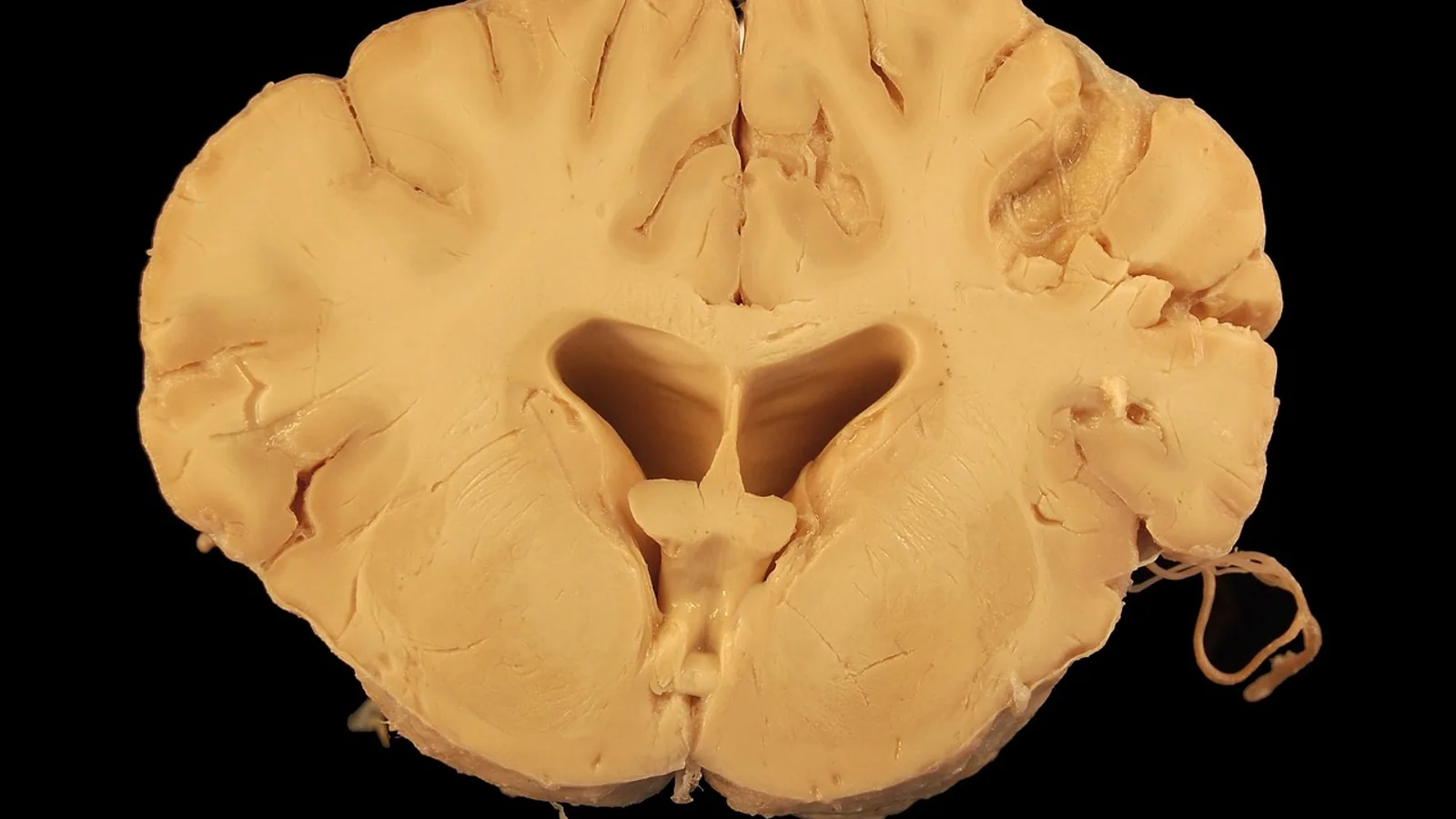
The adrenal cortex is the outer, steroid-producing portion of the adrenal gland, organized into three concentric zones that each manufacture a different class of hormone: mineralocorticoids in the zona glomerulosa, glucocorticoids in the zona fasciculata, and adrenal androgens in the zona reticularis [1][2]. It sits on a separate regulatory track from the adrenal medulla, which produces catecholamines rather than steroids.
This article is educational and is not a substitute for veterinary diagnosis or treatment.
Why the Adrenal Cortex Matters
The adrenal cortex keeps animals alive under two very different kinds of pressure. Aldosterone from the zona glomerulosa defends sodium and potassium balance and therefore blood volume and blood pressure. Cortisol from the zona fasciculata defends glucose supply and modulates the immune and stress response. Adrenal androgens from the zona reticularis contribute to sexual maturation and are increasingly recognized as a marker of adrenal reserve in some species [3][4].
When a cortical zone fails, the clinical picture depends entirely on which zone is lost. A dog that loses the zona glomerulosa develops the classic electrolyte crisis of hypoadrenocorticism. A dog that loses only the zona fasciculata and reticularis can present with lethargy and low albumin but normal electrolytes, a form called atypical hypoadrenocorticism [5]. The zonation of the cortex is therefore not an academic detail. It is the map that explains the bloodwork.
Where Are the Adrenal Glands Located?
The adrenal glands are paired retroperitoneal organs. In most domestic mammals they sit cranial and medial to the corresponding kidney, embedded in the perirenal fat and often partially capped by the renal fascia [2]. In the dog, the right adrenal lies close to the caudal vena cava and the phrenicoabdominal vein, while the left adrenal sits slightly more cranial and lateral. This relationship matters in surgery because the right adrenal is the more dangerous one to approach.
Birds break the rule. The avian adrenal gland is not a single capped organ but a flattened strip of interrenal and chromaffin tissue embedded in the cranial pole of the kidney, running along the caudal vena cava. There is no discrete cortex and medulla the way there is in mammals. The steroidogenic cells (interrenal tissue) and the catecholamine-producing cells (chromaffin tissue) are intermingled. This is why avian adrenal disease does not present with the neat cortical-zone pattern seen in dogs and cats.
Comparative location summary:
- Dog and cat: paired, cranial-medial to each kidney, retroperitoneal.
- Horse and ruminant: similar retroperitoneal position, but the right gland is more firmly attached to the vena cava.
- Pig: left adrenal is elongated and the right is more triangular.
- Bird: interrenal and chromaffin tissue embedded in the cranial kidney, no separate cortex.
Adrenal Gland Histology: The Three Zones
Adrenal histology depends on recognizing three concentric layers around a central medulla [6][7]. From outside in:
- Capsule. A thin connective tissue layer with a mesenchymal cell population that contributes progenitor cells to the cortex [8].
- Zona glomerulosa (zG). The outermost cortical zone, arranged in small rounded clusters or arcs. It produces aldosterone.
- Zona fasciculata (zF). The thickest zone, made of large polyhedral cells in straight cords radiating toward the medulla. It produces cortisol (corticosterone in rodents).
- Zona reticularis (zR). The innermost zone, with irregular anastomosing cords. It produces adrenal androgens such as DHEA and androstenedione.
- Medulla. Neural crest-derived chromaffin cells that secrete catecholamines under sympathetic control [2].
The zone boundaries are not fixed walls. The cortex renews itself continuously from progenitor cells in the outer layers, and newly formed cells migrate inward, differentiating as they go [9][10]. This centripetal migration means the zona reticularis is the oldest cell population in the cortex. That has a practical consequence: in prolonged critical illness, the zona reticularis shows accelerated telomere shortening, consistent with the wear of a long-lived cell population under sustained demand [11].
How Zone-Enriched Cultures Are Made
Researchers can now separate the zones for study. A microdissection approach in mice produces an outer fraction enriched for capsule and zona glomerulosa cells and an inner fraction enriched for zona fasciculata and medullary cells. The outer fraction expresses zona glomerulosa markers such as Cyp11b2, Dab2 and Shh along with Wnt/beta-catenin pathway markers. The inner fraction expresses zona fasciculata markers such as Cyp11b1 and Akr1b7 [8]. This kind of zone-resolved work is how the field confirms that each zone is functionally distinct rather than a gradient.
Zone by Zone: Hormone Output and Regulation
Zona Glomerulosa: Aldosterone
The zona glomerulosa makes aldosterone, the most potent mineralocorticoid produced by the adrenal cortex [1]. Aldosterone is not stored in vesicles. Because steroid hormones are highly lipophilic, they are synthesized and secreted immediately on stimulation rather than packaged for later release [1].
Two stimuli drive aldosterone production:
- Angiotensin II (AngII). A peptide hormone that cannot cross cell membranes, so it acts through G protein-coupled receptors, mainly the AngII type 1 receptor (AT1R). In zona glomerulosa cells, AT1R activation triggers aldosterone synthesis through two pathways: a Gq/11 pathway that activates phospholipase C and raises intracellular free calcium, and a beta-arrestin1/2 pathway that produces sustained ERK activation. Both pathways upregulate and acutely activate the steroidogenic acute regulatory (StAR) protein, which moves cholesterol into mitochondria [1].
- Potassium. Elevated blood potassium (hyperkalemia) directly stimulates aldosterone secretion from zona glomerulosa cells [1].
The practical takeaway is that aldosterone is controlled by the renin-angiotensin-aldosterone system and by potassium, not by ACTH. The zona glomerulosa is largely ACTH-independent.
Zona Fasciculata: Cortisol
The zona fasciculata makes glucocorticoids, principally cortisol in dogs, cats, horses and primates, and corticosterone in rodents [8]. Its dominant regulator is adrenocorticotropic hormone (ACTH) from the pituitary. ACTH binds melanocortin 2 receptors (MC2R) on zona fasciculata cells, and MC2R requires the accessory protein MRAP for function. Spatial transcriptomic work in mice shows that Mc2r and Mrap have discordant spatial distributions with limited co-expression, which suggests MRAP may have roles beyond simply supporting MC2R [12].
Cortisol is the main glucocorticoid in most domestic mammals. It raises blood glucose, supports vascular responsiveness to catecholamines, and suppresses inflammation at pharmacologic doses. Because the zona fasciculata is ACTH-dependent, its output falls when the pituitary fails.
Zona Reticularis: Adrenal Androgens
The zona reticularis produces C19 steroids, chiefly dehydroepiandrosterone (DHEA) and androstenedione [1][3]. These are weak androgens that can be converted peripherally to more potent sex steroids. In humans, the zona reticularis matures at adrenarche, well after the zona glomerulosa and fasciculata are functional [4]. In rodents and many other mammals, the equivalent androgen-producing region is less clearly demarcated, and a transient X-zone occupies the inner cortex in some species [13][12].
The zona reticularis is also ACTH-responsive, but its output is modulated by sex hormones. In the Mongolian gerbil, castration increases the zona fasciculata and causes cell hypertrophy across all zones, while testosterone supplementation increases cell proliferation and cell death and shifts steroidogenic enzyme expression [14]. In the Libyan jird, females have thicker cortices than males, mainly from a larger zona fasciculata, and higher plasma cortisol [15]. These findings confirm that the adrenal cortex is a sexually dimorphic organ in multiple species [9][13].
Summary Table: Zone, Hormone, Regulator, Deficiency
| Zone | Main hormone | Primary regulator | Clinical deficiency state |
|---|---|---|---|
| Zona glomerulosa | Aldosterone | Angiotensin II, potassium | Hyponatremia, hyperkalemia, hypovolemia, collapse (classic Addison) |
| Zona fasciculata | Cortisol | ACTH (via MC2R/MRAP) | Weakness, hypoglycemia, poor stress tolerance, low albumin (atypical hypoadrenocorticism) |
| Zona reticularis | DHEA, androstenedione | ACTH plus sex hormone modulation | Subtle, rarely the sole cause of clinical signs |
| Medulla (for contrast) | Epinephrine, norepinephrine | Sympathetic nervous system | Not part of cortical insufficiency |
Primary vs Secondary Adrenal Insufficiency in Dogs
Adrenal insufficiency is the failure of the cortex to produce adequate steroid hormones. In dogs it comes in two major forms, and the distinction is one of the most commonly tested points in veterinary endocrinology.
Primary Adrenal Insufficiency (Addison Disease)
Primary hypoadrenocorticism is destruction of the adrenal cortex itself. Because the whole cortex is affected, all three zones lose function. The zona glomerulosa is lost along with the others, so aldosterone falls. The result is the classic electrolyte pattern: low sodium, high potassium, and a sodium-to-potassium ratio that drops below the reference range. Dogs present with lethargy, vomiting, diarrhea, weakness, bradycardia, and sometimes collapse.
The ACTH stimulation test is the diagnostic test of choice. In primary disease, the cortisol response to exogenous ACTH is blunted or absent.
Secondary Adrenal Insufficiency (ACTH Deficiency)
Secondary hypoadrenocorticism is failure of the pituitary to produce enough ACTH. The zona glomerulosa does not depend on ACTH, so aldosterone production is preserved and electrolytes stay normal. Only the ACTH-dependent zones, the fasciculata and reticularis, are affected. This is why secondary disease is often called atypical hypoadrenocorticism.
A case report of an 8-year-old pointer illustrates the pattern. The dog had lethargy and hypoalbuminemia, both adrenals were reduced in thickness on ultrasound, and the ACTH stimulation test was abnormal, but electrolytes were normal. After low-dose prednisolone, the dog improved and bloodwork normalized. At autopsy 818 days later, the zonae fasciculata and reticularis were disrupted while the zona glomerulosa remained relatively normal [5]. That case is a clean demonstration that the zona glomerulosa can survive when ACTH is the problem.
Why the Distinction Matters
The distinction guides treatment logic. Primary disease needs both glucocorticoid and mineralocorticoid replacement. Secondary disease needs glucocorticoid replacement alone, because aldosterone is intact. It also guides monitoring. A dog with primary disease can crash from an electrolyte crisis, while a dog with secondary disease is more likely to show vague, chronic signs.
This section deliberately avoids specific drug names and doses. Those decisions belong to the attending veterinarian and depend on the individual patient.
How Adrenal Function Is Tested and Observed
ACTH Stimulation Test
The ACTH stimulation test measures the cortisol response of the zona fasciculata to a synthetic ACTH analog. A normal dog mounts a robust cortisol rise. A dog with primary or secondary hypoadrenocorticism does not. The test is the standard for confirming adrenal insufficiency.
Baseline Cortisol and Electrolytes
A single baseline cortisol is not diagnostic on its own, but a very low value combined with a typical history raises suspicion. Electrolytes separate primary from secondary disease. Normal electrolytes with a failed ACTH stimulation test point to secondary disease [5].
Imaging
Abdominal ultrasound can show reduced adrenal thickness in hypoadrenocorticism [5]. Adrenal size is not a substitute for endocrine testing, but it supports the diagnosis and helps rule out masses.
Histopathology and Immunohistochemistry
Post-mortem adrenal histology shows the zonal pattern directly. In atypical hypoadrenocorticism, the fasciculata and reticularis are disrupted while the glomerulosa is preserved [5]. Immunohistochemistry for zone-specific enzymes such as CYP11B2 (aldosterone synthase) and CYP11B1 (11-beta-hydroxylase) can map functional zones, and this approach has shown that the zona glomerulosa area declines with age in men while the zona fasciculata area increases [16].
Research Models
Transgenic mouse models have been central to understanding how the cortex develops and maintains its zones. Conditional and total knockout strains have revealed the molecular mechanisms controlling zonation and renewal [17][18]. These models also show that adrenal renewal is sex-specific, with testicular androgens inhibiting progenitor cell recruitment [9].
Comparative Notes Across Species
- Dog. Cortisol is the main glucocorticoid. Primary hypoadrenocorticism is well described and often immune-mediated. The right adrenal sits close to the caudal vena cava.
- Cat. Similar cortical architecture. Adrenal disease in cats is more often hyperplastic or neoplastic than primarily deficient.
- Horse. The adrenal cortex produces cortisol and aldosterone in the same zonal pattern. The right adrenal is closely associated with the vena cava.
- Rodent. Corticosterone, not cortisol, is the main glucocorticoid. The zona glomerulosa expresses Cyp11b2, and the zona fasciculata expresses Cyp11b1 [8]. Mice also have a transient X-zone that is prominent in females and nearly absent in males [12].
- Mongolian gerbil. The adrenal morphology resembles that of primates, making it a useful endocrine model. It produces DHEA and steroidogenic enzymes similar to those in humans, and aging causes hypertrophy of the fasciculata and reticularis zones along with lipofuscin accumulation [3].
- Bird. No discrete cortex. Interrenal and chromaffin tissue are intermingled in the cranial kidney. Steroidogenesis still occurs, but the mammalian zone model does not apply cleanly.
Common Misconceptions Students Hold
Misconception 1: The whole adrenal cortex is controlled by ACTH. Only the zona fasciculata and reticularis are ACTH-dependent. The zona glomerulosa is controlled by angiotensin II and potassium [1].
Misconception 2: Adrenal insufficiency always causes electrolyte abnormalities. Secondary hypoadrenocorticism spares the zona glomerulosa, so electrolytes can be normal [5].
Misconception 3: Steroid hormones are stored in granules like peptide hormones. They are not. Steroids are synthesized on demand and secreted immediately because they are lipophilic [1].
Misconception 4: The adrenal medulla and cortex are functionally linked. They share a blood supply and anatomical proximity, but they are embryologically distinct and regulated by different systems. The medulla is neural crest-derived and sympathetic. The cortex is mesodermal and endocrine [2].
Misconception 5: The zona reticularis is the main source of sex steroids in all species. In humans, adrenal androgens are significant. In many domestic species, the gonads are the dominant source, and the zona reticularis contributes a smaller share.
Misconception 6: Adrenal size on ultrasound equals adrenal function. Size can be normal in early disease and reduced in chronic disease. Endocrine testing, not imaging alone, establishes the diagnosis [5].
Clinical Relevance, Limitations and Common Mistakes
The zona map is the fastest way to predict what a failing adrenal gland will do. If the glomerulosa fails, expect an electrolyte crisis. If only the ACTH-dependent zones fail, expect vague chronic signs with normal electrolytes. If the whole gland is destroyed by a tumor or immune process, expect a mixed picture.
Common mistakes in practice include:
- Ordering an ACTH stimulation test but not checking electrolytes, which makes it impossible to separate primary from secondary disease.
- Assuming a normal sodium-to-potassium ratio rules out hypoadrenocorticism. Atypical cases exist [5].
- Forgetting that the zona glomerulosa is not ACTH-dependent, which leads to incorrect predictions about which hormones will fall.
- Treating a dog with glucocorticoid alone when the zona glomerulosa is also lost, which leaves the mineralocorticoid deficit uncorrected.
- Overinterpreting adrenal size on ultrasound without endocrine confirmation.
Limitations of the zonal model exist. Zone boundaries are not always sharp on histology, especially in rodents and birds. The zona reticularis is difficult to isolate cleanly, and its contribution to circulating androgens varies widely by species and age [16]. Individual cases always need a veterinarian who can integrate history, examination, laboratory testing and imaging.
Quick Review
- The adrenal cortex has three zones: glomerulosa (aldosterone), fasciculata (cortisol), reticularis (androgens) [1][2].
- The zona glomerulosa is regulated by angiotensin II and potassium, not ACTH [1].
- The zona fasciculata and reticularis are ACTH-dependent.
- Steroid hormones are synthesized on demand and not stored in vesicles [1].
- Primary adrenal insufficiency destroys all zones and causes electrolyte abnormalities. Secondary ACTH deficiency spares the zona glomerulosa and leaves electrolytes normal [5].
- The adrenal cortex renews from outer progenitor cells that migrate inward and differentiate [9][10].
- The adrenal cortex is sexually dimorphic across species [9][13][12].
Frequently Asked Questions
Where is the adrenal gland located in a dog?
The adrenal glands are paired retroperitoneal organs sitting cranial and medial to each kidney. The right gland lies close to the caudal vena cava [2].
What are the three zones of the adrenal cortex?
The zona glomerulosa, zona fasciculata and zona reticularis. Each produces a different class of steroid hormone [1][2].
What hormone does the zona glomerulosa make?
Aldosterone, the main mineralocorticoid. It is controlled by angiotensin II and potassium [1].
What is the difference between primary and secondary adrenal insufficiency?
Primary disease destroys the whole cortex and causes electrolyte abnormalities. Secondary disease is ACTH deficiency, spares the zona glomerulosa, and leaves electrolytes normal [5].
Why do steroids get secreted immediately instead of being stored?
Steroid hormones are lipophilic and cannot be packaged in vesicles. They are synthesized and released on stimulation [1].
Does the adrenal cortex change with age?
Yes. In humans, the zona glomerulosa area declines with age while the zona fasciculata area increases [16]. Similar age-related changes occur in animal models [3].
Related Articles
- Ferret Adrenal Gland Disease: Diagnosis and Medical Management
- Canine Adrenal Gland Disease: Hyperadrenocorticism and Hypoadrenocorticism
- Adrenal disease in ferrets
- Ferret Adrenal Disease Signs
- Reproductive Hormone Testing in Mares
- Dog Potty Zone
- Antidiuretic Hormone: Function and Regulation
Sources
- Angiotensin II-dependent aldosterone production in the adrenal cortex.
- Adrenal Anatomy and Physiology.
- Aging effects in adrenal cortex of male Mongolian gerbil: A model for endocrine studies.
- Adrenal cortex development and related disorders leading to adrenal insufficiency.
- Atypical hypoadrenocorticism with intact zona glomerulosa of the adrenal cortex after long-term observation: a case report of a dog.
- Pathology Outlines - Anatomy & histology-adrenal cortex, medulla & paraganglia
- Histology of the Adrenal Glands
- Establishment of Zone-Enriched Primary Cultures from the Mouse Adrenal Cortex.
- Adrenal cortex renewal in health and disease.
- Regulation of stem and progenitor cells in the adrenal cortex.
- Accelerated telomere shortening in adrenal zona reticularis in patients with prolonged critical illness.
- Single-cell spatial transcriptomic atlas of the mouse adrenal gland reveals sexual dimorphism in steroidogenic enzyme and hormone receptor expression.
- RNA-Seq Reveals Sub-Zones in Mouse Adrenal Zona Fasciculata and the Sexually Dimorphic Responses to Thyroid Hormone.
- Effects of androgenic modulation on the morphophysiology of the adrenal cortex of male gerbils.
- Sex differences in adrenal cortex beta-catenin immunolocalisation of the Saharan gerbil, Libyan jird (Meriones libycus, Lichtenstein, 1823).
- The Age-Dependent Changes of the Human Adrenal Cortical Zones Are Not Congruent.
- Transgenic Mouse Models to Study the Development and Maintenance of the Adrenal Cortex.
- Adrenal Cortex Development and Maintenance: Knowledge Acquired From Mouse Models.