Adductor Femoris: Anatomy and Function
By Dr. Zubair Khalid, DVM, MS, PhD ·
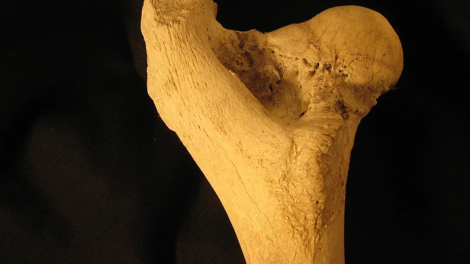
The adductor femoris is a large muscle of the medial thigh that arises from the ventral pelvis and inserts on the caudal surface of the femur, pulling the limb toward the midline and helping to stabilize the hip. In the dog it is a single muscle, homologous to the human adductor magnus, while the human medial thigh contains three separate adductors (longus, brevis, and magnus) that veterinary students must not map one-to-one onto the canine limb.
This distinction matters because the medial thigh is where comparative anatomy, clinical neurology, and surgery intersect. The adductor femoris sits directly over the obturator nerve, it is one of the muscles that keeps the hind limb under the body during the stance phase, and it is a recognized source of lameness and myofascial pain in dogs [1]. A student who confuses the canine adductor femoris with the human adductor longus will misread radiographs, misplace nerve blocks, and misdescribe the muscle in a surgical report. Getting the attachments, the innervation, and the species differences straight is the foundation for everything else.
Defining the Muscle and Its Naming Problem
The adductor femoris is defined by three features: an origin on the pelvic symphysis and ventral pubis, an insertion on the caudal femur, and innervation by the obturator nerve. Any muscle that meets those three criteria in a domestic mammal is part of the adductor group, and in the dog the group is consolidated into one large muscle rather than split into several.
The naming problem is human-centric. Human anatomy textbooks describe a medial thigh compartment with three named adductors: the adductor longus muscle, the adductor brevis, and the adductor magnus. The term musculus adductor longus belongs to that human scheme. When the same term appears in veterinary writing, it usually refers either to a small cranial slip of the canine adductor complex or, in the dog, to a separate muscle that some texts call the adductor longus and others fold into the adductor femoris. The safest reading is this: the canine adductor femoris is the homolog of the human adductor magnus, and the human adductor longus and brevis have no clean one-to-one counterparts in the dog.
Comparative work on the hip adductor group shows how much this pattern varies across mammals. In caviomorph rodents the adductor group is subdivided differently again, and the researchers concluded that a single adductor brevis, made up only of its genicular part, is the characteristic caviomorph arrangement that distinguishes these rodents from other groups [2]. The lesson for veterinary students is that adductor subdivision is a species-specific trait, not a fixed template. You learn the pattern for the species in front of you.
Why the Terminology Confuses Students
Three habits cause most of the confusion.
- Translating human muscle names directly onto the dog. The human adductor longus and adductor brevis are separate muscles with separate origins. The dog does not have that arrangement.
- Assuming "adductor" always means one muscle. In the dog the adductor femoris is one muscle. In the horse the adductor is also a single large muscle. In humans the adductor compartment is three muscles plus the gracilis and pectineus.
- Treating the obturator nerve as a single undifferentiated cable. The obturator nerve divides into branches, and the branch that supplies the periosteal origin of the adductor group has been traced to the anterior division of the obturator nerve in cadaver dissections [3].
Attachments: Origin, Insertion, and the Obturator Nerve
Origin
The adductor femoris originates on the ventral surface of the pelvis. The bony footprint is the pelvic symphysis and the adjacent ventral pubis, with fibers also taking origin from the tendinous arch that spans the obturator foramen. In the dog this origin is broad and fleshy, which is why the muscle forms such a large part of the medial thigh mass.
Insertion
The insertion is on the caudal femur. Fibers converge on the caudal and caudomedial surfaces of the femoral shaft, extending from just distal to the lesser trochanter toward the distal third of the bone. A small portion blends with the medial supracondylar region. Because the insertion is caudal rather than lateral, contraction pulls the femur medially and slightly caudally, which is exactly what an adductor should do.
Innervation
The obturator nerve supplies the adductor femoris. In the dog the obturator nerve arises from spinal cord segments L4 through L6, which places it in the lumbosacral plexus alongside the femoral nerve (L4 to L6) and the sciatic nerve (L6 to S2). The obturator nerve exits the pelvis through the obturator foramen and immediately divides into an anterior and a posterior branch. The anterior branch is the one that carries fibers to the periosteal origin of the adductor group [3]. Blockade of the femoral nerve from the lumbar approach frequently captures the obturator nerve as well, which is a practical point for anyone planning regional anesthesia of the hind limb [4].
Blood Supply
The obturator artery and the medial circumflex femoral artery provide the dominant supply, with contributions from the deep femoral artery. Venous drainage parallels the arterial pattern. This vascular arrangement matters clinically because the muscle is a reliable source of tissue for reconstructive procedures, and experimental work has shown that muscle grafts retain their muscular structure even at long-term follow-up [5].
Summary Table: Key Facts
| Feature | Detail |
|---|---|
| Name in the dog | Adductor femoris (single muscle) |
| Human homolog | Adductor magnus |
| Origin | Pelvic symphysis and ventral pubis |
| Insertion | Caudal femur, from near the lesser trochanter to the distal third |
| Innervation | Obturator nerve, L4 to L6 in the dog |
| Primary action | Adduction of the hind limb |
| Secondary action | Hip stabilization and limb retraction during stance |
| Human muscles with no canine equivalent | Adductor longus, adductor brevis |
Species Comparison Table
| Species | Origin | Insertion | Innervation | Action |
|---|---|---|---|---|
| Dog | Pelvic symphysis and ventral pubis | Caudal femur | Obturator nerve, L4 to L6 | Adduction, hip stabilization, limb retraction |
| Cat | Pelvic symphysis and ventral pubis | Caudal femur | Obturator nerve, L4 to L6 | Adduction, hip stabilization |
| Horse | Ventral pubis and ischium, adjacent to the pelvic symphysis | Caudal and medial femur, with an extensive aponeurotic insertion | Obturator nerve | Adduction, limb retraction, pelvic stabilization |
| Human | Three separate muscles: adductor longus (pubic body), adductor brevis (inferior pubic ramus), adductor magnus (ischial ramus and ischial tuberosity) | Linea aspera and adductor tubercle of the femur | Obturator nerve (adductor magnus also receives a tibial division of the sciatic nerve) | Adduction, medial rotation, and some hip flexion |
The horse deserves a note. The equine adductor is a massive muscle that fills the medial thigh and contributes substantially to the muscle mass that stabilizes the pelvis during the stance phase of each stride. Its aponeurotic insertion gives it a broader attachment than the dog's, which reflects the greater forces involved in supporting a larger body mass.
How the Muscle Works: Step by Step
Understanding the adductor femoris means following what happens from the moment the foot contacts the ground to the moment the limb swings forward.
Step 1: Stance Phase Onset
As the hind foot contacts the ground, the adductor femoris is already active. Comparative work on the hip adductor group in rodents found that these muscles have an important function during the beginning of the stance phase [2]. The same principle applies across quadrupeds: the adductor fires early to keep the limb from splaying outward under the body's weight.
Step 2: Adduction and Limb Positioning
Contraction pulls the caudal femur medially toward the midline. This adduction brings the hind limb under the center of mass, which is the position that allows the limb to bear weight efficiently. Without adequate adduction, the limb drifts laterally and the animal has to compensate with other muscles, which increases energy cost and joint stress.
Step 3: Hip Stabilization
The adductor femoris does not act alone. It works with the pectineus, gracilis, and the external obturator to stabilize the coxofemoral joint from the ventromedial side. A recent ultrasound and MRI study of the canine ventromedial hip region identified six muscles that contribute to coxofemoral joint stability: the pectineus, iliopsoas, adductor longus, adductor magnus et brevis, gracilis, and external obturator [6]. The adductor complex is central to that group.
Step 4: Limb Retraction
As the stance phase progresses, the adductor helps retract the limb, pulling the femur caudally as the body moves forward over the planted foot. This is a secondary action that becomes more important at faster gaits.
Step 5: Swing Phase Relaxation
During the swing phase the adductor relaxes and lengthens to allow the limb to advance. Eccentric control during this phase prevents the limb from swinging too far laterally, which keeps the stride efficient.
Functional Summary
The adductor femoris is a stance-phase muscle. It fires early, holds the limb under the body, stabilizes the hip, and helps retract the limb as the animal moves forward. Its role in adduction is the most obvious, but its role in hip stabilization is arguably more important for normal locomotion.
Comparative and Clinical Relevance
Comparative Anatomy
The adductor femoris is a good example of how muscle homology works across species. The canine muscle is homologous to the human adductor magnus, but the human also has an adductor longus and an adductor brevis that the dog lacks as separate muscles. This is not a case of the dog having "lost" muscles. It is a case of the adductor mass being consolidated differently during evolution.
The caviomorph rodent data reinforce this point. Those rodents have a single adductor brevis that includes only its genicular part, and that muscle forms a sheet with the gracilis that is medial to all other hip adductor muscles [2]. The authors proposed that the adductor brevis and gracilis act synergistically during locomotion, with the adductor brevis reinforcing the multiple functions of the gracilis. Different mammals solve the same mechanical problem with different muscle arrangements.
Clinical Relevance in the Dog
The adductor femoris is clinically important for several reasons.
Myofascial pain. A study of 48 lame dogs with myofascial pain syndromes identified seven trigger points, including one in the adductor-pectineus muscle group [1]. Palpation of these trigger points induced severe pain, and treatment by needling or local anesthetic injection produced excellent results and complete recovery in 34 of the dogs (60 percent) [1]. The adductor-pectineus region is therefore a legitimate target when a dog presents with lameness that has no obvious orthopedic cause.
Nerve blockade. The obturator nerve can be blocked as part of a hind limb anesthesia protocol. A lumbar approach to the femoral nerve often captures the obturator nerve as well, and the signs of obturator blockade are more likely in dogs with long legs [4]. When both the femoral and sciatic nerves are blocked, almost all muscles of the hind limb are inactivated except the pectineus, gracilis, internal obturator, and adductor [4]. That exception list is worth memorizing because it tells you which muscles will still be functional after a combined block.
Surgical anatomy. The obturator nerve's anterior branch supplies the periosteal origin of the adductor group, and resection of that nerve along with an adductor fasciotomy has been used in human patients with chronic groin pull injuries [3]. The anatomical principle, that the nerve to the periosteal origin comes from the anterior obturator division, is directly applicable to veterinary surgical planning.
Imaging. Ultrasound and MRI can reliably identify the adductor longus and the adductor magnus et brevis in the dog, along with the other ventromedial hip muscles [6]. This matters for diagnosing muscle injuries and for planning procedures in the region.
Clinical Relevance in Other Species
In the horse, adductor strains are part of the differential for hind limb lameness, particularly in performance horses that work on tight turns. The large size of the equine adductor and its role in pelvic stabilization make it vulnerable to overuse injury. In the cat, the anatomy is similar to the dog, and the same principles of palpation and imaging apply.
How the Muscle Is Studied and Observed in Practice
Gross Dissection
The classic method is cadaver dissection. You reflect the gracilis and pectineus to expose the adductor femoris, then trace its origin from the pelvic symphysis to its insertion on the caudal femur. The obturator nerve is identified as it exits the obturator foramen and followed into the muscle.
Ultrasound
High-frequency B-mode ultrasonography can identify the adductor longus and adductor magnus et brevis in the dog, and the technique has been validated against MRI and gross dissection [6]. The protocol involves scanning the ventromedial hip region and documenting the appearance and anatomic relationships of each muscle.
MRI
Three-Tesla MRI provides cross-sectional images that confirm the ultrasound findings [6]. The adductor complex is identified by its position relative to the pectineus, gracilis, and external obturator.
Palpation
In the live dog, the adductor-pectineus region can be palpated on the medial thigh. Trigger points in this region are a recognized cause of lameness, and palpation that reproduces the pain is diagnostically useful [1].
Electromyography
Electromyography can confirm that the adductor is active during the stance phase, though this is more of a research tool than a clinical one.
Common Misconceptions
Misconception 1: The dog has an adductor longus, an adductor brevis, and an adductor femoris. The dog has a single adductor femoris. Some texts use "adductor longus" for a cranial slip, but this is not the same as the separate human muscle.
Misconception 2: The obturator nerve only supplies the adductor. The obturator nerve also supplies the pectineus, gracilis, and external obturator. The adductor is one of several targets.
Misconception 3: The adductor only adducts. Adduction is the most visible action, but hip stabilization during stance is at least as important. The muscle fires at the beginning of the stance phase to keep the limb under the body [2].
Misconception 4: The human adductor longus is the same as the canine adductor femoris. They are not homologous in a one-to-one way. The canine adductor femoris corresponds to the human adductor magnus.
Misconception 5: The adductor is not clinically important in lameness. Trigger points in the adductor-pectineus group are a recognized cause of lameness in dogs, and treatment can produce complete recovery in a majority of cases [1].
Quick Review
- The adductor femoris originates on the pelvic symphysis and ventral pubis and inserts on the caudal femur.
- It is innervated by the obturator nerve, which arises from L4 to L6 in the dog.
- In the dog it is a single muscle, homologous to the human adductor magnus.
- Humans have three separate adductors (longus, brevis, magnus), and the term musculus adductor longus is human-centric.
- The muscle adducts the limb and stabilizes the hip during the stance phase.
- Trigger points in the adductor-pectineus region are a recognized cause of lameness in dogs [1].
- Ultrasound and MRI can reliably identify the adductor complex in the ventromedial hip region [6].
This article is educational and is not a substitute for veterinary diagnosis or treatment.
Clinical Relevance, Limitations and Common Mistakes
The adductor femoris is clinically relevant because it is a common site of myofascial pain, a target for nerve blockade, and a landmark for surgical and imaging procedures in the medial thigh. The obturator nerve that supplies it is also the nerve that must be considered when planning regional anesthesia of the hind limb [4].
The limitations are practical. Palpation of the medial thigh is difficult in a tense or painful animal, and trigger points can be missed if the examiner does not know where to look. Ultrasound and MRI require equipment and expertise that are not available in every practice [6]. Nerve blockade carries the risk of incomplete anesthesia if the obturator nerve is not fully captured, and the presence of residual muscle function after a combined femoral and sciatic block is a reminder that the adductor is not always silenced [4].
The most common mistakes are anatomical. Students confuse the canine adductor femoris with the human adductor longus, they assume the dog has three separate adductors, and they forget that the obturator nerve supplies more than just the adductor. Correcting these mistakes starts with the attachments and the innervation.
Individual cases require a veterinarian's assessment. The anatomy described here is the foundation, but the clinical picture in a live animal always includes history, physical examination, and diagnostic imaging.
Frequently Asked Questions
What is the adductor femoris?
The adductor femoris is a large muscle of the medial thigh that originates on the ventral pelvis and inserts on the caudal femur. It adducts the limb and stabilizes the hip during the stance phase.
Is the adductor femoris the same as the human adductor longus?
No. The canine adductor femoris is homologous to the human adductor magnus. The human adductor longus is a separate muscle with its own origin and insertion, and the term musculus adductor longus is human-centric.
What nerve supplies the adductor femoris?
The obturator nerve supplies the adductor femoris. In the dog the obturator nerve arises from spinal cord segments L4 through L6.
Does the dog have an adductor brevis?
The dog has a single adductor femoris rather than separate adductor longus and brevis muscles. Some texts describe a cranial slip as an adductor longus, but this is not the same as the separate human muscle.
What does the adductor femoris do during walking?
The adductor femoris fires at the beginning of the stance phase to keep the limb under the body, then helps retract the limb as the animal moves forward. It also stabilizes the hip joint.
Can the adductor femoris cause lameness in dogs?
Yes. Trigger points in the adductor-pectineus region are a recognized cause of lameness in dogs, and treatment by needling or local anesthetic injection can produce complete recovery in many cases [1].
Related Articles
- Equine Muscular System: Major Muscle Groups and Function
- Equine Larynx Anatomy and Function in Respiration
- Canine Lymphatic System: Anatomy and Function
- Equine Hoof Anatomy: Structures and Function
- Fish Gills: Anatomy, Function, and Common Health Issues
- Muscle Protein Synthesis
Sources
- Trigger points in 48 dogs with myofascial pain syndromes.
- The hip adductor muscle group in caviomorph rodents: anatomy and homology.
- Denervation of the periosteal origin of the adductor muscles in conjunction with adductor fasciotomy in the surgical treatment of refractory groin pull.
- Anatomical possibilities of access to and blockade of m. femoralis in the dog.
- [[The functional and morphological changes in the intestinal sphincter apparatus created by muscle flaps on a fixed vascular-neural pedicle].](https://pubmed.ncbi.nlm.nih.gov/8925052/)
- Establishment of an ultrasonographic approach to the muscular structures of the ventromedial hip region with comparative magnetic resonance imaging in the dog.