Collapsed Windpipe in Dogs: Tracheal Collapse Guide
By Dr. Zubair Khalid, DVM, MS, PhD ·

A collapsed windpipe in dogs, called tracheal collapse, is a progressive airway disease in which the cartilage rings that normally hold the trachea open lose their stiffness and flatten. The result is a narrowed airway, a harsh honking cough, and exercise intolerance that tends to worsen over time. It is most common in small and toy breeds, particularly Yorkshire Terriers and Pomeranians, and it is strongly linked to excess body weight. Diagnosis is confirmed with imaging, most often fluoroscopy or radiography, and treatment ranges from weight loss and a harness instead of a collar all the way to intraluminal stents or extraluminal prosthetic rings for severe, refractory cases.
Owner triage summary: A dog that is coughing but bright, eating, and breathing comfortably at rest can usually be seen by a veterinarian on a routine or next-available appointment. A dog with a blue or gray tinge to the gums or tongue (cyanosis), fainting (syncope), or obvious struggle to breathe needs emergency care immediately. Those three signs mean the airway is critically compromised and cannot wait.
This article is educational and is not a substitute for veterinary diagnosis or treatment.
What Tracheal Collapse Actually Is
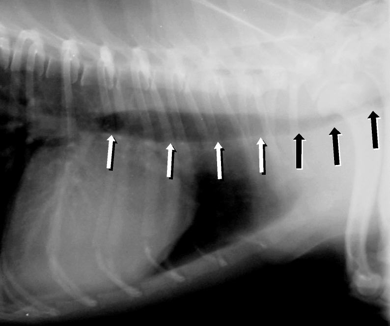

The trachea is the tube that carries air from the larynx to the mainstem bronchi and into the lungs. In a healthy dog, a series of C-shaped cartilage rings sits along the dorsal-to-ventral axis and holds the tube open. A dorsal tracheal membrane connects the free ends of those rings at the top of the tube. When a dog develops tracheal collapse, the cartilage loses rigidity, the rings flatten dorsoventrally, and the dorsal membrane stretches and sags into the airway. The lumen narrows, airflow becomes turbulent, and the lining of the airway becomes inflamed.
Tracheal collapse is described as a chronic and progressively worsening disease, and the severity of the clinical signs a dog shows depends on how much the airway has narrowed [1]. That relationship is not perfectly linear. In a study of 110 small-breed dogs, the grade of collapse seen on fluoroscopy did not correlate with cough severity, but higher body condition scores and lower body weight did correlate with more severe collapse [2]. In other words, a dog can have a dramatic-looking airway on imaging and a modest cough, or the reverse. What matters most is the whole clinical picture, not a single number.
Systemic inflammation is part of the disease. Dogs with tracheal collapse have significantly higher plasma interleukin-8 concentrations than healthy controls, which supports the presence of an ongoing inflammatory state [3]. Yorkshire Terriers were overrepresented in that study population, and the affected dogs were older and had a higher body condition score than the controls [3]. That combination of age, breed, and body condition is the classic profile.
Why Small Breeds and Overweight Dogs Are Hit Hardest
Tracheal collapse is overwhelmingly a disease of small and toy breeds. In a fluoroscopic case series of 110 dogs, the top four breeds represented were the Maltese, Pomeranian, Poodle, and Chihuahua [2]. Yorkshire Terriers are repeatedly identified as overrepresented in affected populations [3]. The most severe collapse in that 110-dog cohort was found at the carinal region, where the trachea splits into the mainstem bronchi [2].
Body condition is one of the few modifiable risk factors. In the same cohort, 97.2 percent of dogs had a body condition score of 4 or higher, and the grade of collapse on fluoroscopy was most strongly related to a high body condition score and low body weight [2]. This is why weight management sits at the foundation of treatment rather than at the margins. Fat deposits around the neck and chest, and the increased respiratory demand that comes with extra body mass, both work against an already narrowed airway.
Airway irritants are the other major aggravator. Smoke, aerosol sprays, strong cleaning fumes, dusty litter, and even a tight collar pressing on the cervical trachea can all trigger coughing episodes. A neck collar pulls directly on the trachea every time the dog pulls on the leash. A body harness distributes that force across the chest instead.
Age matters too. Affected dogs in one study were older than healthy controls [3], and another cohort spanned ages 2 to 19 years with no significant sex difference [2]. Tracheal collapse is usually a disease of middle-aged and older small-breed dogs, though it can appear earlier.
The Four-Grade Classification
Veterinarians grade tracheal collapse by the percentage of the airway lumen that is lost when the trachea collapses. The standard scheme uses five measurement points on imaging: 0 percent, 25 percent, 50 percent, 75 percent, and 100 percent decrease in diameter [4]. Those measurements are grouped into four clinical grades.
- Grade I: less than 25 percent luminal collapse.
- Grade II: 25 to 50 percent collapse.
- Grade III: 50 to 75 percent collapse.
- Grade IV: more than 75 percent collapse.
Grading is done region by region because collapse can occur at different points along the airway. The cervical trachea, the thoracic inlet, the intrathoracic trachea, the carina, and the mainstem bronchi are each assessed separately [4]. A dog can have mild cervical collapse and severe intrathoracic collapse at the same time, and the pattern changes how the dog sounds and how it responds to treatment.
Dynamic collapse is the key concept. The trachea can look normal on a still radiograph and collapse dramatically during the respiratory cycle. Radiography tends to underestimate the degree of collapse in all regions compared with fluoroscopy [4]. That is why many veterinarians use fluoroscopy, which records the airway in motion, or obtain paired inspiratory and expiratory radiographs to catch the airway at its worst.
Grade Versus Clinical Signs and Management
| Grade | Luminal collapse | Typical clinical picture | Typical management focus |
|---|---|---|---|
| I | Under 25 percent | Occasional honking cough, often triggered by excitement, pulling, or irritants. Exercise tolerance usually normal. | Weight optimization, harness instead of collar, irritant avoidance, monitoring |
| II | 25 to 50 percent | More frequent honking cough, mild exercise intolerance, cough with drinking or excitement. | All of grade I plus antitussive therapy and treatment of concurrent airway disease |
| III | 50 to 75 percent | Persistent cough, clear exercise intolerance, intermittent respiratory distress, possible flare-ups. | Medical management with bronchodilators, short courses of corticosteroids for flare-ups, and airway anti-inflammatory support |
| IV | Over 75 percent | Severe obstructive signs, cyanosis, syncope, and asphyxiation episodes are possible. | Aggressive medical management first, then intraluminal stent or extraluminal prosthetic rings when refractory |
This table is a framework, not a rule. Fluoroscopic grade and cough severity do not track together reliably [2], so a grade III dog with a mild cough may need less intervention than a grade II dog with severe, disruptive coughing. The veterinarian treats the patient, not the number.
The Honking Cough and Other Classic Signs
The signature sound of tracheal collapse is a dry, harsh, honking cough that owners often describe as sounding like a goose. It is the single most recognizable feature of the disease. In a series of 75 dogs treated with endoluminal stents, improvement in goose-honking or raspy breathing was reported in 89 percent of dogs after treatment [5]. That is a useful benchmark for how much the cough can improve when the airway is stabilized.
Beyond the cough, owners commonly notice:
- Exercise intolerance. The dog tires faster, lags on walks, or refuses activity it used to enjoy.
- Cough triggered by drinking, excitement, or pulling on the leash. Pressure on the cervical trachea or a sudden change in breathing pattern provokes the reflex.
- Raspy or noisy breathing. Air moving through a narrowed lumen becomes turbulent and audible.
- Gagging or retching at the end of a coughing fit. Some owners mistake this for vomiting or a foreign body.
- Respiratory distress during flare-ups. Labored breathing, increased effort, and a prolonged recovery after exertion.
Certain collapse patterns produce more dramatic presentations. W-shaped tracheal collapse, a specific malformation pattern, is associated with more preoperative stridor and labored breathing than the traditional pattern, and dogs with the W-shaped form were 12.1 times more likely to require preoperative oxygen support [6]. That is a reminder that the shape of the collapse, not just the grade, influences how sick a dog becomes.
What Causes Tracheal Collapse and What Else Looks Like It
The underlying problem is a loss of cartilage rigidity. The cartilage in affected dogs is weaker and more prone to flattening, and the dorsal membrane stretches into the lumen. Chronic inflammation of the tracheal mucosal lining contributes to the progressive nature of the disease [7]. The result is a self-reinforcing cycle: narrowing causes turbulent airflow, turbulent airflow irritates the lining, and irritated lining swells and narrows the airway further.
Several conditions commonly coexist with tracheal collapse and can worsen the clinical picture:
- Bronchial collapse. Collapse often extends beyond the trachea into the mainstem bronchi, which is why the carinal region is assessed during grading [4].
- Pharyngeal collapse. Concomitant pharyngeal collapse increased significantly with cough severity in one study, meaning dogs with worse coughs were more likely to have collapse above the trachea as well [8].
- Laryngeal paralysis. This can appear alongside tracheal collapse and was noted as a postoperative concern in dogs receiving extraluminal prostheses [6].
- Myxomatous mitral valve disease. Heart disease is sometimes diagnosed alongside tracheal collapse, as in a case report of a Maltese dog with both conditions [9].
- Obesity. Higher body condition scores are associated with more severe collapse on fluoroscopy [2].
Differentials a veterinarian will consider include kennel cough and other infectious tracheobronchitis, chronic bronchitis, collapsing mainstem bronchi without tracheal involvement, laryngeal disease, heart failure with a cough, and airway foreign bodies. The honking quality of the cough plus the breed and body condition profile usually points strongly toward tracheal collapse, but imaging confirms it.
How Veterinarians Diagnose It
Diagnosis starts with a physical examination and a careful history. The veterinarian will ask about the character and triggers of the cough, exercise tolerance, any fainting episodes, and exposure to irritants. They will listen to the airway and may be able to provoke a cough with gentle pressure on the cervical trachea.
Imaging is the cornerstone. Fluoroscopy allows the veterinarian to watch the airway collapse dynamically during breathing, and it is the reference method for grading in many practices [4]. Radiography is widely available and sensitive for detecting collapse in the cervical and thoracic inlet regions, with the best negative predictive value in those areas, but it underestimates the degree of collapse everywhere [4]. Inspiratory and expiratory radiographs improve accuracy by capturing the airway at different points in the respiratory cycle [4].
Tracheoscopy (also called bronchoscopy) lets the veterinarian see the airway directly and grade the collapse visually. It requires anesthesia and can irritate the airway, so it is used selectively [10]. It is also the tool used to assess stent position and to look for granulation tissue after stent placement [11].
Pulmonary function testing offers a noninvasive alternative. Tidal breathing flow-volume loop analysis differentiated dogs with tracheal collapse from healthy controls and helped separate mild-to-moderate from severe disease using three indices [10]. This kind of testing is not available everywhere, but it shows that objective, anesthesia-free assessment is possible.
Artificial intelligence is entering the picture. A deep learning model trained on lateral cervicothoracic radiographs was able to screen for tracheal collapse across normal, grade 1 to 2, and grade 3 to 4 categories with 98.30 percent sensitivity, 99.20 percent specificity, and 98.90 percent accuracy [1]. Agreement between the model and radiologists was excellent [1]. This kind of tool is a screening aid, not a replacement for a veterinarian, but it points toward faster triage in general practice.
Blood markers are an active area of research. Plasma interleukin-8 is elevated in dogs with tracheal collapse compared with controls [3]. Serum MMP-9 was significantly higher in dogs with the most severe cough grade [8]. These markers are not yet standard clinical tools, but they reflect the inflammatory nature of the disease and may eventually help monitor progression.
The Treatment Ladder, Step by Step
Treatment escalates in a defined order. Most dogs start at the bottom of the ladder and only move up when the current step stops controlling the signs.
Step 1: Weight Loss and Body Condition Management
Weight optimization is the first and most important intervention. Higher body condition scores are associated with more severe collapse on fluoroscopy [2], and affected dogs as a group have higher body condition scores than healthy controls [3]. In a cohort of 110 dogs, clinical symptoms improved in 86.6 percent of cases following interventions that included weight reduction and environmental changes [2]. That is a large effect from a step that carries no drug side effects.
Weight loss in a small-breed dog should be guided by the veterinarian, with a measured target weight and a controlled calorie plan. Crash dieting is not appropriate. The goal is a lean body condition maintained for life, not a short-term drop.
Step 2: Harness Instead of Collar
Switching from a neck collar to a body harness removes direct pressure from the cervical trachea. This is a simple, permanent change. Every pull on a collar transmits force to the windpipe, and for a dog whose windpipe is already collapsing, that force can trigger a coughing fit. A well-fitted harness that does not restrict the chest is the standard recommendation.
Step 3: Irritant Avoidance
Remove smoke, aerosol sprays, scented candles, strong cleaning products, and dusty bedding from the dog's environment. These do not cause tracheal collapse, but they reliably worsen coughing in a dog whose airway is already inflamed. Environmental change was one of the interventions associated with clinical improvement in the 110-dog cohort [2].
Step 4: Antitussives
Antitussive (cough-suppressant) medication reduces the cough reflex and breaks the cycle in which coughing irritates the airway and provokes more coughing. These are prescription medications and the dose depends on the individual dog. Follow the label and your veterinarian's instructions exactly. Do not give human cough preparations to a dog.
Step 5: Bronchodilators
Bronchodilators relax the smooth muscle in the lower airway and can ease breathing when bronchial collapse or concurrent lower airway disease is part of the picture. Theophylline is a bronchodilator with anti-inflammatory effects that has been studied specifically in tracheal collapse. In a retrospective series of 47 small-breed dogs, theophylline-based therapy produced clinical improvement in 46 of 47 dogs (97.9 percent), with a mean coughing score that decreased after treatment [12]. Higher intrathoracic collapse grades required higher final theophylline doses [12]. Theophylline is sometimes chosen when corticosteroids are a concern because of their side effects [12].
Step 6: Corticosteroids for Flare-Ups
Corticosteroids reduce airway inflammation and are typically reserved for flare-ups rather than used continuously, because long-term use carries side effects. They are most useful when a dog has a sudden worsening of cough or respiratory effort. The dose, duration, and tapering schedule are decisions for the veterinarian. Follow the prescription label.
Step 7: Adjunctive Anti-Inflammatory Support
Some dogs benefit from adjunctive nutritional support aimed at airway inflammation. A marine lipid extract combining Perna canaliculus and Euphausia superba was studied as an add-on to standard medication in 41 dogs with confirmed tracheal collapse. Dogs receiving the supplement showed significant improvement in exercise tolerance by day 14 and significant reductions in the percentage change in tracheal luminal diameter at the cervical, thoracic inlet, and intrathoracic regions by day 14 [13]. Both groups improved in cough scores, with no statistical difference between them [13]. A separate study of the same extract found reductions in serum malondialdehyde, interleukin-6, and tumor necrosis factor-alpha after five weeks of supplementation alongside standard treatment [7]. These are adjuncts, not replacements for the core steps.
Step 8: Intraluminal Stents for Refractory Cases
When medical management no longer controls severe signs, an intraluminal stent can be placed inside the trachea to hold it open. Stents are most often considered for grade IV collapse that has not responded to medication. In a series of 12 dogs with grade IV collapse and severe obstructive respiratory failure, Dumon silicone stent placement reduced obstructive dyspnea and asphyxiation episodes, and survival after placement ranged from 97 to 1,310 days with a median of 810 days [14]. Complications occurred in 9 of 12 dogs and included granulation tissue growth, incoercible cough, stent migration, and stent deformation [14].
Metallic stents are also widely used. In a series of 75 dogs receiving self-expanding metallic stents, 93 percent survived to hospital discharge and median survival time was 1,005 days [5]. Improvement in goose-honking or raspy breathing was reported in 89 percent of dogs and improvement in dyspnea in 84 percent at final follow-up [5]. Major complications requiring additional stent procedures occurred in 47 percent of dogs over the follow-up period, with stent fracture and tissue ingrowth the most common [5]. A double-wire woven nitinol stent resolved respiratory signs immediately in all 10 dogs in one case series, with mild to moderate stent fractures in five dogs and no stent migration [15]. A cross-and-hook braided stent in 22 dogs achieved a median survival time of 879 days at a median follow-up of 990 days, with loss or mild improvement of cough in all dogs [16].
Granulation tissue is a known consequence of stenting. A randomized trial of 111 dogs tested whether mycophenolate mofetil reduced granulation tissue after stent placement and found no statistically significant difference in granulation tissue grade at one, three, or six months compared with controls [11]. The authors concluded that mycophenolate can be safely given to these patients, but it did not prevent granulation tissue in that study [11].
Step 9: Extraluminal Prosthetic Rings and Other Surgical Support
Extraluminal prostheses are placed around the outside of the trachea rather than inside the lumen. A continuous extraluminal tracheal prosthesis was studied in 69 dogs with grade IV collapse, 45 with the traditional pattern and 24 with the W-shaped pattern. All dogs were discharged after placement. The 36-month postoperative survival rates were 75.7 percent for traditional collapse and 90.9 percent for W-shaped collapse [6]. Postoperative laryngeal paralysis occurred in three dogs in the traditional group and two in the W-shaped group, and recurrent collapse occurred in one traditional and two W-shaped dogs [6]. Seven of the eight dogs with postoperative complications required surgical intervention or intraluminal stent placement [6].
Extraluminal support can also be used to rescue a failed stent. In a case report of a 4-year-old Yorkshire Terrier with a completely fractured endoluminal stent, a parallel loop line prosthesis was placed as external support. It restored a near-normal tracheal contour and stabilized the fractured stent without adding more material inside the airway, and no further deformation was seen over several months [17].
The Treatment Decision Path
The following flowchart shows how a veterinarian moves from diagnosis through the treatment ladder.
flowchart TD
    A[Honking cough or exercise intolerance] --> B[Veterinary examination]
    B --> C[Imaging confirms collapse]
    C --> D[Assign grade and regions]
    D --> E{Grade and clinical severity}
    E --> F[Mild signs]
    E --> G[Moderate signs]
    E --> H[Severe or refractory signs]
    F --> I[Weight loss harness irritant control]
    G --> J[Add antitussives bronchodilators and flare up steroids]
    H --> K[Intraluminal stent or extraluminal rings]
    I --> L[Recheck and adjust]
    J --> L
    K --> L
When Tracheal Collapse Is an Emergency
Three signs turn a manageable chronic disease into an emergency.
Cyanosis. A blue, gray, or purple tinge to the gums, tongue, or lips means the blood is not getting enough oxygen. This is a critical airway compromise.
Syncope. Fainting or collapsing episodes happen when the brain is briefly starved of oxygen during a severe collapse event. A dog that has fainted needs immediate evaluation.
Respiratory distress. Labored breathing, an obvious struggle to move air, a prolonged recovery after exertion, or a dog that cannot settle into a comfortable breathing pattern all indicate that the airway is critically narrowed.
Dogs with W-shaped collapse are more likely to need oxygen support before any procedure, with a 12.1 times higher odds of requiring preoperative oxygen management than dogs with traditional collapse [6]. Severe obstructive respiratory failure is the presentation that leads to stent placement in the first place [14].
If your dog shows any of these signs, go to an emergency veterinary hospital. Do not wait to see if it passes. Do not attempt to manage it at home.
Unsafe Home Remedies and Common Mistakes
Several well-intentioned owner behaviors make tracheal collapse worse.
- Using a neck collar on a dog with tracheal collapse. Every pull transmits force to the cervical trachea. Switch to a harness.
- Giving human cough syrups or cold medications. These are not formulated for dogs and some ingredients are toxic. Only use medication prescribed by your veterinarian.
- Delaying weight loss. Owners often wait until the cough is severe before addressing body condition. The evidence links higher body condition scores to more severe collapse [2], so weight management belongs at the start.
- Ignoring environmental irritants. Smoke and aerosols are easy to overlook but reliably provoke coughing.
- Assuming a normal resting radiograph rules out collapse. Radiography underestimates collapse in all regions [4], and dynamic collapse can be invisible on a still image.
- Stopping prescribed medication during a good period without veterinary guidance. Flare-ups often follow premature discontinuation.
Prevention and Long-Term Management
Tracheal collapse cannot be prevented in a dog that is genetically predisposed, but the severity of the disease and the frequency of flare-ups can be influenced.
Keep the dog lean for life. This is the single most impactful long-term measure. Higher body condition scores track with more severe collapse on imaging [2], and weight reduction was part of the intervention package that improved symptoms in 86.6 percent of dogs in one cohort [2].
Use a harness permanently. This is not a temporary measure. It is a lifelong change for any dog with tracheal collapse.
Control the environment. Keep the home free of smoke and aerosol irritants.
Treat concurrent conditions. Bronchial collapse, pharyngeal collapse, laryngeal paralysis, and heart disease all affect how a dog with tracheal collapse feels. Managing them together produces better results than managing the trachea alone.
Keep recheck appointments. Dogs on medical management need periodic reassessment to adjust therapy. Dogs with stents need monitoring for granulation tissue, stent fracture, and migration, which are the most common complications [5]. Recheck tracheoscopy after stent placement is typically performed at intervals such as one, three, and six months [11].
Understand the trajectory. Tracheal collapse is progressive. Even after stent placement, disease progression is inevitable, though substantial improvement in respiratory function can be achieved for months to years [14]. The goal of treatment is quality of life and control of signs, not a cure.
Prognosis
Prognosis depends on grade, collapse pattern, concurrent disease, and how well the dog responds to each step of the treatment ladder.
Dogs managed medically from the early grades often do well for long periods. In the theophylline study, the symptom-free period with therapy averaged 189.7 days with a wide range from 0 to 720 days, and there was no statistically significant correlation between symptom-free period and age, sex, or fluoroscopic grade [12]. That wide range reflects how variable the disease is.
Dogs that reach the point of stenting have a median survival time of about 1,005 days in one large series [5] and about 810 days in a silicone stent series [14]. Extraluminal prostheses in grade IV dogs produced 36-month survival rates of 75.7 percent for traditional collapse and 90.9 percent for W-shaped collapse [6]. These are meaningful survival times for a disease that would otherwise be fatal in its severe form.
Limitations and When to Contact a Veterinarian
This article describes the general patterns of tracheal collapse. Individual dogs vary in grade, collapse pattern, concurrent disease, and response to treatment, and only a veterinarian who has examined your dog can make treatment decisions.
Contact a veterinarian promptly if your dog develops any of the following:
- A new or worsening honking cough that lasts more than a few days.
- Coughing that interferes with sleep, eating, or normal activity.
- Exercise intolerance that is getting worse over weeks.
- Any episode of fainting or collapse.
- Labored breathing, a prolonged recovery after exertion, or a dog that cannot settle.
- Blue, gray, or purple gums, tongue, or lips.
- A sudden severe worsening of breathing after a period of stability.
Go to an emergency hospital immediately for cyanosis, syncope, or respiratory distress. These are not signs to monitor at home.
Frequently Asked Questions
What does a collapsed windpipe sound like in a dog?
It sounds like a dry, harsh honking cough, often compared to a goose honk. It is the most recognizable sign of tracheal collapse and often worsens with excitement, drinking, or pulling on a leash.
Which dog breeds are most likely to have tracheal collapse?
Small and toy breeds are most affected, especially Yorkshire Terriers and Pomeranians. Maltese, Poodle, and Chihuahua are also commonly represented in affected populations.
Can a collapsed windpipe be cured?
There is no cure. Tracheal collapse is a chronic, progressive disease, and treatment focuses on controlling signs, improving quality of life, and slowing the impact of the disease over time.
How is the grade of tracheal collapse determined?
The veterinarian measures the percentage of the airway lumen that is lost during collapse, usually with fluoroscopy or paired inspiratory and expiratory radiographs. The measurements are grouped into grade I through grade IV.
Does my dog need surgery for tracheal collapse?
Most dogs do not. Surgery and stenting are reserved for severe, grade IV collapse that no longer responds to medical management. The treatment ladder starts with weight loss, a harness, irritant control, and medication.
Is a harness really better than a collar?
Yes. A neck collar pulls directly on the cervical trachea and can trigger coughing. A body harness distributes force across the chest instead and removes that trigger.
Can I give my dog human cough medicine?
No. Human cough preparations are not formulated for dogs and some ingredients are toxic. Only give medication prescribed by your veterinarian.
When should I rush my dog to the emergency vet?
Go immediately if your dog has blue or gray gums, faints, or struggles to breathe. These signs mean the airway is critically compromised and the dog needs oxygen and emergency care right away.
Related Articles
- Why Is My Dog coughing up white foam? Kennel Cough vs Tracheal Collapse
- Chihuahua Health Problems: Hypoglycemia Open Fontanel and Tracheal Collapse
- Yorkshire Terrier Health Problems: Tracheal Collapse Liver Shunt and Hypoglycemia
- Dog Honking Cough After Drinking Water: Tracheal Collapse vs Kennel Cough & Triage
- Dog Reverse Sneeze Or Collapsed Trachea
- Tracheal Mite Monitoring and Colony Differentials
- Medication for Collapsing Trachea in Dogs: Drug Guide
Sources
- Automatic classification and grading of canine tracheal collapse on thoracic radiographs by using deep learning.
- A retrospective study of tracheal collapse in small-breed dogs: 110 cases (2022-2024).
- Assessment of the circulating inflammatory mediator interleukin-8 in dogs with tracheal collapse.
- Fluoroscopic and radiographic evaluation of tracheal collapse in dogs: 62 cases (2001-2006).
- Short-, intermediate-, and long-term results for endoluminal stent placement in dogs with tracheal collapse.
- Long-term outcomes of dogs with W-shaped or traditional tracheal collapse treated with a continuous extraluminal tracheal prosthesis: A retrospective study.
- Polyunsaturated Fatty Acid EAB-277(®) Supplementation Improved Heart Rate Variability and Clinical Signs in Tracheal Collapse Dogs.
- Assessment of MMP-9 and clinical characteristics in dogs with tracheal collapse based on cough severity and fluoroscopic findings: a cross-sectional study.
- Use of a double-wire woven uncovered nitinol stent for the treatment of refractory tracheal collapse in a dog: a case report
- Tidal breathing flow-volume loop analysis for the diagnosis and staging of tracheal collapse in dogs.
- The Effects of Mycophenolate on the Formation of Granulation Tissue Post-operatively in Canine Tracheal Stent Patients (2014-2020).
- A retrospective study of theophylline-based therapy with tracheal collapse in small-breed dogs: 47 cases (2013-2017).
- Adjunctive Therapy With a Perna canaliculus and Euphausia superba Extract (EAB-277(TM)) for Clinical Signs of Tracheal Collapse in Dogs: A Prospective, Randomized and Placebo-Controlled Exploratory Trial.
- Dumon silicone stents can improve respiratory function in dogs with grade IV tracheal collapse: 12 cases (2019-2023).
- Double-wire woven nitinol stent for treating dogs with refractory tracheal collapse: A case series.
- Surgical outcomes in dogs with tracheal collapse treated with a novel cross-and-hook braided endoluminal stent
- Tracheal External Support Using a Parallel Loop Line Prosthesis for Tracheal Stent Fracture in a Dog: A Case Report.