Puppy Strangles: Juvenile Cellulitis Guide
By Dr. Zubair Khalid, DVM, MS, PhD ·
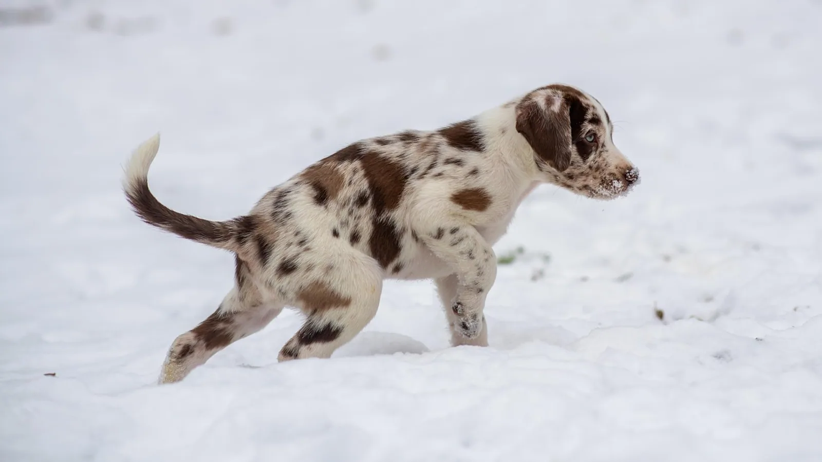
Puppy strangles is a sudden, painful swelling of a young puppy's face, muzzle, and eyelids that is caused by sterile inflammation rather than infection. The medical name is juvenile cellulitis, and it is treated with immunosuppressive glucocorticoids, often alongside antibiotics for secondary bacterial infection.
If your puppy woke up this morning with a puffy, swollen face, crusty pustules around the nose and eyes, and golf-ball-sized lumps under the jaw, this condition should be near the top of your veterinarian's list. The good news is that most puppies recover fully when treated promptly. The critical catch is that the steroids must be tapered slowly over several weeks. Stopping them early causes the disease to flare right back up.
This article is educational and is not a substitute for veterinary diagnosis or treatment.
What Is Strangles in Dogs?
Puppy strangles is the common name for juvenile cellulitis, also called juvenile pyoderma [1]. Despite the alarming nickname, it has nothing to do with the bacterial disease called strangles in horses. In dogs, the word "strangles" refers to the dramatic enlargement of the lymph nodes under the jaw, which can make a puppy look and feel as though something is constricting the throat.
Juvenile cellulitis is a sterile, inflammatory skin disease. "Sterile" means no bacteria, fungus, or virus can be cultured from the affected tissue. Investigators who studied a litter of affected dogs looked for bacterial, fungal, and viral agents in the lymph nodes and found none [2]. They also tried to transfer the disease by inoculating newborn puppies with tissue from affected dogs, and that failed too [2]. The trigger remains unknown.
The word "cellulitis" simply describes inflammation of the deeper layers of skin and the tissue beneath it. That is why the swelling is so pronounced. This is different from the common human skin infection also called cellulitis, which is caused by bacteria and treated with antibiotics alone. In juvenile cellulitis, antibiotics do not fix the underlying problem.
Which Puppies Get It
Juvenile cellulitis is most often seen in puppies between 1 and 4 months of age [2]. Most veterinary reports describe puppies under 6 months, and typical ages in published cases include 7 weeks, 8 weeks, and 10 weeks [3][4][5]. The disease has also been confirmed in an 8-month-old dog, which was the first report of its age at the time [6].
Breed predispositions are not strongly established for puppies, but several goldens, Labradors, Australian shepherds, and cocker spaniels appear in the case literature [3][4][7][5]. Adult dogs can develop a nearly identical condition, and in a large review of 90 adult cases, Havanese, Australian shepherds, Irish setters, Dachshunds, bichon frises, and Maltese dogs were significantly over-represented [8]. Whether similar puppy breed patterns exist has not been confirmed in large studies.
The Classic Presentation
The hallmark of puppy strangles is an acutely swollen face in a young puppy [4]. Owners often describe the puppy as looking like it has been stung by a bee, or as if the muzzle has puffed up overnight.
The swelling concentrates around the muzzle, eyelids, lips, and chin. Within a day or two, small pustules and pimples appear in the same areas. These quickly rupture, weep, and form thick yellow-brown crusts [1]. The skin becomes red, painful, and sometimes ulcerated. In severe cases, the swelling can close the eyes.
Lymph nodes under the jaw (mandibular nodes) and in front of the shoulders (prescapular nodes) become noticeably enlarged. In one review of 15 affected dogs, mandibular lymphadenopathy was present in 14 of them, and in five dogs the swollen nodes were the only finding, with no obvious skin lesions at all [1]. This point matters because a puppy can be quite sick from lymph node swelling alone, leading owners to think the problem is a sore throat or a dental issue.
Other common findings include:
- Otitis externa. The ear canals can become swollen, painful, and crusted. Some dogs have a hemorrhagic discharge from the ears [9][5].
- Fever and lethargy. Several puppies have been pyrexic, lethargic, and off their food [4][1][5]. In one review, eight of 15 dogs were lethargic, but fever and anorexia were inconsistent findings [1].
- Joint pain and lameness. Four dogs in one series showed pain when their joints were manipulated [1]. In a Labrador puppy, the first sign was a hindlimb lameness that progressed rapidly to the classic facial disease [5]. Joint aspirates from three of those dogs showed suppurative arthritis with no bacteria and no growth on culture [1].
- Neurologic signs. Three 7-week-old English cocker spaniel littermates developed juvenile cellulitis with hindlimb paresis, and both the skin lesions and the weakness improved with treatment [7].
Symptoms typically progress over 24 to 72 hours, which is why a puppy that seemed fine at breakfast can look dramatically worse by evening.
Why the Lymph Nodes Swell
The lymph nodes are not infected. They are reacting to inflammation in the skin they drain. In affected dogs, the same pyogranulomatous inflammation found in the facial skin also appears in the mandibular and superficial cervical lymph nodes [2]. Investigators found this inflammatory process even in a lymph node located far from the visibly affected skin, and in one littermate with no visible skin lesions or lymph node enlargement at all, histology still showed a milder version of the same inflammation in a mandibular node [2]. That finding explains why the disease behaves like a systemic problem rather than a purely local skin issue.
What the Skin Looks Like Under the Microscope
The diagnosis rests on the clinical picture plus supportive laboratory findings, not on any single test.
Cytology
Cytology from a pustule or an aspirate of an abscess typically shows large numbers of neutrophils with no visible bacteria [1]. Neutrophils are the white blood cells that rush into inflamed tissue. Their presence without organisms supports a sterile inflammatory process. In one review of aspirated lymph nodes, three dogs showed suppurative lymphadenitis with many neutrophils, again with no bacteria [1].
Biopsy
A skin biopsy is the most definitive supportive test. It shows pyogranulomatous inflammation, meaning a mix of neutrophils and macrophages, in the skin and in the draining lymph nodes [2]. The predominant inflammatory cell identified by light microscopy, electron microscopy, and immunohistochemical staining is an epithelioid macrophage [2]. When the inflammation extends into the fat layer beneath the skin, the term panniculitis applies. Biopsy findings of granulomatous dermatitis and panniculitis are classic for this disease.
Culture
Bacterial and fungal cultures of intact pustules and lymph nodes are negative in uncomplicated cases [1]. Coagulase-positive Staphylococcus species were isolated from draining lesions in two dogs, and those dogs likely had secondary bacterial infection on top of the sterile disease [1]. That distinction drives the treatment plan: antibiotics treat the secondary infection, but glucocorticoids treat the disease itself.
flowchart TD
    A[Puppy under 6 months] --> B[Acute facial swelling]
    B --> C[Pustules and crusts]
    C --> D[Enlarged jaw lymph nodes]
    D --> E{Veterinarian examination}
    E --> F[Cytology shows neutrophils]
    E --> G[Biopsy shows granulomatous dermatitis]
    F --> H[Start glucocorticoid therapy]
    G --> H
    H --> I[Add antibiotics if infected]
    I --> J[Taper steroids slowly over weeks]
    J --> K{Lesions resolved}
    K --> L[Gradual steroid withdrawal]
    K --> M[Relapse if stopped early]
    M --> H
Differential Diagnoses
Several skin diseases in puppies look like juvenile cellulitis at first glance. The clinical presentation, cytology, and biopsy findings help tell them apart.
| Condition | Typical appearance | Key distinguishing features |
|---|---|---|
| Juvenile cellulitis (puppy strangles) | Acute facial swelling, pustules, crusts, and enlarged jaw lymph nodes in puppies under 6 months | Sterile pyogranulomatous inflammation with macrophages on biopsy, no organisms on culture, responds to glucocorticoids |
| Demodicosis | Patchy hair loss, scaling, and pustules, often on the face and paws | Mites are visible on skin scraping or hair pluck, pustules typically contain bacteria and mites |
| Pyoderma | Pustules, crusts, and collarettes, commonly on the trunk or in skin folds | Bacteria are seen on cytology and grow on culture, responds to antibiotics alone |
| Angioedema | Rapid, soft swelling of the muzzle, eyelids, or ears, often after an insect sting or allergen exposure | Swelling is not pustular, resolves quickly with antihistamines or supportive care, no lymph node enlargement |
| Eosinophilic furunculosis | Sudden crusted plaques and ulcers on the muzzle, usually after insect bites | Biopsy shows eosinophilic inflammation rather than pyogranulomatous dermatitis, no lymphadenopathy |
The single most useful clue is the combination of facial pustules, dramatic lymph node enlargement, and a young age. When that pattern is present, juvenile cellulitis is the leading diagnosis until proven otherwise.
Treatment: Glucocorticoids Plus Antibiotics
The cornerstone of treatment is immunosuppressive glucocorticoid therapy, usually prednisolone or prednisone [6][4][5]. Doses are set by the treating veterinarian based on the puppy's weight, severity, and response. Never adjust or stop a steroid dose without veterinary guidance.
The standard approach is to start at an immunosuppressive dose and then taper gradually over roughly 3 to 4 weeks. The taper is the most important part of the plan. If the steroid is withdrawn too quickly, the lesions come right back. Owners who stop treatment early because the puppy looks better almost always see a relapse within days. Your veterinarian will typically schedule a recheck partway through the taper to confirm the skin is truly quiet before reducing the dose further.
Antibiotics are added when the skin is secondarily infected, which is common because the pustules break open and bacteria colonize the damaged surface. Coagulase-positive Staphylococcus species have been isolated from draining lesions, so your veterinarian may choose an antibiotic based on culture and sensitivity results [1]. Antibiotics alone will not resolve the disease. In one 8-month-old dog, a four-week course of antibiotics before diagnosis had already failed [6].
Other Medications
Most puppies improve quickly on glucocorticoids. In the English cocker spaniel littermates with hindlimb paresis, treatment combined cyclosporine A with prednisolone, and both the skin lesions and the weakness improved [7]. This combination is reserved for cases that are not responding to steroids alone or that have severe or atypical features. Long-term cyclosporine carries its own monitoring requirements, so it is a decision for the veterinarian, not something to start at home.
Topical care for the skin and ears can make the puppy more comfortable and reduce crust buildup, but it does not replace systemic treatment.
What Recovery Looks Like
Owners often see significant improvement within the first week. In a 7-week-old golden retriever, inflammation had dropped markedly and pustules were healing one week after treatment started [4]. In the 8-month-old crossbred dog, complete resolution occurred with glucocorticoid therapy [6]. The adult literature, which may be relevant to how the disease behaves, reported a median time to remission of 28 days and a median treatment duration of 60 days in 90 adult dogs with the histopathologic diagnosis [8]. Puppies often respond faster, but the principle is the same: keep treating until the skin is fully normal, then taper slowly.
Relapse: The Most Common Mistake
The single most common reason treatment fails is stopping steroids too early. The skin may look normal while inflammation is still active in the deeper layers and the lymph nodes. If the drug is withdrawn at that point, the pustules and swelling return, sometimes worse than the first episode.
If a relapse happens, treatment usually needs to restart, and the new taper may be longer than the first one. This is not a sign that the diagnosis was wrong or that the puppy is difficult to treat. It is a predictable consequence of ending immunosuppression before the disease has gone quiet.
Rechecks during the taper are not optional. They are how your veterinarian confirms that the deeper inflammation has resolved and that the dose can safely drop.
Practical Care at Home
Beyond medication, a few practical steps help puppies recover comfortably:
- Keep the face clean and dry. Gently remove crusts with a soft cloth dampened with warm water or a veterinarian-recommended cleanser. Do not scrub open sores.
- Do not squeeze pustules. Rupturing them spreads bacteria and worsens crusting.
- Feed soft food if the mouth is sore. Swollen lips and jaw nodes can make chewing painful.
- Limit rough play with other dogs. The skin is fragile while it heals, and the immune system is suppressed by treatment.
- Keep all recheck appointments. The taper schedule depends on what your veterinarian sees, not on how the puppy looks at home.
- Watch for new lesions. If new pustules appear or the swelling returns, call your veterinarian rather than waiting for the next scheduled visit.
What Is Still Uncertain
The cause of juvenile cellulitis is unknown. Investigators looked for bacterial, fungal, and viral agents and found none, and they could not transfer the disease to newborn puppies by inoculation [2]. The tendency to affect littermates and puppies in the same age range suggests an infectious trigger, but no organism has been identified [2]. Some researchers have proposed an immune-mediated mechanism, where the puppy's developing immune system attacks its own skin and lymph nodes. That remains a hypothesis rather than a proven fact.
The relationship between juvenile cellulitis and adult-onset sterile granulomatous dermatitis and lymphadenitis is also not fully understood. Adult dogs can develop a condition that looks identical under the microscope, with breed predispositions and outcomes documented in a large case series [8][9][10]. Whether these are the same disease at different ages or two related conditions is an open question.
One case report described a 3-month-old puppy that developed juvenile cellulitis and then, within a week of starting antibiotics, nonsteroidal anti-inflammatory drugs, and eye ointment, developed skin lesions consistent with pemphigus foliaceus [11]. The pemphigus-like lesions resolved when the drugs were withdrawn, while the original juvenile cellulitis required glucocorticoids to resolve [11]. This appears to be a drug reaction rather than part of the disease itself, but it is a reminder that new lesions during treatment deserve veterinary attention.
Limitations and When to Contact a Veterinarian
This article describes patterns reported in the veterinary literature. Every puppy is an individual, and only a veterinarian who examines your dog can diagnose and treat juvenile cellulitis.
Contact a veterinarian promptly if your puppy develops any of the following:
- Sudden swelling of the face, muzzle, or eyelids
- Pustules, pimples, or crusts around the nose, eyes, lips, or chin
- Noticeably enlarged lymph nodes under the jaw or in front of the shoulders
- Pain when the mouth or jaw is touched, or reluctance to eat
- Redness, swelling, or discharge from the ear canals
- Fever, lethargy, or loss of appetite
- Lameness or reluctance to walk
- New skin lesions appearing during treatment, or return of old lesions after steroids are reduced
- Any difficulty breathing, which requires emergency care
Do not start, stop, or change a steroid dose without veterinary direction. Sudden withdrawal can cause a flare and, in some cases, a serious medical crisis.
Frequently Asked Questions
What is strangles in dogs?
Strangles in dogs is a nickname for juvenile cellulitis, also called puppy strangles. The name comes from the dramatic swelling of the lymph nodes under the jaw, not from any connection to the horse disease of the same name.
Is puppy strangles contagious to other pets or to people?
There is no evidence that juvenile cellulitis spreads to other animals or to humans. Investigators could not culture bacteria, fungi, or viruses from affected tissue, and they could not transfer the disease to newborn puppies by inoculation [2].
How is juvenile cellulitis diagnosed?
Diagnosis is based on the clinical picture in a young puppy, supported by cytology showing neutrophils without bacteria and by biopsy showing granulomatous dermatitis and panniculitis [1][2]. Cultures are typically negative in uncomplicated cases [1].
Can antibiotics alone cure puppy strangles?
No. Antibiotics treat secondary bacterial infection but do not resolve the underlying sterile inflammation. The disease itself requires immunosuppressive glucocorticoid therapy [6][4][5].
How long does treatment last?
Most puppies are treated for roughly 3 to 4 weeks with a gradual steroid taper. Treatment duration in adult dogs with the same histopathologic diagnosis had a median of 60 days, and the disease lasted longer in some individuals [8]. Your veterinarian will adjust the schedule based on your puppy's response.
Why does the disease come back when steroids are stopped?
The deeper inflammation can still be active even when the skin looks normal. Stopping steroids before that inflammation resolves allows the pustules and swelling to return. The taper must be slow and guided by rechecks.
Can a puppy older than 6 months get juvenile cellulitis?
Yes, though it is uncommon. Cases have been reported in an 8-month-old dog and in adult dogs, where the condition is called sterile granulomatous dermatitis and lymphadenitis [6][8][9][10].
What other conditions look like puppy strangles?
Demodicosis, pyoderma, angioedema, and eosinophilic furunculosis are the main look-alikes. Skin scraping, cytology, and biopsy findings usually separate them clearly from juvenile cellulitis.
Related Articles
- Nursery Tank Design for Juvenile Fish Rearing
- Equine Strangles: Clinical Signs, Diagnosis, and Biosecurity
- Strangles in Horses: Streptococcus equi Isolation and Treatment Protocol
- Equine Strangles: Outbreak Investigation, Isolation, and Return to Herd
- Equine Strangles: Streptococcus equi subsp. equi Diagnostic Testing and Outbreak Management
- Puppy Training Guide Uk
Sources
- Juvenile cellulitis in dogs: 15 cases (1979-1988).
- Clinicopathologic characterization of canine juvenile cellulitis.
- Hypertrophic osteodystrophy preceding canine juvenile cellulitis in an Australian shepherd puppy.
- Juvenile cellulitis in a 7-week-old golden retriever dog.
- Juvenile cellulitis in a puppy.
- Juvenile cellulitis in an 8-month-old dog.
- Combination of cyclosporin A and prednisolone for juvenile cellulitis concurrent with hindlimb paresis in 3 English cocker spaniel puppies.
- Sterile granulomatous dermatitis and lymphadenitis (juvenile cellulitis) in adult dogs: a retrospective analysis of 90 cases (2004-2018).
- Dermatitis and lymphadenitis resembling juvenile cellulitis in a four-year-old dog.
- A dermatosis resembling juvenile cellulitis in an adult dog.
- Pemphigus foliaceus-like drug reaction in a 3-month-old crossbreed dog treated for juvenile cellulitis.