Eschar Wound: What the Black Crust Means and When to Worry
By Dr. Zubair Khalid, DVM, MS, PhD ·
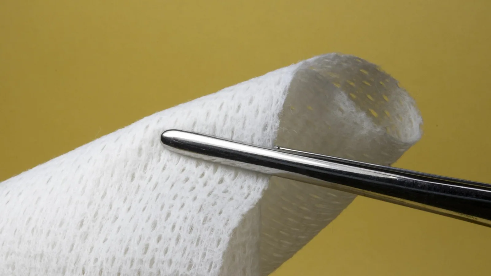
An eschar wound is a wound covered by a dry, dark, leathery crust of dead tissue. That black or brown shell is not dirt and it is not a normal scab. It is necrotic tissue that has dried and hardened over the surface of the injury, and whether it helps or harms your pet depends on what sits underneath it.
The short answer: a stable, dry eschar on intact skin can act as a natural bandage while new skin forms beneath it. An eschar that is wet, foul-smelling, spreading, or sitting over an infected wound is a problem that needs veterinary removal. The sections below explain how eschar forms, how to tell it apart from slough and scabs, and how veterinarians decide whether to debride or leave it alone.
This article is educational and is not a substitute for veterinary diagnosis or treatment.
What Eschar Actually Is
Eschar is a dense layer of necrotic tissue. The cells in that layer are dead, and the tissue has lost its blood supply. As the dead tissue loses moisture, it contracts into a firm, dry, dark plaque that can feel like leather or cardboard. The color ranges from tan to deep brown to black depending on how much blood was in the tissue before it died and how long it has been drying.
The word describes a physical state, not a single disease. Eschar can form over a burn, a pressure sore, a crush injury, a frostbite injury, or a surgical incision that lost its blood supply. In each case the underlying cause is the same: tissue died, and the dead layer dried out on the surface.
Veterinary and human wound literature treats eschar as a barrier to healing when it is thick and adherent. A chronic wound frequently develops eschar, and that dense necrotic layer both impedes healing and gives bacteria a protected place to build biofilm [1]. Biofilm is a community of bacteria wrapped in a slimy matrix that resists antimicrobial drugs, which is one reason an eschar-covered wound can look quiet while infection smolders underneath [1].
Eschar also forms as part of normal healing in some species. In a study of wild black bears, researchers created small full-thickness skin wounds during early denning and re-examined the sites two to three months later. The wounds healed with evidence of initial eschar and scab formation, complete restoration of the epidermis and dermis, and regrowth of hair follicles, all while the bears were in a mildly hypothermic hibernating state [2]. That is a useful reminder that eschar is not automatically a sign of failure. Sometimes it is the first stage of a wound closing.
How Eschar Forms
Step 1: Tissue Loses Its Blood Supply
Eschar starts with ischemia, which means the tissue is not getting enough blood. Pressure, heat, cold, chemical injury, or a blocked vessel can all cut off perfusion. A study of amputation stumps found that lower suture-line perfusion correlated with more eschar formation, and diabetic patients had lower stump perfusion and increased eschar volume [3]. Poor perfusion is the common thread.
Step 2: The Tissue Dies
Once perfusion drops far enough, cells die. This is necrosis. The dead tissue stays in place because nothing removes it yet. Inflammatory cells move in at the border between dead and living tissue, but the dead layer itself has no blood supply to support that cleanup.
Step 3: The Dead Layer Dries and Hardens
Exposed dead tissue loses water and contracts. Collagen and other structural proteins cross-link and stiffen. The result is the dry, leathery plaque that gives eschar its name. A burn eschar is largely made of denatured collagen, fibrin, and elastin, which is why debridement research focuses on enzymes that can digest those specific proteins [4][5].
Step 4: The Body Tries to Separate the Dead Layer
Underneath a stable eschar, the body can mount a healing response. In a minipig model of chemical burn, a well-developed eschar covered skin that showed significant wound healing beneath it, including a hyperplastic epidermis and a new multilayered basal epithelium [6]. The eschar stayed on top while new skin was being built below. This is the scenario where leaving eschar alone makes sense.
Eschar vs Slough vs Scab: How to Tell Them Apart
These three terms get used loosely, and owners often confuse them. They describe different tissues with different implications.
| Feature | Eschar | Slough | Scab |
|---|---|---|---|
| Appearance | Dark tan, brown, or black | Yellow, cream, gray, or green | Dark red to brown to black |
| Texture | Dry, hard, leathery, adherent | Wet, soft, stringy, gelatinous | Thin, brittle, crusty |
| Depth | Full-thickness dead tissue | Partial-thickness dead tissue and debris | Superficial dried serum, blood, and cells |
| Attachment | Firmly stuck to wound bed | Loosely attached, can be wiped | Sits on top of intact or healing skin |
| Odor | Often none if dry and stable | Frequently foul | None |
| Infection risk | High if wet or undermined | High, slough feeds bacteria | Low |
| Typical management | Leave if stable and dry, debride if infected or over extremities | Usually debrided | Leave to fall off on its own |
Eschar is the thick, dry, dark plaque described above. It represents full-thickness necrosis.
Slough is the wet cousin of eschar. It is yellow, stringy, and soft, and it wipes away more easily. Slough is still dead tissue, but it has not dried out. A wound with slough is usually a wet, draining, likely infected wound.
A scab is a thin crust of dried serum, blood, and dead surface cells over a healing wound. Scabs form on superficial injuries and fall off on their own. The bear study used the terms eschar and scab together for the early crust on healing biopsy sites, which reflects how the two overlap in the earliest stage of a small wound [2].
The practical takeaway: a dry black plaque on a quiet wound is eschar. A wet yellow film on a draining wound is slough. A thin dark crust on a scrape is a scab.
Why Eschar Matters for Healing
Eschar changes wound biology in three ways.
It blocks the surface. New skin cells have to migrate across the wound bed to close it. A hard, adherent eschar is a physical barrier that stops that migration. It also prevents topical treatments from reaching the tissue that needs them, because many drugs penetrate necrotic tissue poorly [7].
It shelters bacteria. The dense necrotic layer protects biofilm from antimicrobial agents [1]. A wound that looks sealed can be quietly colonized, and the infection can extend into healthy tissue at the margins.
It can hide what is underneath. A stable eschar can cover a healing wound, as seen in the minipig burn model [6]. It can also cover a pocket of pus, a deeper burn, or spreading necrosis. You cannot tell the difference from the surface alone.
When to Leave Eschar Alone
The general principle in wound care is that stable, dry, intact eschar on a wound with good perfusion can be left in place as a biological cover. This is sometimes called a dry eschar or a stable eschar. It is most reasonable when:
- The eschar is dry, hard, and firmly adherent
- There is no odor, drainage, or redness at the margins
- The wound is not on a limb with poor circulation
- The patient is not showing signs of systemic illness
- The eschar is on a pressure point where the tissue beneath is intact
There is a real caution here. A review of wounds that failed treatment at an advanced wound center identified misapplication of debridement principles, particularly removal of stable eschar, as one of the recurring correctable errors [8]. In other words, aggressive removal of an eschar that was doing its job can set healing back. The decision is not "always remove" or "never remove." It is a judgment about what is underneath.
When Eschar Needs to Be Removed
Debridement is the medical removal of dead tissue. Eschar usually needs debridement when any of the following are true:
- The eschar is wet, soft, or separating at the edges, which suggests infection or liquefaction
- There is foul odor, purulent drainage, or spreading redness
- The wound is on an extremity with marginal blood supply, where eschar can act like a tourniquet as it contracts
- The eschar is over a joint or a site that needs to move
- The patient has fever, lethargy, or loss of appetite
- The eschar is circumferential around a limb
Early eschar removal is widely accepted in burn care. Removing eschar within 72 hours improves burn wound outcomes by reducing bacterial colonization, infection, and length of hospital stay [9]. That timing principle comes from burn medicine, but the underlying logic, that dead tissue feeds infection and delays closure, applies to any eschar wound.
How Veterinarians Remove Eschar
There are several approaches, and the choice depends on the wound, the species, and the equipment available.
Surgical or Sharp Debridement
This is direct removal of the eschar with a scalpel, scissors, or a dermatome. It is fast and thorough. The downside is that it is invasive, can be painful, and can damage healthy tissue at the margins [1]. In a study comparing a hydrosurgical debridement system with conventional instruments on pig skin burn models, the hydrosurgery system removed significantly less tissue (2.88 g versus 13.81 g) for the same eschar excision, meaning it was more precise and preserved more viable tissue [10]. Precision matters because you want to remove dead tissue and keep living tissue.
Enzymatic Debridement
Enzymatic debridement uses proteolytic enzymes to digest the eschar. These enzymes break down the structural proteins in the dead layer, including collagen, fibrin, and elastin, and can also disrupt biofilm [1][4]. A recombinant serine protease from Bacillus species degraded up to 50 percent of an artificial wound eschar in laboratory testing, and a cream formulation maintained its activity for up to 24 hours [5]. Bromelain-based enzymatic debridement is an established option in burn care and can reduce blood loss and the need for skin grafting compared with surgical excision [9]. A fast-track protocol using short pre-soaking achieved adequate debridement in all treated wounds with no wound infections or early safety signals in a small matched-pair study [11].
Hydrosurgical Debridement
Hydrosurgery uses a high-pressure stream of saline to cut and remove dead tissue while sparing viable tissue. In the pig skin model, it was more precise than conventional instruments for eschar excision [10].
Autolytic and Moist Wound Healing
Autolytic debridement lets the body's own enzymes break down dead tissue under a moist dressing. A case report on a neonatal scalp defect used staged moist wound dressings (silver, foam, and hydrogel) to control inflammation, manage exudate, and promote granulation, achieving complete epithelialization with minimal scarring [12]. Moist wound healing works by mimicking physiological conditions to support cell migration and tissue regeneration [12].
Topical Alternatives
A case report described a traumatic amputation stump with ischemic eschar that was managed noninvasively with a honey-based salve and burdock leaf dressings. Over five months of follow-up, the eschar resolved completely and the skin re-epithelialized, and surgical debridement was avoided [13]. This is a single case, so it is not a general recommendation, but it shows that noninvasive options exist for some wounds.
Adjuncts Under Study
Researchers have tested microneedles combined with a chitosan-based gel to improve drug penetration into burn eschar, since many drugs penetrate necrotic tissue poorly. The gel plus microneedle produced the best results for increased drug flux and faster recovery in animal studies [7]. Other experimental work has examined agents such as rosuvastatin and mesenchymal stromal cells for burn and frostbite wounds [14][15]. These are research directions, not standard care.
Species Differences: Reptiles and Birds
Mammalian wound healing is the best-studied model, but reptiles and birds handle dead tissue differently, and owners of these species should not assume the same rules apply.
Reptiles are ectotherms, and their healing rate depends heavily on their preferred optimal temperature zone. A reptile kept too cool will heal slowly, and dead tissue may persist or progress rather than separate cleanly. The bear study is a useful parallel here: even mildly hypothermic body or limb temperatures can retard healing processes in mammals, yet hibernating bears still resolved wounds while maintaining mildly hypothermic states [2]. The lesson is that temperature and metabolic state shape how dead tissue behaves, and a reptile outside its thermal optimum is a poor healer.
Birds have thin skin, minimal subcutaneous fat, and a high metabolic rate. Dead tissue on a bird can dry into a firm crust quickly, and birds are prone to self-trauma at wound sites. A bird with an eschar-like lesion needs a veterinarian who works with avian patients, because the risk of underlying infection and the tolerance for debridement differ from mammals.
For both groups, the core advice is the same: do not soak, pick, or peel an eschar at home. Let an exotics or avian veterinarian assess it.
Myths and Common Questions
Myth: A black crust always means the wound is infected. Not true. A dry, stable eschar can be sterile and can cover a wound that is healing underneath [6]. Infection is suggested by wetness, odor, drainage, and redness at the margins, not by color alone.
Myth: You should pick eschar off to let the wound breathe. Removing stable eschar can damage the tissue beneath and set healing back [8]. Debridement is a medical decision.
Myth: Eschar and slough are the same thing. They are not. Eschar is dry and leathery. Slough is wet and stringy. The management differs.
Myth: A scab and an eschar are the same. A scab is a thin superficial crust. An eschar is full-thickness dead tissue. A scab falls off on its own. An eschar may need help.
Question: Can eschar heal on its own? Sometimes. In the bear study, wounds healed with initial eschar formation and went on to complete epidermal and dermal restoration [2]. In the minipig burn model, a well-developed eschar covered skin that was actively healing [6]. Stable eschar can resolve without intervention.
Question: Does eschar always need surgery? No. Enzymatic, hydrosurgical, autolytic, and topical approaches exist, and some wounds are managed conservatively [13][12][9].
Practical Implications for Owners
If you see a dark crust on your pet's wound, do the following:
- Do not pick, peel, or soak it. You can introduce infection or damage healing tissue.
- Photograph it with a coin or ruler for scale, and note the date.
- Check for red flags: wetness, odor, drainage, spreading redness, swelling, heat, pain, fever, lethargy, or loss of appetite.
- Note the location. Eschar on a limb, over a joint, or around a digit is more concerning than eschar on a flat, well-perfused area.
- Contact your veterinarian with your photos and observations. They can decide whether to debride or monitor.
For reptiles and birds, contact an exotics or avian veterinarian rather than a general practitioner if one is available.
Limitations and When to Contact a Veterinarian
This article describes general principles. Every wound is individual, and only a veterinarian who can examine your pet can judge whether an eschar should be removed.
Contact a veterinarian promptly if you see any of the following:
- Foul odor from the wound
- Wet, yellow, or green tissue on the wound surface
- Pus, cloudy drainage, or bleeding under the eschar
- Redness, swelling, or heat spreading beyond the wound margins
- The eschar is expanding or the wound is getting larger
- The eschar is circumferential around a limb, digit, or tail
- Your pet is limping, reluctant to move, or guarding the area
- Fever, lethargy, vomiting, diarrhea, or loss of appetite
- The wound is on a bird, reptile, or other exotic species
- The wound has not improved in 48 to 72 hours
- Your pet is diabetic, immunocompromised, or has known poor circulation
Seek emergency care if the wound is bleeding heavily, if there is exposed bone or deep tissue, if your pet is collapse-prone or unresponsive, or if you suspect a burn or chemical injury.
Frequently Asked Questions
What does a black crust on a wound mean?
A black crust on a wound is usually eschar, which is a dry layer of dead tissue. It can be a stable protective cover or a sign of necrosis that needs veterinary removal, depending on whether it is dry and quiet or wet and infected.
Is eschar the same as a scab?
No. A scab is a thin, superficial crust of dried serum and cells. Eschar is a thick, leathery layer of full-thickness dead tissue. Scabs fall off on their own, while eschar may need debridement.
How do I tell eschar from slough?
Eschar is dry, hard, and dark. Slough is wet, soft, yellow or gray, and stringy. Slough usually indicates a draining, likely infected wound and is typically debrided.
Should I remove eschar from my pet's wound at home?
No. Do not pick, peel, or soak eschar at home. Removal is a medical decision, and taking off a stable eschar can damage healing tissue and delay recovery.
When does eschar need to be debrided?
Eschar needs debridement when it is wet, foul-smelling, spreading, over an extremity, or accompanied by signs of infection or systemic illness. Stable, dry eschar on a well-perfused wound can often be left alone.
Can eschar heal without surgery?
Sometimes. Enzymatic debridement, hydrosurgery, moist wound dressings, and topical agents are all options, and some stable eschars resolve on their own. Your veterinarian will choose the approach based on the wound.
Do reptiles and birds get eschar the same way as dogs and cats?
Not exactly. Reptile healing depends on temperature, and birds have thin skin and fast metabolism. Both need a veterinarian experienced with those species, because the rules for debridement differ.
How long should I wait before calling the vet about an eschar wound?
Call right away if there is odor, drainage, spreading redness, or systemic signs. If the wound is dry and quiet, monitor for 48 to 72 hours and call if it has not improved or if it changes.
Related Articles
- Bearded Dragon Black Beard: What It Means and When to Worry
- Dog Ear Infection with Black Discharge: What It Means
- Dog Throwing Up White Foam: What It Means and When to Worry
- Dog Vomiting White Foam: What It Means and When to Worry
- Black Cat
- Bearded Dragon Black Beard Meaning
- Dark Patches on Dog Skin: Causes and When to Worry
- Maggots in Wounds: Risks and Medical Maggot Therapy
- Mean Square Displacement: Formula and Examples
Sources
- Protease-driven approaches for wound eschar debridement and biofilm disruption: current advances, future prospects, and limitations.
- Wound healing during hibernation by black bears (Ursus americanus) in the wild: elicitation of reduced scar formation.
- Amputation stump perfusion is predictive of post-operative necrotic eschar formation.
- Effects of a New Enzymatic Debrider (SN514-066b) on Eschar Protein Digestion, Burn Wound Debridement, and Healthy Skin Irritation.
- Debridement efficacy of serine protease and formulated cream by In Vitro assessment against artificial wound eschar.
- Skin remodeling and wound healing in the Gottingen minipig following exposure to sulfur mustard.
- Corticosteroid-loaded chitosan-based in-situ forming gel combined with microneedle technology for improvement of burn eschar wound healing.
- Managing Complex Wounds After Treatment Failure: Patterns Identified in an Advanced Wound Healing Center.
- Eschar removal by bromelain based enzymatic debridement (Nexobrid(®)) in burns: An European consensus.
- [[Application effects of the hydrosurgical debridement system in eschar excision and debridement in ex vivo porcine skin burn wound models].](https://pubmed.ncbi.nlm.nih.gov/42706053/)
- Fast-track enzymatic debridement enables earlier eschar removal without compromising early outcomes in deep burns: A retrospective matched-pair analysis.
- Moist wound healing theory in neonatal aplasia cutis congenita: a case report.
- Honey-Based Salve and Burdock Leaf Dressings as an Alternative to Surgical Debridement of a Traumatic Wound Eschar.
- Experimental beneficial effect of rosuvastatin on burn wound healing in a rat model.
- Local injection of bone-marrow derived mesenchymal stromal cells alters a molecular expression profile of a contact frostbite injury wound and improves healing in a rat model.