Myiasis Disease: Fly Strike and Larval Infestation
By Dr. Zubair Khalid, DVM, MS, PhD ·
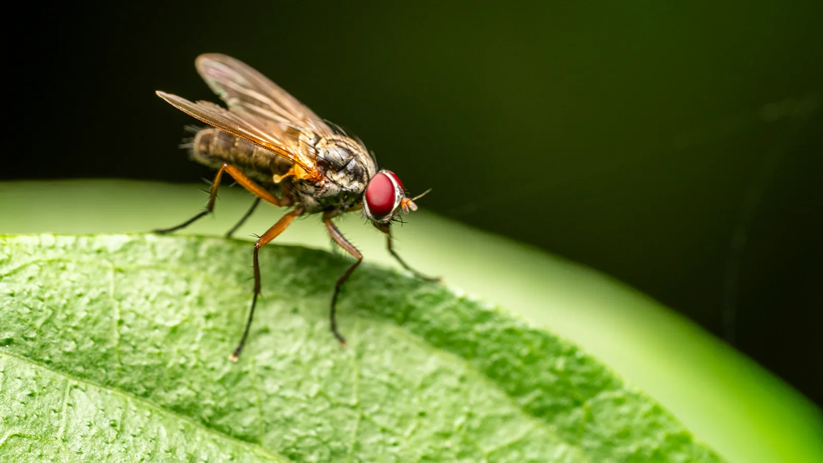
Myiasis disease is the infestation of living vertebrate tissue by the larvae of flies in the order Diptera. The larvae, commonly called maggots, feed on the host's tissue or on necrotic debris within a wound, and the resulting damage ranges from a minor skin lesion to a rapidly fatal systemic illness. Fly strike is the everyday name for the cutaneous form, and it is one of the few parasitic diseases in which the environment, the host's hygiene, and the fly population all have to line up before an animal is at risk.
This article covers the obligate, facultative, and accidental categories of myiasis, the main agents by species and host, the risk factors that owners and keepers can actually control, and the treatment pathway that veterinarians follow. It is written for US pet owners, sheep producers, and small-animal and exotic-animal keepers who need to recognize the condition early and act quickly.
This article is educational and is not a substitute for veterinary diagnosis or treatment.
What Myiasis Disease Actually Is
Myiasis is defined as a parasitic infestation of the tissues and organs of living vertebrates by dipterous larvae [1]. The female fly deposits eggs or first-stage larvae on the host's tissue, and the larvae that hatch go through three instars before they drop off to pupate in the soil. Each instar is larger and more destructive than the last, which is why the severity of a strike can escalate within days.
Two features separate myiasis from other parasitic infestations. First, the larvae are not passive passengers. They have mouth hooks and, in some species, rings of spines that anchor them in tissue and rasp at the wound bed. Second, the infestation is a moving target. A wound that is clean on Monday can be a deep, malodorous cavity by Thursday if the weather is warm and the fly population is high.
The larvae feed on living tissue, on necrotic tissue, or on both, depending on the species. Obligate myiasis flies require a living host to complete their life cycle. Facultative flies normally breed in carrion or decaying organic matter but will opportunistically colonize a wound on a living animal. Accidental myiasis occurs when eggs or larvae are ingested or deposited on a body surface where they cannot develop, and it usually resolves without treatment once the larvae are passed or removed.
The Three Categories of Myiasis
Obligate Myiasis
Obligate parasites cannot complete their life cycle without a living host. The classic examples are the New World screwworm, Cochliomyia hominivorax, and the Old World screwworm, Chrysomya bezziana. Both deposit eggs at the edges of wounds, and the larvae burrow into healthy tissue rather than feeding on dead material. This is the most dangerous category because the larvae actively enlarge the wound and can invade deeper structures.
Oestrus ovis, the sheep nasal bot fly, is also an obligate parasite, but it occupies a different niche. The adult fly deposits first-stage larvae around the nostrils of sheep, and the larvae migrate into the nasal sinuses and frontal sinuses, where they develop. Elephantoloemus indicus causes obligatory cutaneous myiasis in Indian elephants, forming warbles in the skin [2].
Facultative Myiasis
Facultative flies normally develop in carrion, feces, or decaying vegetation, but they will lay eggs on a wound or on soiled skin if the opportunity arises. Lucilia sericata, Lucilia cuprina, Calliphora vicina, and Chrysomya species fall into this group. The Australian sheep blowfly, L. cuprina, is the principal cause of blowfly strike in Australian sheep, and strike is endemic across the sheep-producing areas of that country [3]. In Great Britain, L. sericata is the primary causative species in pet rabbits [4] and the most frequent primary agent of northern European ovine cutaneous myiasis [5].
Facultative myiasis is the category owners are most likely to encounter. It is also the category where husbandry has the greatest effect, because the flies need a predisposing factor such as a wound, urine or fecal soiling, or moisture trapped in the coat.
Accidental Myiasis
Accidental myiasis happens when a fly deposits eggs or larvae on a host in a way that does not support development. Larvae may be ingested with food, may enter the gastrointestinal tract and be passed in the stool, or may be deposited on intact skin and fail to establish. These cases are usually self-limiting, but they can cause confusion when larvae are found in a stool sample or on a body surface.
The Main Agents by Species and Host
The table below summarizes the principal myiasis agents, their predilection sites, and the host and environmental factors that raise risk. It is organized by host so that owners and keepers can find their animal quickly.
| Host | Main agent | Predilection site | Key risk factors |
|---|---|---|---|
| Sheep | Lucilia cuprina | Breech, body | Fecal soiling, skin wrinkles, warm humid weather, fleece moisture [3] |
| Sheep | Lucilia sericata | Wounds, body | Wounds, soiled fleece, warm season [5][6] |
| Sheep | Chrysomya megacephala, C. albiceps | Wounds | Open wounds, autumn and summer peaks [6][7] |
| Sheep | Oestrus ovis | Nasal and frontal sinuses | Grazing near fly activity, warm months |
| Cattle, dogs, cats, wildlife (Americas) | Cochliomyia hominivorax | Wounds, body orifices | Any break in the skin, warm climate |
| Dogs, cats (tropics) | Chrysomya bezziana | Ear canals, perineum, paws, tail | Fight wounds, exudative lesions [8] |
| Dogs (Africa) | Cordylobia anthropophaga | Skin, ears | Warm humid conditions, outdoor exposure [9][10] |
| Dogs, cats (Europe) | Lucilia sericata, Calliphora vicina | Wounds, perineum | Wounds, soiling, moisture [11] |
| Rabbits | Lucilia sericata | Perineal area | Age over five years, entire females, perineal soiling [4] |
| Rodents and rabbits | Cuterebra species | Subcutaneous swellings | Outdoor housing, warm months |
| Reptiles (box turtles) | Sarcophagid larvae | Cutaneous and subcutaneous tissue | Bog habitat, poor body condition [12][13] |
| Deer | Dermatobia hominis | Skin | Spring and summer, herd exposure [14] |
| Elephants | Elephantoloemus indicus | Skin warbles | Captive housing in endemic regions [2] |
Sheep Blowfly Strike
Blowfly strike is the most economically important myiasis in livestock. In Australia, L. cuprina is the principal agent, and surveys of sheep farmers between 2003 and 2019 showed that breech strike was reported at a higher rate than body strike in every survey year [3]. Reported annual incidence of breech strike ranged from 2.2 to 4.7 percent of sheep nationally, while body strike ranged from 1.0 to 7.1 percent, with younger sheep more affected [3].
The reason breech strike dominates is anatomical. Fecal soiling and urine staining around the breech create the moist, protein-rich substrate that Lucilia females seek. Skin wrinkles trap moisture and delay drying, which is why the Mules operation, a surgical procedure that removes skin from the breech area, has been used as a preventative measure. The use of mulesing did not change between 2011 and 2018 in the Australian surveys, but the use of pain relief at mulesing of replacement ewe lambs increased substantially, from 59 percent in 2011 to 87 percent in 2018 [3].
Preventative chemical treatment has become the dominant control strategy. The use of fixed-time routine preventative treatments rose from 43 percent in 2003 to 66 percent in 2018, with dicyclanil and cyromazine the main insecticides used for control since 2011 [3]. A deterministic model of strike management found that not employing prophylactic treatment is the lowest-cost strategy only where strike risk is low. In all other circumstances, prophylactic treatment costs less than doing nothing, because the deaths associated with strike outweigh the cost of prevention [15]. Lamb treatment has a particularly large effect, since lambs are the most abundant age class and face the highest risk during the period of greatest fly abundance [15].
Screwworm Myiasis in the Americas and Tropics
Cochliomyia hominivorax is the New World screwworm and the most feared obligate agent in the Americas. It deposits eggs at the edges of wounds, and the larvae feed on living tissue. Any wound can attract the fly, including castration sites, ear notches, and bite wounds. Chrysomya bezziana is the Old World screwworm and occupies the same ecological niche in tropical and subtropical regions of Africa, Asia, and the Middle East.
A study of 76 canine and feline cases in Malaysia found that intact male mixed-breed dogs and intact male domestic short-hair cats with suspected fight-related wounds were most commonly presented with exudative and ulcerative lesions associated with screw-worm myiasis [8]. The most common anatomical sites in dogs were the external ear canals, followed by the perineum and the medial canthus of the eye. In cats, the paws and tail were most often affected [8]. This pattern reflects the underlying cause: fight wounds create the break in the skin that the fly needs, and intact animals fight more.
Cuterebra in Rodents and Rabbits
Cuterebra species, the rodent and rabbit bot flies, cause a different form of myiasis. The adult fly deposits eggs near the burrow or nest, and the larvae enter the host through the skin or a body opening. They develop in a subcutaneous warble, a swelling with a small breathing pore, and then drop out to pupate. These lesions are usually solitary and less destructive than screwworm infestations, but they can become secondarily infected, and a larva that migrates to an abnormal site such as the eye or the central nervous system can cause serious disease.
Oestrus ovis in Sheep
Oestrus ovis is the sheep nasal bot fly. The adult fly is viviparous, depositing first-stage larvae around the nostrils rather than laying eggs. The larvae crawl into the nasal cavity and migrate to the sinuses, where they grow and cause irritation, nasal discharge, and head shaking. The condition is seasonal and tied to fly activity, and it is managed through the same seasonal treatment planning that producers use for cutaneous strike.
Myiasis in Reptiles and Exotic Species
Myiasis is not limited to mammals. Eastern box turtles on Cape Cod were found with fly larvae in cutaneous and subcutaneous tissue in 30.5 percent of sampled animals, and every turtle found in bogs had evidence of larval infection [12]. Turtles with myiasis had lower body condition index, lower total white blood cell counts, lower packed cell volume, lower total solids, higher erythrocyte sedimentation rate, and lower calcium and phosphorus than unaffected turtles [12]. A separate case in an ornate box turtle used computed tomography to identify gas-filled sinus tracts containing larvae before surgical extraction [13]. These findings show that myiasis in reptiles is a marker of underlying debility, not just a surface problem.
Dermatobia hominis, the human bot fly, caused enzootic cutaneous myiasis with 100 percent morbidity in a herd of sambar deer in Brazil [14]. The skin lesions were characterized by eosinophilic and granulomatous chronic active severe necrohemorrhagic dermatitis associated with the botfly [14]. This is a reminder that myiasis can affect entire groups when environmental conditions and host density align.
How the Life Cycle Drives the Disease
The myiasis life cycle has four stages: egg or first-stage larva, three larval instars, pupa, and adult fly. The stage that matters clinically is the larval stage, because that is when tissue damage occurs. The female fly's choice of oviposition site determines whether the infestation becomes a minor nuisance or a life-threatening emergency.
For facultative species such as Lucilia, the female is attracted to moisture, ammonia, and the smell of decomposing organic matter. A soiled fleece, a urine-stained perineum, or a wound with exudate all provide that signal. For obligate species such as Cochliomyia and Chrysomya bezziana, the female is attracted specifically to the edges of fresh wounds, and the larvae then feed on living tissue rather than waiting for necrosis to develop.
The immune response of the host also shapes the outcome. A study comparing Merino ewes genetically selected for resistance to breech strike with non-selected ewes found that larval challenge triggered an infiltration of leukocytes, predominantly neutrophils, at the feeding sites [16]. Lymphocytes expressing T cell markers for CD4, CD1, CD8, T19, gamma-delta T cell, and the B cell marker CD45R increased significantly compared with control sites, and the pro-inflammatory cytokines IL-1 alpha, IL-6, and IL-17a, along with the chemoattractants IL-8 and MIP-1 alpha, were significantly elevated [16]. This suggests that resistance to strike is partly an immune phenomenon, which is why breeding for resistance has become a recognized component of control programs [3].
The following flowchart shows the decision path from a suspicious lesion to definitive treatment.
flowchart TD
    A[Animal with skin lesion or soiling] --> B{Wound or soiled area}
    B --> C[Inspect for larvae]
    C --> D{Larvae present}
    D --> E[Do not attempt manual removal of embedded larvae]
    E --> F[Contact veterinarian same day]
    F --> G[Debridement of wound]
    G --> H[Species appropriate antiparasitic treatment]
    H --> I[Address underlying cause]
    I --> J[Recheck and monitor for recurrence]
    D --> K[No larvae but risk factors present]
    K --> L[Preventative fly control and hygiene]
Recognizing Fly Strike in Your Animal
The earliest sign of cutaneous myiasis is often behavioral rather than visible. An affected animal may stamp its feet, bite or scratch at a specific spot, become restless, or separate itself from the group. In sheep, a struck animal may be seen nibbling at the affected area or holding its tail in an abnormal position.
As the infestation progresses, the lesion becomes visible. The wound is typically moist, malodorous, and enlarging, with a dark, necrotic center and a rim of inflamed tissue. Larvae may be visible at the edges or within the wound. In rabbits, the perineal area is overwhelmingly the site of blowfly strike lesions, accounting for 52.4 percent of recorded cases in a British study of 243 consultations [4]. Other body areas and the head were less commonly affected, and in 39.9 percent of consultations the affected area was not specified [4].
The outcome of untreated strike is severe. In the same rabbit study, 44.7 percent of rabbits presenting with blowfly strike were recorded as euthanized or died [4]. This figure is a strong argument for early recognition and for treating predisposing conditions before strike season begins.
Predisposing Conditions in Companion Animals
In dogs and cats, cutaneous myiasis is usually secondary to an underlying problem. A review of cases in Italy described a cat with Calliphora vicina myiasis and a dog with Lucilia sericata myiasis that occurred in the absence of lesions, which is unusual and highlights that not every case has an obvious wound [11]. The authors emphasized the importance of identifying predisposing conditions and risk factors rather than treating the larvae alone [11].
In Africa, Cordylobia anthropophaga, the tumbu fly, is a principal cause of nonmigratory cutaneous myiasis in domestic animals. A cross-sectional study in Kitui County, Kenya found an overall prevalence of canine cutaneous myiasis of 45 percent, with all larvae identified as C. anthropophaga [10]. Puppies under six months appeared more affected, although the difference was not statistically significant [10]. A separate study in Chad found C. anthropophaga, Chrysomya bezziana, and Chrysomya species in dogs, with a mean infestation of 2.28 larvae per animal and a range of 1 to 24 [9].
Treatment: Debridement Plus Antiparasitic Therapy
The treatment of cutaneous myiasis has two essential components. The first is mechanical removal of the larvae and debridement of devitalized tissue. The second is species-appropriate antiparasitic treatment. Neither component alone is sufficient, because larvae that remain in the wound continue to cause damage, and debridement alone does not address larvae that are still developing or prevent reinfestation.
Why Manual Removal Alone Is Not Enough
Owners sometimes try to remove larvae by squeezing or picking at the wound. This is risky. Larvae have mouth hooks and spines that anchor them in tissue, and a larva that is partially removed can rupture, releasing bacteria and antigenic material into the wound. In obligate myiasis, the larvae are embedded in living tissue, and attempting removal without analgesia and proper instrumentation causes pain and may drive the larva deeper.
Veterinary debridement involves clipping and cleaning the surrounding area, removing necrotic tissue, and extracting larvae under appropriate restraint or anesthesia. In a case of a complex scalp wound with myiasis, initial debridement and mechanical removal of larvae resulted in a large wound that required a combined wound approach with negative pressure wound therapy and a skin graft [17]. That case illustrates the principle that the wound left behind after larval removal often needs its own management plan.
Antiparasitic Treatment
Antiparasitic treatment for myiasis is species-specific and must be selected by a veterinarian. The choice depends on the fly species, the host species, the extent of the infestation, and the presence of secondary infection. In a herd of sambar deer with enzootic cutaneous myiasis caused by Dermatobia hominis, systemic treatment with oral ivermectin at 0.08 percent was effective in 93.7 percent of deer after fourteen days [14]. That result is specific to that host, that parasite, and that formulation, and it should not be generalized to other species or other agents.
For dogs with Chrysomya bezziana myiasis in Thailand, removal of fly larvae together with specific treatment cured all cases in the study [18]. The authors did not report a single drug protocol that would apply universally, which reflects the reality that treatment is tailored to the case.
Antibiotic coverage is often part of the plan because myiasis wounds are contaminated and can be secondarily infected. In a case of furuncular myiasis in a traveler, timely larval removal and prophylactic antibiotics were used to minimize complications [19]. The decision to use antibiotics, and which antibiotic to use, is a clinical judgment based on the wound and the host.
Addressing the Underlying Cause
Treating the larvae without treating the reason the fly was attracted guarantees a recurrence. In sheep, that means managing fecal soiling, treating diarrhea, controlling internal parasites, and considering breeding for resistance [3][16]. In rabbits, it means addressing perineal soiling, dental disease that causes drooling, and obesity that prevents normal grooming. In dogs and cats, it means treating the fight wounds, skin infections, and other conditions that created the break in the skin [8].
Prevention and Control
Prevention is organized around three levers: reduce fly attraction, reduce host susceptibility, and reduce fly abundance.
Reducing fly attraction means keeping the animal clean and dry. For sheep, that means managing breech soiling and avoiding prolonged fleece moisture. For rabbits, it means daily checks of the perineal area and prompt veterinary care for any condition that causes soiling. For dogs and cats, it means cleaning and covering wounds and treating skin disease promptly.
Reducing host susceptibility includes breeding for resistance where that is feasible. The Australian surveys documented increased use of breeding for resistance as a control measure [3], and the immune study in Merino ewes supports the biological basis for that approach [16].
Reducing fly abundance includes chemical control and environmental management. The Australian data show that fixed-time routine preventative chemical treatments are now the dominant strategy, with dicyclanil and cyromazine the main insecticides used [3]. The modeling work confirms that prophylactic treatment is cost-effective whenever strike risk is more than low, and that treating lambs is particularly valuable because they are the most abundant and most vulnerable age class [15].
Climate is the wild card. A species distribution model for Lucilia sericata in Great Britain predicted that the range of elevated temperatures expected under climate change scenarios will increase the risk of strike and elongate the blowfly season [5]. The model did not predict strike throughout the winter even under the most rapid warming scenario, but the lengthening of the risk period has direct implications for when preventative treatment should start and stop [5].
Limitations and When to Contact a Veterinarian
Myiasis is a clinical diagnosis that requires a veterinarian to identify the larvae, assess the depth and extent of tissue damage, and select appropriate treatment. Individual cases vary widely, and the information in this article cannot replace an examination.
Contact a veterinarian immediately if you see any of the following:
- Larvae in a wound, in the coat, or around a body opening
- A wound that is enlarging, malodorous, or discharging
- An animal that is restless, biting or scratching at one spot, or separating from the group
- Perineal soiling in a rabbit, especially in an animal over five years old or an entire female
- A subcutaneous swelling with a visible pore in a rodent or rabbit
- Any sign of lethargy, loss of appetite, or collapse in an animal with a skin lesion
- A sheep with breech or body soiling during warm, humid weather
Do not wait to see whether the lesion improves on its own. The rabbit data show that nearly half of affected animals died or were euthanized, and the speed of progression in obligate myiasis means that hours matter [4].
Frequently Asked Questions
What is myiasis disease?
Myiasis disease is the infestation of living vertebrate tissue by the larvae of flies. The larvae feed on living or necrotic tissue, and the severity depends on the fly species, the site, and how quickly treatment begins [1].
Is fly strike the same as myiasis?
Fly strike is the common name for cutaneous myiasis, the form that affects the skin. It is the most common form of myiasis, but larvae can also infest other organs and body cavities [20].
Which flies cause myiasis in sheep?
Lucilia cuprina is the principal cause of blowfly strike in Australian sheep, and Lucilia sericata is the most frequent primary agent in northern Europe [3][5]. Chrysomya species and Oestrus ovis also affect sheep [6][7].
Can myiasis affect indoor pets?
Yes. Blowfly strike occurs in pet rabbits, and Lucilia sericata is the primary causative species in Great Britain [4]. Indoor housing reduces but does not eliminate risk, especially if the animal has a wound or perineal soiling.
How is cutaneous myiasis treated?
Treatment requires debridement of the wound and removal of larvae, plus species-appropriate antiparasitic treatment selected by a veterinarian [18][14]. Antibiotics may be needed if the wound is secondarily infected [19].
Can I remove the maggots myself?
No. Larvae anchor themselves in tissue and can rupture if squeezed, and obligate species are embedded in living tissue. Removal should be done by a veterinarian with appropriate restraint, analgesia, and instrumentation [17].
Why do some animals get fly strike and others do not?
Risk depends on fly abundance, host susceptibility, and climate. Fecal soiling, skin wrinkles, wounds, and warm humid weather all raise risk, and genetic resistance plays a role in sheep [3][5][16].
Is myiasis contagious between animals?
Myiasis is not transmitted directly from one animal to another. The fly is the vector, so an affected animal indicates that flies are active in the environment and that other animals may also be at risk [15].
Related Articles
- Melophagus ovinus in Sheep: Sheep Ked, Wingless Fly Ectoparasite Infestation, Fleece Damage, and Anemia
- Gasterophilus intestinalis in Horses: Equine Bot Fly Larvae Infestation of Stomach Walls and Control Strategies
- Larval Rearing Protocols for Marine Finfish
- Fly and Pest Control for Horses
- Dairy Farm Fly Control
- Bunostomum phlebotomum Cattle Hookworm: Percutaneous Larval Infection and Dermatitis
- Dog and Flies: Fly Strike and Bite Relief
- Sand Fly: Biology, Diseases, and Veterinary Importance
- Do Fleas Fly? Flea Movement and Jumping Explained
Sources
- [[Cutanous myiasis caused by Sarcophaga spp. larvae in a diabetic patient].](https://pubmed.ncbi.nlm.nih.gov/24819275/)
- Elephantoloemus indicus Austen, 1930 (Diptera: Calliphoridae) as the cause of cutaneous myiasis in captive Indian elephants from Assam, India.
- Australian surveys on incidence and control of blowfly strike in sheep between 2003 and 2019 reveal increased use of breeding for resistance, treatment with preventative chemicals and pain relief around mulesing.
- Risk factors for cutaneous myiasis (blowfly strike) in pet rabbits in Great Britain based on text-mining veterinary electronic health records.
- Modelling the impact of climate change on spatial patterns of disease risk: sheep blowfly strike by Lucilia sericata in Great Britain.
- Unravelling the role of Calliphoridae (Insecta: Diptera) as a causative agent of myiasis in sheep with Chrysomya megacephala (Fabricius) as the first documented agent of myiasis in ruminants from Kashmir Himalaya, India.
- Molecular identification of Chrysomya albiceps (Diptera: Calliphoridae) in a case of primary cutaneous myiasis in sheep at southern Brazil.
- Canine and feline cutaneous screw-worm myiasis in Malaysia: clinical aspects in 76 cases.
- Cutaneous myiasis by Calliphoridae dipterans in dogs from Chad.
- Prevalence, Etiology, and Risk Factors Associated with Occurrence of Canine Cutaneous Myiasis in Kitui County, Kenya.
- Cutaneous myiasis in cats and dogs: Cases, predisposing conditions and risk factors.
- CUTANEOUS MYIASIS AND ITS RELATIONSHIP TO WELLNESS IN EASTERN BOX TURTLES (TERRAPENE CAROLINA CAROLINA) IN CAPE COD, MASSACHUSETTS.
- COMPUTED TOMOGRAPHY OF CUTANEOUS MYIASIS IN AN ORNATE BOX TURTLE (TERRAPENE ORNATA ORNATA).
- High morbidity cutaneous enzootic myiasis by Dermatobia hominis (Diptera: Oestridae) in sambar deer (Rusa unicolor).
- Sheep blowfly strike: the cost of control in relation to risk.
- Characterising the innate immune response in breech strike resistant and non-selected sheep to the sheep blowfly (Lucilia cuprina).
- A complex scalp wound following a myiasis infestation: cranial burr holes and negative pressure wound therapy.
- First report of canine cutaneous myiasis caused by Chrysomya bezziana Villeneuve (Diptera: Calliphoridae) in Thailand.
- Cutaneous myiasis in a Serbian traveller returning from Kenya.
- Wound myiasis by housefly in a patient with pemphigus vulgaris in Riyadh, Saudi Arabia.