Dermatobia hominis: Human Bot Fly Lifecycle
By Dr. Zubair Khalid, DVM, MS, PhD ·
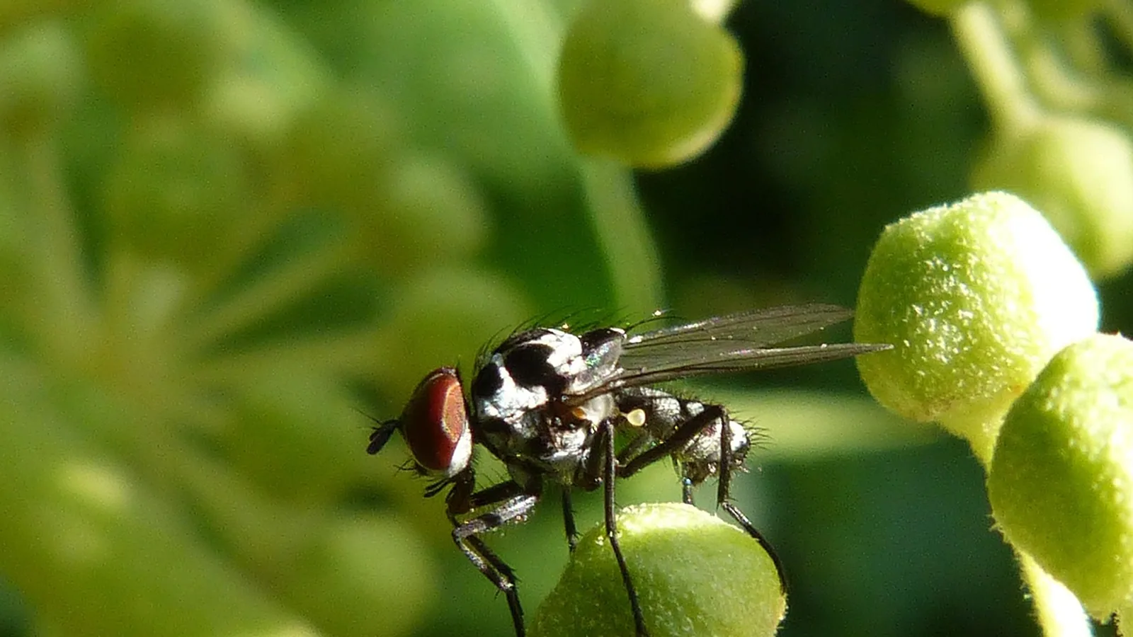
Dermatobia hominis is a Neotropical bot fly whose larvae develop inside a boil-like swelling under the skin of mammals, including cattle, dogs, cats, and people. The lifecycle is unusual because the adult female never lays eggs directly on the host. She catches a blood-feeding insect such as a mosquito or tick, glues her eggs to its body, and releases it to deliver her offspring.
That single behavior explains most of what owners observe. Infestations appear without any obvious fly strike, lesions show up as firm nodules with a central pore, and the parasite can travel far outside its native range inside animals and people who move between countries.
This article is educational and is not a substitute for veterinary diagnosis or treatment.
The Short Answer
A female D. hominis captures a blood-feeding arthropod mid-flight, attaches a batch of eggs to its abdomen, and lets it go. When that carrier lands on a warm mammal to feed, body heat triggers the eggs to hatch. First-instar larvae enter through skin or mucous membranes, molt twice inside a subcutaneous nodule, and drop to the ground as third-instar larvae to pupate in soil. The full parasitic phase in cattle runs roughly seven to ten weeks, and each nodule holds one larva with a breathing pore open to the air.
Why the Human Bot Fly Is Not a Typical Myiasis Fly
Furuncular myiasis is the clinical name for the boil-like lesion that D. hominis produces. The term "human bot fly" is common in the medical literature, but the parasite is not restricted to people. Cattle are the most economically important hosts in Latin America, where D. hominis, the horn fly Haematobia irritans, and the New World screwworm Cochliomyia hominivorax are considered the three most significant flies affecting livestock production [1].
Infestation has been documented across a wide host range in the Neotropics. Domestic dogs are commonly affected, and furuncular myiasis from D. hominis is described as the second most common skin disease in dogs living in tropical climates of Central and South America [2]. Cats are infested less often, but cases exist. A Brazilian Shorthair cat from rural Rio de Janeiro state developed two nodules, one over the sacrum and one on the tail, and the lesions initially resembled a tumor or an abscess [3]. Five additional cats in Rio de Janeiro state were diagnosed with the same condition [4].
Wild mammals are affected too. Furuncular myiasis was recorded for the first time in wild jaguars in the Brazilian Amazon, with lesions on the rear thighs and tail [5]. A herd of sambar deer in Rio de Janeiro showed 100 percent morbidity from enzootic cutaneous myiasis during spring and summer, and no other wild herbivore species in the same area was affected [6].
The pattern is consistent. D. hominis is a generalist parasite of warm-blooded mammals in the Neotropics, and any mammal in that region can develop nodules.
The Full Lifecycle, Stage by Stage
The lifecycle has two distinct phases. The free-living phase involves the adult fly, the egg, and the pupa. The parasitic phase covers the three larval instars inside the host.
Stage 1: The Adult Female and Egg Transport
Adult D. hominis are free-living flies that do not feed on blood. The female's reproductive strategy is the defining feature of the species. Instead of depositing eggs on a host, she intercepts a blood-feeding arthropod, most often a mosquito or a tick, and cements her eggs to its body. The carrier is then released.
This behavior is called phoresy, and it turns an unrelated insect into a delivery vehicle. The eggs ride on the vector until it lands on a mammal to take a blood meal. The warmth of the host's body and the proximity of skin trigger hatching.
Stage 2: Entry of the First-Instar Larva
First-instar larvae (L1) emerge on or near the host and enter the skin. Experimental work in cattle showed that L1 larvae penetrate actively through the skin and migrate toward the subcutaneous tissues rather than waiting in a wound [7].
Entry does not require a pre-existing break in the skin. Larvae can also enter through mucous membranes. Once inside, each larva settles in the subcutaneous tissue and begins to feed.
Stage 3: Development Through Three Instars
Inside the host, the larva passes through three instars. The first instar establishes the nodule. The second instar enlarges it. The third instar (L3) is the mature, barrel-shaped larva that eventually exits.
The developmental timeline has been measured in several ways. In a mouse model, larvae implanted at four days (L1), six days (early L2), twelve days (L2), or twenty days (L3) after original infestation completed development on a schedule similar to larvae that stayed in their original host. The L3 that emerged from implanted hosts developed into pupae and fertile adults, and their offspring were used to maintain the parasite in the laboratory [8]. That work confirms the three instars are distinct, viable stages rather than arbitrary divisions.
Stage 4: The Furuncular Nodule and the Breathing Pore
Each larva sits in its own furuncular lesion, a firm, boil-like nodule with a small central opening. The larva maintains this opening as a breathing pore, and the pore connects the larva to the outside air. This is why a nodule may show a tiny central punctum, sometimes with a droplet of serosanguinous discharge [9] or intermittent air bubbles at the surface [10].
The nodule is not a passive cavity. The larva produces soluble antigens along a fistulous tract, and those antigens appear to help maintain the tract by interfering with normal wound healing [7]. The lesion also carries a characteristic bacterial community. In one study of 102 furuncular lesions in cattle, the significant bacteria recovered were Staphylococcus aureus, S. epidermidis, S. warneri, Bacillus subtilis, and Escherichia coli [11]. The same study found S. aureus, B. subtilis, and several other species associated with larvae and pupae, including Proteus species and Moraxella phenylpyruvica [11].
Stage 5: Exit and Pupation
When the third instar is mature, it leaves the host and falls to the ground. Pupation occurs in soil. The pupa is the transition stage between the parasitic larva and the free-living adult fly. Once the adult emerges, the cycle can begin again with a new egg batch and a new carrier arthropod.
The table below summarizes each stage, its host or substrate, and the approximate duration where the literature supports a figure.
| Lifecycle stage | Host or substrate | Approximate duration |
|---|---|---|
| Adult female and egg transport | Free-living, eggs carried on a blood-feeding arthropod | Not fixed. Eggs remain on the carrier until it feeds on a mammal |
| First instar (L1) entry | Mammalian skin or mucous membrane | Entry is immediate on hatching. L1 present in the host from day 0 |
| Second instar (L2) | Subcutaneous furuncular nodule | Develops after the first molt. Early L2 documented at about day 6 post-infestation in a mouse model [8] |
| Third instar (L3) | Subcutaneous furuncular nodule | Mature L3 documented at about day 20 post-infestation in the same model [8] |
| Exit and pupation | Soil | Larva drops from the nodule, pupates in soil, then emerges as an adult |
The mouse model gives relative timing for the instars [8]. Cattle treatment studies describe the parasitic phase in weeks rather than days, and the Merck Veterinary Manual describes D. hominis hypodermiasis as a grub infestation of cattle skin [12].
A Visual Map of the Cycle
The flow below traces the cycle from egg transport to the next generation of adult flies.
flowchart TD
    A[Adult female fly] --> B[Captures blood feeding arthropod]
    B --> C[Glues eggs to carrier]
    C --> D[Carrier lands on mammal]
    D --> E[Body heat triggers hatching]
    E --> F[Larva enters skin or mucosa]
    F --> G[First instar forms nodule]
    G --> H[Second instar enlarges nodule]
    H --> I[Third instar matures in nodule]
    I --> J[Larva exits and drops to soil]
    J --> K[Pupates and emerges as adult]
    K --> A
Why the Nodule Is Easy to Misread
Owners and even clinicians can mistake a D. hominis nodule for something else. The cat case from Brazil is a clear example. The lesions looked like a neoplasm or an abscess, and the correct diagnosis only became clear when digital compression expelled the larvae along with purulent material [3]. The authors concluded that D. hominis belongs on the differential diagnosis list for cats with nodules [3].
A dog case from Colombia followed a similar pattern. A two-year-old German Shepherd had a single furuncular lesion over the sacral area that resembled an inflammatory papule with a central punctum and serosanguinous discharge. Pressure around the wound revealed a larva inside the tissue, and surgical removal followed [9].
The takeaway is practical. A firm nodule with a central pore in an animal with outdoor access in an endemic region should raise suspicion of D. hominis, not just abscess or tumor.
Hosts and Species Differences
Cattle are the primary production animal affected, and the economic impact is substantial. D. hominis causes reduced productivity, veterinary expenses, and decreased animal welfare in Latin American cattle operations [1]. Control programs in Argentina, Brazil, Colombia, Mexico, and Uruguay rely heavily on insecticidal products, though resistant fly populations have emerged and reduced their effectiveness [1].
Dogs are frequent hosts. In one study, 25 naturally infested dogs each received a single oral dose of afoxolaner, and all larvae removed 24 hours later were dead, giving 100 percent larvicidal efficacy against D. hominis larvae [2]. Another study of ten dogs treated with sarolaner found no live larvae 24 hours after treatment, also 100 percent efficacy [13]. A third study of 12 dogs treated with lotilaner reported 80.6 percent efficacy six hours after a single oral dose, with the surviving larvae showing hypomobility [14].
Cats are less commonly infested, but the condition is real. Five cats treated with topical fluralaner had dead larvae within 24 hours in three cats and within 48 hours in two, with spontaneous expulsion in two cats and manual expression needed in the other three [4]. The earlier cat case resolved with mechanical removal and antiseptic use [3].
Wild mammals including jaguars and sambar deer are also affected [5][6]. In the sambar deer herd, systemic ivermectin at 0.08 percent was effective in 93.7 percent of affected deer after fourteen days [6].
Myiasis in Animals: What the Nodule Does to Tissue
The lesion is not just a cosmetic problem. Histology from the sambar deer herd showed eosinophilic and granulomatous chronic active severe necrohemorrhagic dermatitis associated with the botfly, plus moderate surrounding fibroplasia [6]. After treatment, the skin showed eosinophilic and granulomatous chronic severe dermatitis associated with degenerated botfly and severe fibroplasia [6]. Leukocyte profiles shifted as well, with significant decreases in relative neutrophils and eosinophils and an increase in monocytes after treatment [6].
In cattle, the immune response to first-instar larvae involves large numbers of eosinophils, which the authors identified as the most important cell in mediating damage to D. hominis larvae. Immunoglobulins G and M bound only to dead or molting larvae, which may reflect a mechanism that protects living larvae from the host immune response [7].
That immune evasion helps explain why nodules persist. The larva is not simply tolerated. It actively manages the local environment to stay alive.
The Relationship With Screwworm Myiasis
A common claim is that D. hominis lesions predispose cattle to New World screwworm myiasis. One study tested this directly. Only 5.2 to 7.4 percent of gravid C. hominivorax females laid eggs in offered furuncular lesions. Of 3,242 eggs laid on botfly lesions, only 82 reached second instar and all died. The lesions served as a food source in 81.3 percent of tested cases, but the authors concluded that furuncular lesions do not serve as a predisposing factor for screwworm myiasis in the tropics [15]. The proposed reasons included lesion pH, associated microflora, and the structure of the furuncle itself [15].
This matters for owners because it corrects a widespread assumption. A bot fly nodule is not automatically an open door for screwworm.
Diagnosis in Animals
Diagnosis is usually clinical. A veterinarian identifies a firm nodule with a central pore, often on the back, flanks, tail head, or limbs. Visualization of larval motility through the lesion orifice was used to confirm diagnosis in the sarolaner dog study [13]. In the cat case, diagnosis was confirmed when digital compression expelled the larvae [3].
In people, diagnosis is sometimes supported by microscopy and molecular confirmation. Two imported cases in Romania used larval morphology and mitochondrial cytochrome c oxidase subunit I sequencing to confirm D. hominis, with 97.14 to 99.33 percent identity to reference sequences [10]. That level of testing is not routine in veterinary practice, but it shows how confirmation works when morphology alone is ambiguous.
Extraction Risks and Why Larval Rupture Matters
Manual extraction is the traditional approach, and it carries a specific hazard. If the larva ruptures during removal, its contents spill into the surrounding tissue. That can trigger a severe inflammatory response, and in sensitized hosts it can progress to anaphylaxis. This is the main reason veterinarians prefer to kill the larva in place before removing it, or to use controlled techniques that avoid crushing the grub.
The literature supports pre-treatment with larvicidal drugs to make removal safer and less traumatic. The sarolaner study noted that medications to kill the larva before extraction can reduce inflammation and discomfort and provide a less traumatic larval removal [13]. The same logic applies to afoxolaner, lotilaner, and fluralaner, all of which showed larvicidal activity in dogs or cats [14][2][13][4].
Occlusion of the breathing pore is another technique. In the Romanian human cases, petroleum jelly applied to the breathing pore facilitated mechanical extraction of one larva per lesion [10]. The principle is that blocking the pore forces the larva to move toward the surface, where it can be removed more cleanly.
Owners should not attempt extraction at home. Squeezing a nodule can crush the larva, and the resulting reaction can be serious.
Prevention and Control in Endemic Regions
Prevention in animals relies on reducing exposure to carrier arthropods and on strategic use of parasiticides. Because the eggs arrive on mosquitoes and ticks, vector control is part of bot fly control. The integrated approach described for Latin American cattle operations combines chemical and non-chemical methods, including mechanical, environmental, biological, and genetic strategies, to slow the development of insecticide resistance [1].
Resistance is a real concern. A study in Brazil documented D. hominis resistance to doramectin in cattle with a history of clinical parasitism after avermectin use [16]. That finding changes how producers should think about routine avermectin use.
For dogs and cats in endemic areas, the isoxazoline class has both preventive and treatment roles. Afoxolaner, sarolaner, lotilaner, and fluralaner all demonstrated larvicidal activity against D. hominis in naturally infested animals [14][2][13][4]. Pet owners in endemic regions should discuss year-round parasite prevention with their veterinarian.
Travelers and relocated animals can carry the parasite into non-endemic countries. Cases have been imported into the United Kingdom from Belize [17], into Saudi Arabia in people with no travel history outside the country [18], and into Romania from Peru [10]. The Saudi cases suggested domestic cattle as the source and raised the possibility that the parasite may be endemic in that region [18]. This means a nodule in a pet with no travel history is not automatically something else.
What Is Still Uncertain
Several gaps remain. The precise duration of each instar in natural cattle infestations is not fixed in the literature, and much of the timing data comes from experimental models [8]. The role of specific bacterial species in the furuncular lesion is described but not fully explained [11]. The reasons furuncular lesions fail to attract screwworm flies are proposed but not settled [15]. Susceptibility differences between host species, such as why sambar deer were affected while other wild herbivores in the same area were not, need more study [6].
Limitations and When to Contact a Veterinarian
This article is educational and is not a substitute for veterinary diagnosis or treatment. Individual animals need individual assessment.
Contact a veterinarian promptly if you see any of the following:
- A firm nodule with a central pore, especially in an animal with outdoor access in an endemic region or with recent travel to Latin America.
- Visible movement inside a skin lesion.
- A nodule that is painful, growing, or draining.
- Signs of systemic illness such as fever, lethargy, or loss of appetite alongside a skin nodule.
- Any lesion in a pet that has traveled to or from Central or South America, the Caribbean, or another tropical region.
Do not squeeze, cut, or attempt to remove a suspected bot fly larva at home. Larval rupture can cause a severe reaction.
Frequently Asked Questions
What is the Dermatobia hominis lifecycle?
A female fly glues eggs to a blood-feeding arthropod, the carrier delivers them to a mammal, larvae enter the skin, develop through three instars in a nodule, then drop to soil to pupate.
How do bot fly larvae get into a dog or cat?
They hatch from eggs carried by a mosquito, tick, or other blood-feeding arthropod that lands on the animal. The larvae then enter through skin or mucous membranes.
Can indoor pets get Dermatobia hominis?
Indoor pets are at lower risk, but any animal exposed to mosquitoes or ticks in an endemic region can be infested. Outdoor access is a common factor in reported cases.
Is a bot fly nodule dangerous?
The nodule itself is usually localized, but it causes pain, inflammation, and tissue damage. Secondary bacterial involvement is common, and rupture during extraction can cause a severe reaction.
Why should you not squeeze a bot fly larva out?
Squeezing can rupture the larva and release its contents into the tissue, which can trigger intense inflammation and, in sensitized hosts, anaphylaxis.
Can Dermatobia hominis affect cattle?
Yes. Cattle are a major host, and the parasite causes production losses, veterinary costs, and reduced welfare in Latin American herds.
Does a bot fly lesion attract screwworm flies?
Research suggests it does not. One study found furuncular lesions did not serve as a predisposing factor for screwworm myiasis.
Can Dermatobia hominis spread outside Latin America?
Yes. Imported cases have been reported in the United Kingdom, Romania, and Saudi Arabia, usually linked to travel or animal movement.
Related Articles
- Gasterophilus intestinalis in Horses: Equine Bot Fly Larvae Infestation of Stomach Walls and Control Strategies
- Fly and Pest Control for Horses
- Dairy Farm Fly Control
- Method validation lifecycle stages
- Draschia megastoma in Horses: Stomach Bots, Gasterophilus Granulomas, and Diagnosis
- Chicken Flight Capabilities: Why They Fly and How to Manage It
- Viral Gastroenteritis Viruses in Animals and Humans
- Human Leukocyte Antigen (HLA) and MHC Explained
Sources
- Management Practices for the Control of Haematobia irritans, Dermatobia hominis, and Cochliomyia hominivorax in Cattle Across Latin America: A Sustainable, Collective Approach.
- Efficacy of Afoxolaner (NexGard®) in the treatment of furuncular myiasis caused by Dermatobia hominis fly (Diptera: Cuterebridae) in naturally infested dogs.
- Furuncular myiasis caused by Dermatobia hominis in a domestic cat - case report.
- Furuncular myiasis caused by Dermatobia hominis in five cats and efficacy of topical fluralaner for its treatment.
- Furuncular Myiasis by Dermatobia hominis (Diptera: Oestridae) in Wild Jaguars in the Amazon Rainforest.
- High morbidity cutaneous enzootic myiasis by Dermatobia hominis (Diptera: Oestridae) in sambar deer (Rusa unicolor).
- Histological and immunological reaction of cattle skin to first-instar larvae of Dermatobia hominis.
- Implantation of human bot fly larvae in host skin.
- First report of furuncular myiasis in a domestic dog caused by Dermatobia hominis (Linnaeus, 1781) in Colombia.
- Imported Furuncular Myiasis in a Non-Endemic Setting: Two Case Reports of Dermatobia hominis Infection in Romania and a Review of Reports from Southeast and Eastern Europe.
- The associated microflora to the larvae of human bot fly Dermatobia hominis L. Jr. (Diptera: Cuterebridae) and its furuncular lesions in cattle.
- Dermatobia hominis Hypodermiasis - Integumentary System - Merck Veterinary Manual
- Effectiveness of sarolaner in the clinical management of furuncular myiasis in dogs naturally infested with Dermatobia hominis (Diptera: Cuterebridae).
- Effectiveness of lotilaner on furuncular myiasis in dogs naturally infested with Dermatobia hominis (Diptera: Cuterebridae).
- The role of botfly myiasis due to Dermatobia hominis L.Jr. (Diptera:Cuterebridae) as a predisposing factor to New World screwworm myiasis (Cochliomyia hominivorax coquerel) (Diptera:Calliphoridae).
- First report of Dermatobia hominis resistant to doramectin in cattle.
- Cutaneous myiasis due to Dermatobia hominis: a report of six cases.
- Cutaneous myiasis due to Dermatobia hominis in Saudis.