Platelet-Rich Plasma Therapy for Pets
By Dr. Zubair Khalid, DVM, MS, PhD ·
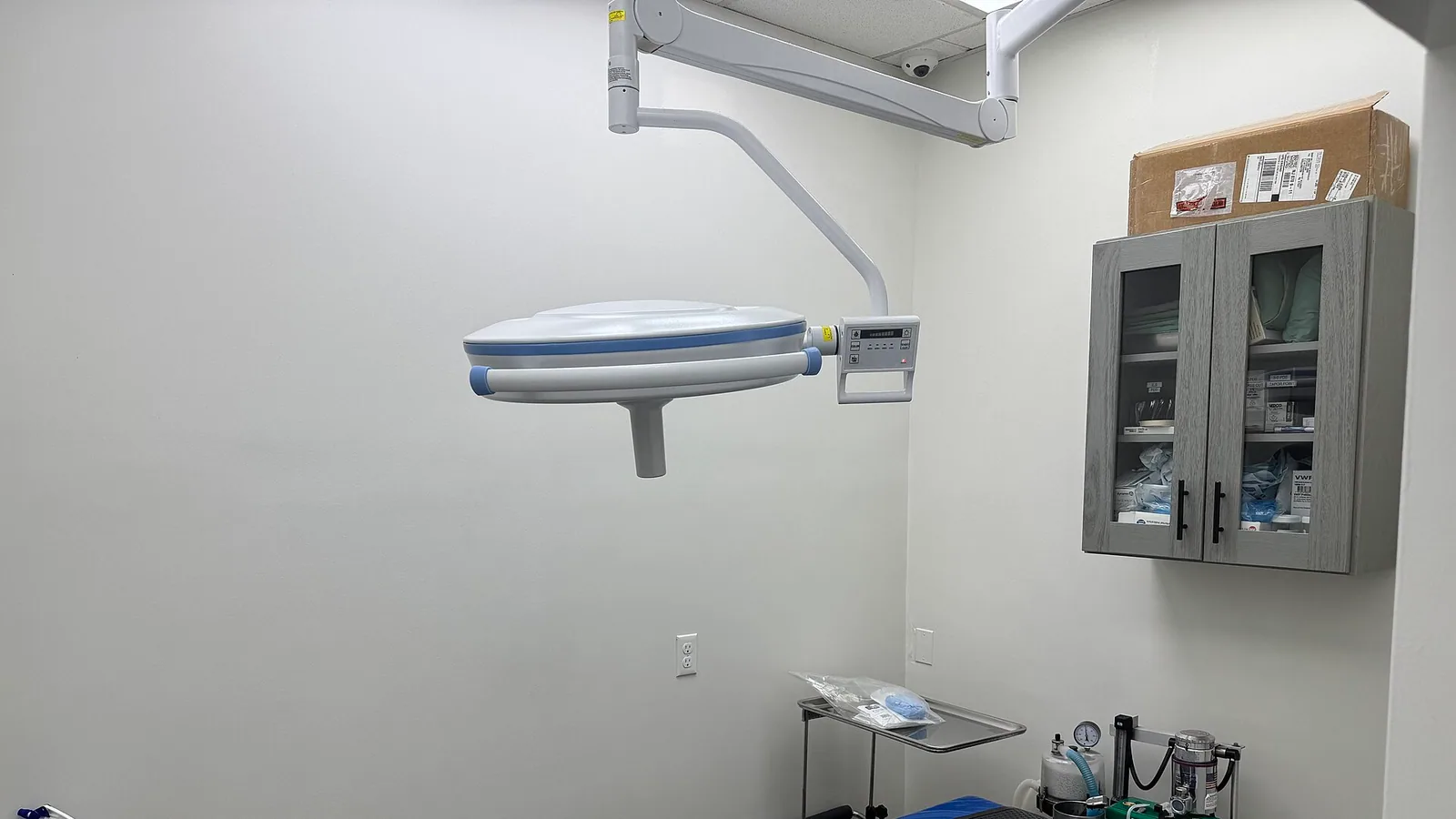
Platelet-rich plasma (PRP) therapy is a regenerative medical treatment that uses a patient's own blood to concentrate platelets and growth factors, which are then injected into damaged joints, tendons, or ligaments. For pets suffering from osteoarthritis, tendonitis, or ligament damage, PRP offers a treatment option that goes beyond simply managing pain. By directly modulating the inflammatory environment and promoting tissue repair, PRP aims to restore function and improve quality of life. While much of the rigorous clinical research on PRP has been conducted in equine medicine, the biological principles may be conserved, but clinical benefits must be demonstrated in dogs and cats. This article provides a comprehensive, source-grounded overview of PRP therapy for pets, covering the underlying science, clinical evidence, procedure details, safety, and what pet owners can expect.
At a Glance: PRP Therapy for Pets
PRP is often compared to traditional treatments for orthopedic conditions, such as non-steroidal anti-inflammatory drugs (NSAIDs) and corticosteroids. The table below summarizes the key differences.
| Feature | PRP Therapy | Corticosteroids (e.g., Triamcinolone) | NSAIDs (e.g., Carprofen) |
|---|---|---|---|
| , - | , - | , - | , - |
| Primary Goal | Regenerate tissue, modulate inflammation [4] | Rapid pain relief, reduce inflammation | Reduce pain and inflammation |
| Mechanism | Growth factors, cytokines, antiproteases [5], [6] | Inhibits inflammatory cascade | Inhibits COX enzymes |
| Cartilage Effects | Protective, promotes healing [4], [7] | May impair cartilage metabolism [7] | Variable |
| Onset of Action | Gradual, weeks [3] | Rapid (days) | Rapid (hours to days) |
| Duration of Effect | Potentially long-lasting [1] | Short to medium | Short, requires daily dosing |
| Common Side Effects | Transient synovitis, low infection risk [2] | Cartilage damage, joint infection risk | GI, kidney, liver concerns |
Understanding the Science Behind PRP
To understand how PRP works, it helps to know a little about the biology of blood and healing.
What is Platelet-Rich Plasma?
PRP is a blood-derived product with a platelet concentration significantly higher than baseline. Platelets are small cell fragments that play a central role in hemostasis (stopping bleeding) and tissue healing. They contain alpha-granules packed with bioactive proteins, including growth factors such as platelet-derived growth factor-BB (PDGF-BB) and transforming growth factor-beta 1 (TGF-β1) [8], [6]. When platelets are activated, they release these growth factors, which recruit stem cells, stimulate cell proliferation, and promote angiogenesis (the formation of new blood vessels) [3].
The Role of Growth Factors and Cytokines
Beyond growth factors, PRP contains a complex milieu of cytokines and other signaling molecules. For instance, equine autologous platelet concentrate (APC) has been shown to contain high concentrations of interleukin-1 receptor antagonist (IL-1Ra), a naturally occurring anti-inflammatory molecule that blocks the activity of the pro-inflammatory cytokine IL-1β [6]. This high IL-1Ra to IL-1β ratio is a key mechanism by which PRP exerts its anti-inflammatory effects [6], [9].
The Importance of Antiproteases
PRP also contains antiproteases such as alpha-2-macroglobulin (A2M) and alpha-1-antitrypsin (A1AT) [5]. These antiproteases inhibit the activity of matrix metalloproteinases (MMPs) and other proteases that break down cartilage and tendon tissue during inflammation and injury. By neutralizing these destructive enzymes, A2M and A1AT help protect the extracellular matrix and preserve tissue integrity [5].
How PRP is Prepared and Classified
The preparation of PRP involves collecting a sample of the patient's blood, typically into a citrate anticoagulant tube, and then processing it to separate the platelets from other blood components [10]. The most common method is double-spin centrifugation. The first spin separates red blood cells from the plasma and buffy coat. The second spin concentrates the platelets [8], [10].
Double-Spin Centrifugation
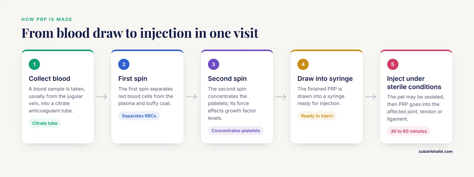
A study evaluating manual double-spin protocols in horses found that while cellular composition was similar across different first-spin protocols, the second-spin force significantly affected growth factor concentrations. PDGF-BB increased from 2629 pg/mL at 200 x g to 3467 pg/mL at 1000 x g, while TGF-β1 increased from 13,364 pg/mL to 16,998 pg/mL [8]. This highlights the importance of standardized preparation protocols to ensure a consistent and potent product. Another study described a simple double centrifugation tube method using 120 x g for 5 minutes followed by 240 x g for 5 minutes, which yielded a platelet concentration 2.41 times higher than whole blood [10].
Leukocyte-Rich vs. Leukocyte-Poor PRP
PRP can be classified based on its leukocyte (white blood cell) content. Leukocyte-rich PRP (L-PRP) contains higher concentrations of white blood cells, while leukocyte-poor PRP (P-PRP) is filtered to remove them. Equine APC was found to be a leukocyte-rich PRP with a median leukocyte concentration of 51.60 x 10^3 cells/μL compared to 7.43 x 10^3 cells/μL in whole blood [6]. Interestingly, a systematic review found that both L-PRP and P-PRP exhibited comparable efficacy in treating equine osteoarthritis [2].
Fresh vs. Freeze-Dried PRP
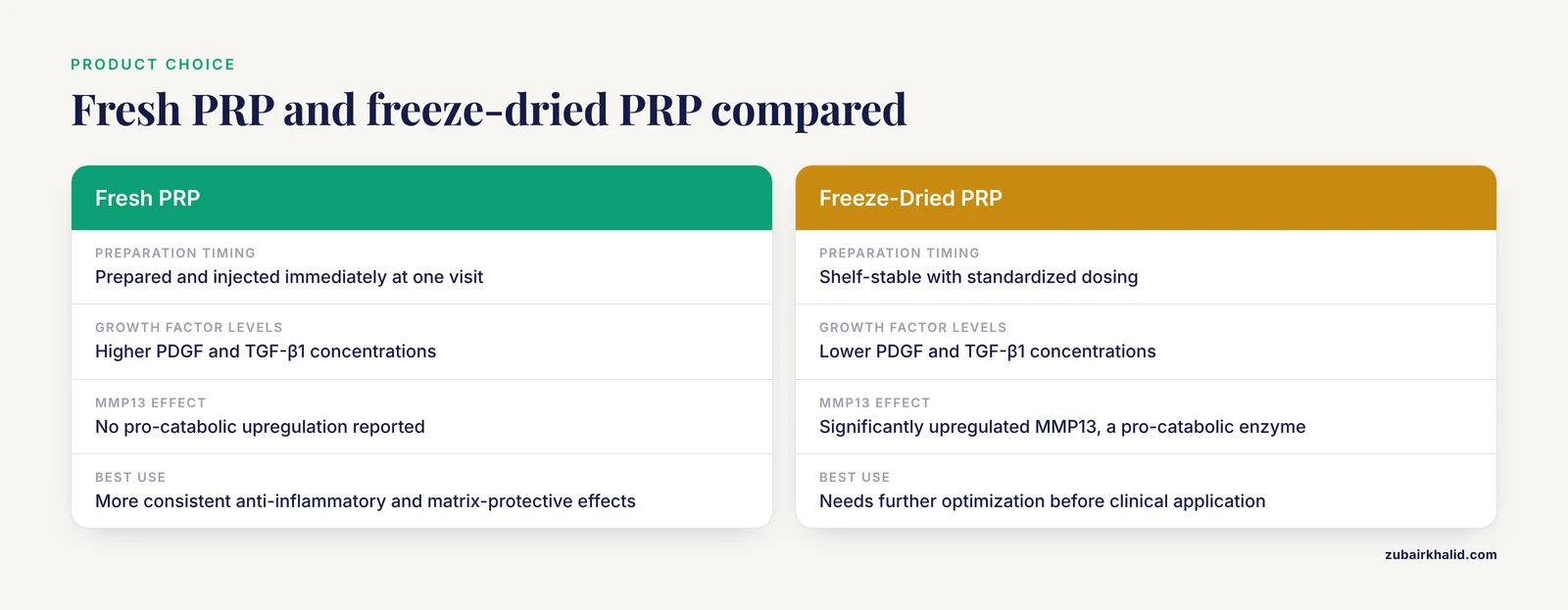
PRP is also available in fresh and freeze-dried (lyophilized) formulations. Fresh PRP (F-PRP) is prepared and injected immediately. Freeze-dried PRP (FD-PRP) offers the advantage of shelf-stability and standardized dosing [4], [11]. However, a pilot study comparing the two found that F-PRP had more consistent anti-inflammatory and matrix-protective effects, while FD-PRP had lower concentrations of PDGF and TGF-β1 and significantly upregulated MMP13, a pro-catabolic enzyme [4]. This suggests that fresh PRP may be more biologically potent, and freeze-dried products require further optimization before clinical application [4].
Activation Methods
Historically, PRP was often activated with bovine thrombin or calcium chloride before injection. However, a systematic review found that activation with bovine thrombin may be pro-inflammatory and is unnecessary [2]. Modern PRP protocols often rely on endogenous activation, which occurs naturally when the PRP comes into contact with tissue collagen. A study on Dickkopf-1 (Dkk-1), a protein involved in bone metabolism, found that bovine thrombin created a greater release of Dkk-1 from platelets than a freeze-thaw cycle, but the clinical significance of this is still under investigation [12].
Evidence for PRP in Osteoarthritis
Osteoarthritis (OA) is a leading cause of lameness and chronic pain in animals [1], [2]. It is characterized by progressive degeneration of articular cartilage, synovial inflammation, and subchondral bone changes. Traditional treatments, such as NSAIDs and corticosteroids, primarily manage pain and inflammation but do not address the underlying disease process. PRP has emerged as a potential disease-modifying therapy for OA.
Systematic Reviews and Meta-Analyses
A systematic review and meta-analysis of PRP for equine joint disease evaluated 21 publications and found that the overall estimated effect was consistent with a significant improvement in the PRP treatment group compared to the control group, with an odds ratio of 15.32 [1]. This provides strong quantitative evidence for the efficacy of PRP in treating joint disease.
Another systematic review, specifically focusing on equine OA, analyzed 11 studies and concluded that intra-articular PRP injections are generally safe, with transient synovial inflammation occurring mainly when PRP was activated with bovine thrombin [2]. The review also noted that both L-PRP and P-PRP formulations exhibited comparable efficacy, and that multiple injections could improve outcomes [2].
PRP Combined with Corticosteroids
Combining PRP with corticosteroids is an area of active research. A study investigating the combined use of triamcinolone acetonide (TA) and PRP in horses with metacarpophalangeal joint OA found that PRP prevented TA-induced chondrocyte death in vitro [7]. In vivo, the TA+PRP group had improved flexion assessments and lameness scores two weeks after treatment, while the TA-only group did not [7]. This suggests that combining PRP with corticosteroids may enhance chondrocyte viability and provide better long-term therapeutic outcomes [7].
Fresh PRP vs. Freeze-Dried PRP in OA
A pilot study using an equine cartilage-synovium explant co-culture model compared the effects of fresh PRP (F-PRP) and freeze-dried PRP (FD-PRP) on interleukin-1β-induced inflammation [4]. PRP treatments reduced inflammatory responses, with significant downregulation of COX-2 expression and PGE2 production. F-PRP, particularly at a 25% concentration, showed the most consistent inhibition of nitric oxide production and significantly reduced glycosaminoglycan release, indicating a matrix-protective effect [4]. In contrast, FD-PRP significantly upregulated MMP13, a pro-catabolic enzyme, suggesting a potential negative effect on cartilage [4].
Evidence for PRP in Tendon and Ligament Injuries
Tendon and ligament injuries are common causes of lameness in athletic animals [3]. These injuries heal slowly and often result in scar tissue that is biomechanically inferior to the original tissue, leading to a high risk of re-injury. PRP has gained popularity as a treatment to improve the quality of healing and reduce recurrence.
Clinical Outcomes
A systematic review of PRP for equine tendon and ligament injuries evaluated 22 studies and found that PRP demonstrated a favorable safety profile, with no severe adverse effects reported [3]. Clinical outcomes included improved lameness scores, ultrasonographic tissue organization, and return-to-work rates [3]. However, the review also highlighted variability in PRP formulations and activation methods, which contributed to inconsistent results [3].
The Role of Extracellular Vesicles
The mechanism by which PRP improves tendon healing is complex. Proteomic profiling of equine PRP and PRP-derived extracellular vesicles (EVs) revealed that these products are enriched in proteins related to cellular waste disposal and inhibition of lipid metabolism [13]. When applied to inflamed tenocytes, PRP and PRP-EVs significantly influenced the levels of 18 proteins, including a decrease in collagen type 1 alpha 1 chain abundance and an increase in sequestosome 1, a protein involved in inflammation and autophagy [13]. These findings suggest that PRP and its EVs actively modulate the tendon cell environment to promote repair [13].
PRP and Stem Cell Combinations
Sequential application of PRP and mesenchymal stem cells (MSCs) is another promising approach. A prospective, randomized, double-blind, placebo-controlled trial in horses with naturally occurring tendinopathies found that horses treated with muscle-derived MSCs after PRP pretreatment showed significant improvements in clinical and ultrasonographic parameters, whereas no significant changes were observed in the placebo group [14]. This suggests that PRP can be used as a primer to enhance the effects of subsequent stem cell therapy [14].
Safety and Drug Interactions
PRP is an autologous therapy, meaning it uses the patient's own blood, which minimizes the risk of allergic reactions or disease transmission. The safety profile of PRP is well-documented in the veterinary literature, [3]. A safety study of leucoreduced allogeneic pooled freeze-dried PRP in healthy equine joints also reported no significant systemic or local adverse effects compared to a saline placebo [11].
PRP and NSAIDs
A common clinical question is whether PRP can be used in conjunction with other medications. A study investigating the influence of a systemic NSAID (flunixin meglumine) on equine plasma and platelet-rich fibrin (PRF) found no significant differences in growth factor and cytokine concentrations between the NSAID-treated group and the control group [15]. Similarly, another study found that systemic flunixin meglumine did not significantly affect the concentrations of the antiproteases A2M and A1AT in plasma or PRF [5]. These findings suggest that concurrent NSAID use may not negatively impact the quality of PRP.
PRP and Sedatives
Sedatives are commonly used during PRP collection and injection. A study evaluating the effects of xylazine and detomidine on PRP and autologous protein solution (APS) found that these sedatives did not acutely alter the concentrations of most bioactive proteins [16]. The only exception was a decrease in IL-8 concentration in PRP after xylazine administration [16].
PRP and Laser Therapy
High-intensity laser therapy (HILT) is sometimes used alongside PRP. An in vitro study found that HILT applied to equine PRP and APS did not significantly increase or decrease the concentrations of select cytokines and growth factors [17]. This suggests that HILT does not enhance or degrade the bioactive components of PRP [17].
The PRP Procedure: What Pet Owners Should Expect
The PRP procedure begins with a thorough veterinary examination, including a lameness evaluation, palpation, and diagnostic imaging such as ultrasound or radiographs [3]. This is essential to confirm the diagnosis and determine if PRP is the appropriate treatment.
Initial Consultation and Diagnostics
During the initial consultation, the veterinarian will take a detailed history and perform a physical examination. They may recommend blood work to rule out underlying health issues. Diagnostic imaging, such as radiographs (X-rays) or ultrasound, is often necessary to assess the extent of joint damage, tendon tears, or ligament injuries [3].
The Injection Procedure
If PRP is recommended, the procedure typically involves the following steps:
- A blood sample is collected from the patient, usually from the jugular vein.
- The blood is placed in a specialized centrifuge and processed to concentrate the platelets [8], [10].
- The PRP is then drawn into a syringe for injection.
- The patient may be sedated to ensure comfort and safety during the procedure.
- The PRP is injected directly into the affected joint, tendon, or ligament under sterile conditions [2], [3].
The entire process typically takes 30 to 60 minutes. Most patients tolerate the procedure well and can go home the same day.
Recovery and Follow-Up
After the injection, the patient may experience mild, transient synovitis (inflammation of the joint lining) [2]. This usually resolves within a few days. The veterinarian may recommend restricted activity for a period of time to allow the PRP to work.
Follow-up examinations are crucial to monitor progress. In one study, horses were evaluated at 1, 2, and 4 weeks post-treatment. In another, clinical and ultrasonographic parameters were evaluated at 4 and 8 weeks [14]. Recheck timing will vary based on the patient's condition and the veterinarian's recommendations.
Unsafe Home Remedies and Myths
There are no safe or effective home remedies for PRP therapy. PRP must be prepared and injected by a trained veterinarian under strict sterile conditions. Attempting to prepare or administer PRP at home carries a significant risk of infection, tissue damage, and improper dosing.
Another myth is that activating PRP with bovine thrombin is necessary for it to work. In fact, a systematic review found that activation with bovine thrombin may be pro-inflammatory and is unnecessary [2]. Modern PRP protocols often rely on endogenous activation, which occurs naturally when the PRP comes into contact with tissue collagen.
Prevention and Long-Term Management
PRP is a powerful tool, but it is not a standalone cure. Long-term management of orthopedic conditions requires a multimodal approach. This includes:
- Weight management to reduce stress on joints.
- Controlled, low-impact exercise to maintain muscle mass and joint mobility.
- Physical therapy and rehabilitation.
- Nutritional support with joint supplements (e.g., omega-3 fatty acids, glucosamine).
- Regular veterinary check-ups to monitor progress and adjust the treatment plan.
Prognosis
The prognosis for pets undergoing PRP therapy depends on several factors, including the specific condition being treated, the severity of the injury, the patient's overall health, and adherence to the recommended treatment plan.
The meta-analysis of equine studies reported a significant improvement in the PRP treatment group compared to controls, with an odds ratio of 15.32 [1]. This translates to a substantially higher likelihood of improvement with PRP compared to no treatment.
For tendon and ligament injuries, PRP has been shown to improve lameness scores, ultrasonographic tissue organization, and return-to-work rates [3]. However, it is important to have realistic expectations. PRP is a regenerative therapy that works gradually over weeks to months, and multiple injections may be needed to achieve optimal results [2].
Emergency Red Flags
While PRP is generally safe, it is important to be aware of potential complications. Contact your veterinarian immediately if you notice any of the following signs after a PRP injection:
- Severe swelling, heat, or redness at the injection site.
- Acute worsening of lameness or refusal to bear weight on the affected limb.
- Fever, lethargy, or loss of appetite.
- Drainage or discharge from the injection site.
These signs could indicate an infection or a severe inflammatory reaction that requires prompt veterinary attention.
Limitations and When to Contact a Veterinarian
PRP is not a substitute for surgical intervention in cases of complete tendon rupture, severe joint instability, or fractures. In these cases, surgery is necessary to stabilize the injury, and PRP may be used as an adjunct to promote healing.
Individual results vary, and PRP may not be effective for every patient. If your pet is not showing signs of improvement within 4 to 6 weeks after treatment, or if their condition worsens, contact your veterinarian to discuss alternative or additional treatment options.
This article is educational and is not a substitute for veterinary diagnosis or treatment. Always consult with a qualified veterinarian to determine the best course of action for your pet.
Species Differences: What PRP Evidence in Dogs and Cats Actually Shows
The clinical trials and systematic reviews cited throughout this article were performed in horses [1], [2], [3]. Equine research dominates the PRP literature because of the economic value of performance horses and the ease of blood collection. Dogs and cats are not small horses. Their platelet biology, joint volumes, and healing responses differ, and those differences matter when interpreting evidence.
Canine and Feline Evidence
Published data on PRP in dogs and cats are far more limited. Most canine studies are small, uncontrolled, or based on experimental models rather than naturally occurring disease. Feline evidence is even scarcer. The equine systematic reviews [2], [3] and the meta-analysis reporting an odds ratio of 15.32 for improvement [1] cannot be extrapolated to dogs and cats as if they were equivalent patients. An odds ratio from horses describes horses; it does not predict a pet's response.
Platelet Handling Differs by Species
Equine PRP preparation protocols, including double-spin centrifugation parameters [8], [10], were validated in equine blood. Canine and feline platelets have different sizes, densities, and activation thresholds, so a protocol that concentrates equine platelets effectively may produce a weaker or more activated product in a dog or cat. Commercial PRP kits and centrifuge tubes are often marketed for veterinary use, but not all are validated for feline blood, which is harder to process because of the small sample volume and platelet clumping. Freeze-dried allogeneic PRP products have been tested in horses [11], but their safety and efficacy in dogs and cats are not established by the same evidence.
Diagnosis Comes First
PRP is injected into a specific joint, tendon, or ligament. If the diagnosis is wrong, the injection is wasted. A veterinarian must first perform a lameness examination and imaging such as ultrasound or radiographs to confirm which structure is injured [3]. Only then can a PRP plan be tailored to the individual pet.
Owners should ask their veterinarian whether the PRP system being used has been validated for the species being treated, and should expect a frank discussion about the limits of the evidence.
Patient Selection and Contraindications for PRP
Patient selection starts with a confirmed diagnosis, not a guess based on a limp. The veterinarian reviews history, performs a lameness examination and palpation, and uses imaging such as ultrasound or radiographs to identify the damaged joint, tendon, or ligament and judge severity [3]. PRP is most reasonable for mild to moderate osteoarthritis or incomplete tendon and ligament injuries, where tissue is still present and healing can be supported. A complete rupture, fracture, or severely unstable joint needs surgical stabilization first. PRP may be an adjunct in those cases, not a replacement.
Overall health also matters. The pet must be stable enough for blood collection, possible sedation [16], and injection. Blood work and a medication review help flag anemia, low platelet count, clotting disorders, or organ disease. Systemic NSAID use did not reduce PRP quality in the cited studies [15], [5], but owners should not start, stop, or change medications without veterinary advice.
When PRP is not appropriate
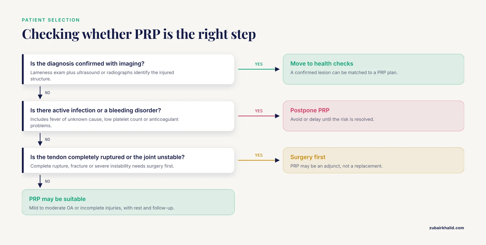
Avoid or postpone PRP when:
- Active infection is present at the injection site or systemically, including fever of unknown cause.
- A bleeding disorder, low platelet count, or anticoagulant problem makes blood collection or injection risky.
- The joint is unstable, a fracture is present, or a tendon is completely ruptured.
- The diagnosis is uncertain or imaging has not identified a treatable lesion.
- The owner cannot provide the required rest and follow-up.
When to seek re-evaluation
Contact the veterinarian if lameness worsens after injection, swelling or fever develops, or the pet does not improve within the expected timeframe. A changing diagnosis, new injury, or poor response should prompt reassessment rather than repeated injections.
Evaluating a PRP Clinic: Protocol, Cost, and Realistic Expectations
PRP is not a standardized product. Two clinics can quote very different prices for the same joint injection because they differ in preparation, handling, and follow-up. Judge cost against what is actually delivered, not just the injection itself.
What Drives Cost and Variability
- Platelet concentration. Double-spin protocols vary, and the second-spin force affects growth factor concentrations [8]. One simple tube method yielded a platelet concentration 2.41 times that of whole blood [10]. Ask whether the clinic states a target concentration.
- Fresh versus freeze-dried product. Fresh PRP is prepared and injected at one visit. Freeze-dried PRP is shelf-stable, but pilot data showed lower PDGF and TGF-β1 concentrations and upregulation of a pro-catabolic enzyme compared with fresh PRP [4]. A lower price may reflect a different product.
- Sterile technique and guidance. Joint and tendon injections require clipping, aseptic preparation, and often ultrasound guidance. These are standards, not optional extras.
- Sedation. Sedation is used for comfort and safety. Common alpha-2 sedatives did not acutely alter most bioactive proteins in PRP [16].
- Follow-up. Rechecks are part of treatment. Studies assessed horses at 1, 2, and 4 weeks [7] and at 4 and 8 weeks [14]. A quote that excludes rechecks is incomplete.
Questions to Ask Before Booking
- How is the PRP prepared, and what platelet concentration do you target?
- Is this fresh or freeze-dried PRP, and why?
- Will the injection be image-guided, and what sterile precautions are used?
- How many injections are planned, and which rechecks are included?
- What improvement do you expect, and over what timeline?
When to Seek a Second Opinion
Seek another veterinary opinion if the clinic cannot describe its protocol, promises a guaranteed cure, skips diagnostic imaging, or has no follow-up plan. PRP works gradually, and multiple injections may be needed [2]. A clinic that explains its preparation and sets realistic timelines is a better sign than one selling PRP as a generic injection.
Related Guides
- ITP in Cats: Immune-Mediated Platelet Destruction Explained: Explains immune-mediated platelet destruction, a key cause of low platelet count
- Platelet Indices in Veterinary Interpretation: Context for interpreting platelet parameters before PRP candidacy assessment
- Fluid Therapy During Anesthesia in Exotic Species
Frequently Asked Questions
Is PRP safe for pets?
Yes, PRP is considered very safe. Because it uses the patient's own blood, the risk of allergic reactions or disease transmission is extremely low. Studies report a favorable safety profile with no severe adverse effects, [3].
How long does a PRP injection take?
The entire process, from blood draw to injection, typically takes about 30 to 60 minutes. This includes the time needed for blood collection, centrifugation, and the injection procedure itself.
How many PRP injections will my pet need?
The number of injections varies depending on the condition and the patient's response. Some protocols use a single injection, while others recommend multiple injections to achieve optimal results [2].
Is PRP a replacement for surgery?
No. PRP is a regenerative therapy that helps heal tissues, but it is not a replacement for surgery in cases of complete tendon tears, severe joint instability, or fractures. In some cases, PRP may be used alongside surgery to enhance healing.
Can PRP be used with other medications?
Yes. Studies have shown that PRP can be safely combined with certain medications. For example, combining PRP with triamcinolone showed better long-term outcomes than triamcinolone alone [7]. Systemic NSAIDs do not appear to negatively affect PRP composition [15], [5].
What is the success rate of PRP for arthritis?
A meta-analysis of equine studies found a significant improvement in the PRP treatment group compared to controls, with an odds ratio of 15.32 [1]. This indicates a substantially higher likelihood of improvement with PRP.
Are there side effects of PRP?
The most common side effect is transient synovitis, or inflammation of the joint lining, which usually resolves within a few days. This occurs mainly when PRP is activated with bovine thrombin [2].
How soon will I see improvement in my pet?
Improvement is typically gradual. Some studies show improvements in lameness and flexion scores as early as 1 to 2 weeks post-injection [7], with later improvements at 4 to 8 weeks [14].
Sources
- A systematic review and meta-analysis of the efficacy of platelet-rich plasma products for treatment of equine joint disease.
- Platelet-Rich Plasma in Equine Osteoarthritis: A Systematic Review of Clinical and Experimental Evidence.
- Efficacy of Platelet-Rich Plasma in the Treatment of Equine Tendon and Ligament Injuries: A Systematic Review of Clinical and Experimental Studies.
- Anti-Inflammatory and Metabolic Effects of Fresh Versus Freeze-Dried Platelet-Rich Plasma on Equine Osteoarthritis in an Ex Vivo Cartilage-Synovium Explant Co-Culture System: A Pilot Study.
- The Impact of Systemic Treatment With Flunixin Meglumine on the Two Antiproteases Alpha-2-Macroglobulin (A2M) and Alpha-1-Antitrypsin (A1AT) in Equine Plasma and Platelet-Rich Fibrin (PRF).
- Equine autologous platelet concentrate is a leukocyte-rich platelet-rich plasma with a high interleukin-1Ra-to-interleukin-1β ratio and high transforming growth factor-β1 concentration.
- The Combined Use of Triamcinolone and Platelet-Rich Plasma in Equine Metacarpophalangeal Joint Osteoarthritis Treatments: An In Vivo and In Vitro Study.
- Influence of Manual Double-Spin Preparation Protocols on the Cellular and Growth Factor Composition of Equine Platelet-Rich Plasma.
- Allogeneic Platelet-Rich Gel Supernatant Reprograms the Cytokine and Growth Factor Microenvironment in an Equine In Vitro Inflammatory Tendon System.
- A Simple Double Centrifugation Tube Method to Obtain Platelet-rich Plasma from Equine Blood.
- Safety study of leucoreduced allogeneic pooled freeze-dried platelet-rich plasma in healthy equine joints.
- Dickkopf-1 is present in equine platelet-rich plasma and correlates to platelet concentration.
- Proteomic profiling reveals the potential role of allogenic equine platelet-rich plasma and extracellular vesicles in modulating tendon inflammation and repair.
- Sequential Application of Autologous Platelet Rich Plasma and Muscle-Derived Mesenchymal Stem Cells for Acute Tendon Injuries in Horses: Early Clinical and Ultrasonographic Outcomes in a Randomized, Double-Blind Controlled Study.
- The influence of a systemic non-steroidal anti-inflammatory drug on growth factors and cytokines in equine plasma, solid platelet-rich fibrin (PRF), and liquid PRF.
- Single-dose intravenous α2-agonists do not acutely alter anti-inflammatory bioactive protein profiles but reduce interleukin-8 in equine autologous protein solution and platelet-rich plasma.
- The in vitro application of high-intensity laser therapy on equine platelet-rich plasma and autologous protein solution does not alter select cytokine and growth factor concentrations.