Foot and Mouth Disease in Cattle: Signs and Control
By Dr. Zubair Khalid, DVM, MS, PhD ·
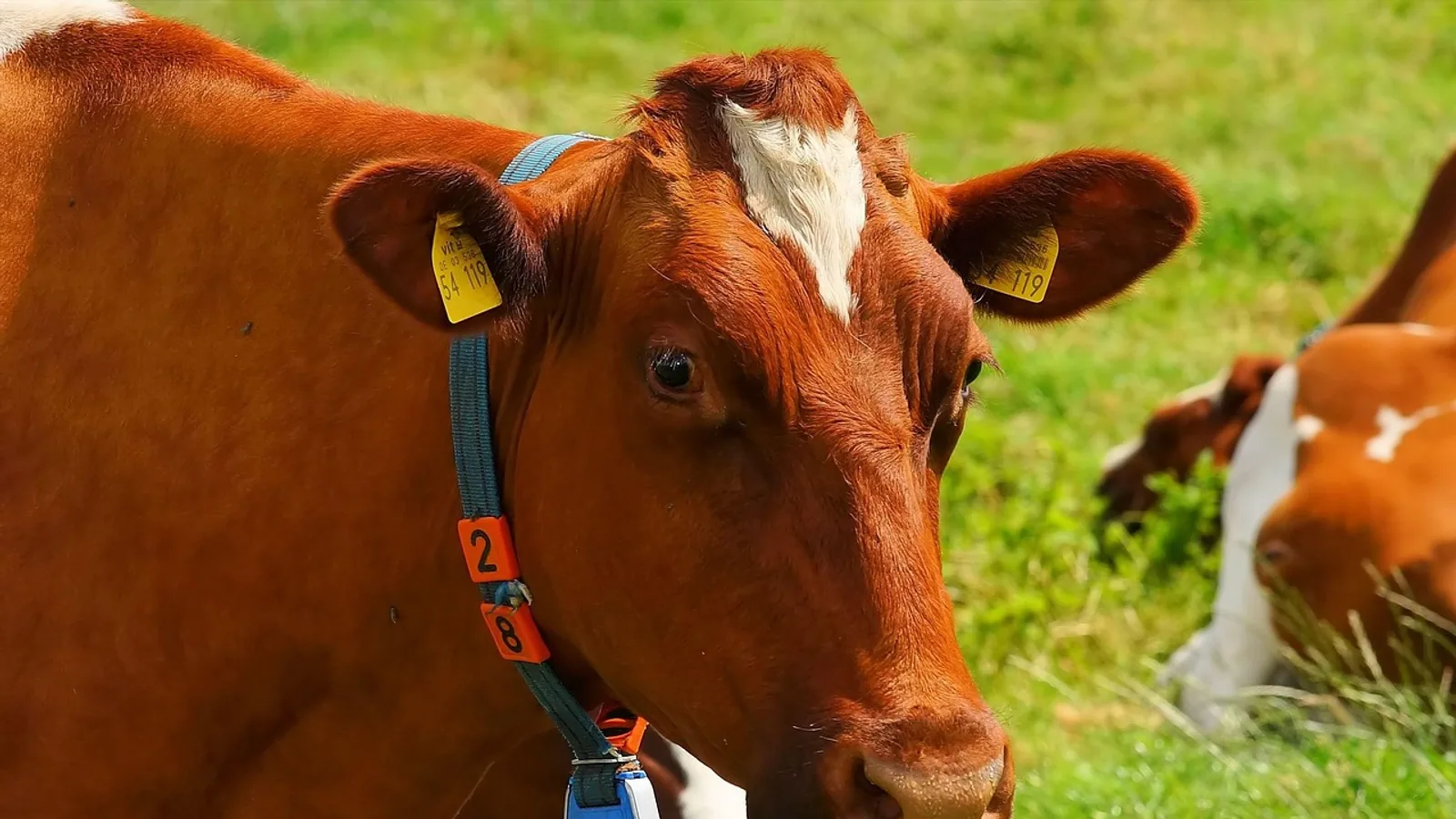
Foot and mouth disease (FMD) in cattle is a highly contagious viral infection of cloven-hoofed animals caused by a picornavirus of the genus Aphthovirus. It produces vesicles (fluid-filled blisters) on the tongue, dental pad, teats, and the interdigital space between the hooves, together with drooling, lameness, fever, and a sudden drop in milk yield. FMD is a reportable foreign animal disease in the United States. If you see blisters in a cow's mouth or between the hooves, stop all animal movement, isolate the affected animals, and call your state veterinarian or the USDA immediately. Do not wait for a laboratory result before reporting.
This article is educational and is not a substitute for veterinary diagnosis or treatment.
At a Glance: FMD Signs, Differentials, and Control Actions
| Category | Key features in cattle | Action required |
|---|---|---|
| Core clinical signs | Vesicles on tongue, dental pad, gums, teats, and interdigital space. Drooling, lameness, fever, milk drop | Isolate the animal and the group. Report immediately |
| Transmission | Direct contact, aerosol, fomites (people, vehicles, equipment), animal movement | Stop all movement on and off the premises |
| Top differentials | Vesicular stomatitis, bluetongue, bovine viral diarrhea, foot rot, digital dermatitis, chemical or thermal burns | Report any vesicular lesion. Do not assume a benign cause |
| Immediate control | Notification, quarantine, movement ban | Notify the state veterinarian or USDA within hours, not days |
| Longer-term control | Culling or vaccination per national policy, disinfection of people and equipment | Follow the incident command plan for the jurisdiction |
| Vaccine caveat | Seven serotypes with no cross-protection. Serotype must match the vaccine | Serotype-specific vaccine selection is a national decision |
What Causes Foot and Mouth Disease in Cattle?
The cause is foot and mouth disease virus (FMDV), a small, non-enveloped RNA virus in the family Picornaviridae, genus Aphthovirus. The Merck Veterinary Manual describes FMD as one of the most contagious viral diseases of animals and notes that it affects all cloven-hoofed species, including cattle, pigs, sheep, goats, and buffalo [1].
FMDV has seven distinct serotypes: O, A, C, SAT1, SAT2, SAT3, and Asia 1. Infection with one serotype does not protect against the others. This is the single most important fact for vaccine planning. A vaccine built against serotype O will not protect a herd exposed to serotype A, SAT1, or Asia 1. Field studies repeatedly confirm that multiple serotypes can circulate in the same country at the same time. In Nigeria, surveillance of cattle outbreaks between 2017 and 2020 identified three serotypes, O/EA-3 and O/WA, A/Africa/G-IV, and SAT2/VII, and the authors recommended those specific serotypes for vaccine production in the country [2]. A separate Nigerian study of cattle, sheep, goats, and pigs found serotype O most common at 74.9% of positive samples, followed by A at 56.3% and SAT2 at 35.8% [3]. In Syria, an outbreak in six cattle farms in early 2026 was caused by SAT-1, the first molecularly confirmed detection of that serotype in the country [4]. In Sudan, samples collected from 2019 to 2022 yielded serotypes A and O, with A detected in 2019, 2021, and 2022 and O predominating in 2020 [5].
The practical consequence is that "FMD vaccine" is not a single product. Serotype matters for vaccine choice, and a country's vaccine must be matched to the serotypes circulating in that region at that time. Egypt's response to the emergence of SAT1 illustrates this: a monovalent inactivated SAT1 vaccine was prepared specifically for the newly introduced serotype, and vaccinated cattle and buffaloes developed protective antibody levels by 14 days after vaccination, with immunity lasting up to 32 weeks in cattle and buffaloes and 28 weeks in sheep [6].
Virus stability and why it spreads so easily
FMDV is stable across a wide pH range and survives well in cool, moist organic material, which is why contaminated manure, bedding, milk, and hides are efficient fomites. It is destroyed by high temperature and by acidic or alkaline conditions outside its stable range. This stability is the biological reason disinfection and movement control work, and also the reason they must be applied thoroughly rather than casually.
How FMD Spreads Between Cattle and Between Farms
Transmission of FMD occurs by four main routes, and outbreak control targets all four.
Direct contact. Infected and susceptible animals in the same pen or pasture transmit virus through saliva, nasal secretions, vesicular fluid, milk, and semen. Cattle are highly susceptible by the respiratory route.
Aerosol. Under the right conditions of temperature, humidity, and airflow, FMDV can travel as an aerosol over considerable distances. This is a major reason regional zoning and rapid detection matter in outbreak models [7].
Fomites. People, vehicles, boots, clothing, milking equipment, feed, and bedding move virus between premises without any animal moving. In a New England dairy modeling study, reducing indirect transmission through enhanced biosecurity improved outbreak control, and combining regional zoning with earlier detection produced synergistic reductions in outbreak impact beyond either measure alone [7].
Animal movement. Movement of infected but not yet diagnosed cattle is the classic driver of long-distance spread. A quantitative risk assessment of the China-Laos live-cattle trade interface identified illegal live-cattle trade as the dominant modeled pathway for FMDV introduction, with habitat suitability for the virus most strongly associated with wind speed seasonality and distance to roads [8].
Preclinical transmission changes everything
Cattle can transmit FMDV before they look sick. An experimental study demonstrated that infected cattle were capable of transmitting infection at least 24 hours before clinical signs developed [9]. When that preclinical transmission was included in between-farm simulations, the number of affected farms rose by 33.7% compared with models that assumed transmission only after signs appeared [9].
This finding has direct practical meaning. A herd that looks completely normal today can already be infectious. That is why control depends on movement bans and biosecurity rather than on visual inspection alone, and why early passive surveillance and rapid reporting shorten outbreaks [7].
The carrier state
Vaccination protects cattle against clinical disease but does not always prevent persistent infection. In an experimental study in which vaccinated cattle were challenged with homologous serotype O or serotype A virus, the overall incidence of the carrier state was 22.5% [10]. Carriers showed a slower rise in antigen-specific and neutralizing antibody titers after challenge, with significantly lower antibody titers during the early infection stage, and viral genomes and particles were found mainly in nasopharyngeal tissues including the nasopharynx, soft palate, and adjacent lymph nodes and tonsils [10]. Neither viral serotype nor vaccine dose significantly influenced final carrier incidence in that study [10].
Clinical Signs of Foot and Mouth Disease in Cattle
The incubation period in cattle is typically short, on the order of two to seven days, though it varies with dose and route. Signs develop in a recognizable sequence.
Early signs
- Fever, often above 104°F (40°C)
- Depression, reduced feed intake, and a sudden, sharp drop in milk yield
- Stiff gait or reluctance to move
- Increased salivation that progresses to obvious drooling
The milk drop is often the first thing a dairy producer notices, sometimes before any lesion is visible. Treat an unexplained herd-wide milk drop with fever as a reportable event until proven otherwise.
Vesicular lesions
The hallmark lesion is a vesicle, a thin-walled blister filled with clear to straw-colored fluid. Vesicles appear on:
- The tongue, especially the dorsum and margins
- The dental pad and gums
- The inner lips and buccal mucosa
- The teats and udder skin
- The interdigital space and coronary band of the hooves
Vesicles rupture within about 24 hours, leaving raw, painful erosions. In the mouth, ruptured vesicles on the tongue and dental pad cause profuse drooling and smacking of the lips. On the feet, ruptured vesicles cause severe lameness, and animals may kneel or refuse to stand. On the teats, lesions make milking painful and predispose to mastitis.
Sequelae
After the acute phase, cattle may lose condition, develop secondary bacterial infection of eroded tissue, and suffer permanent hoof deformities from coronary band damage. Cows that were milking often do not return to full production in that lactation. FMD also causes a systemic inflammatory response and oxidative stress, with measurable changes in acute phase proteins, cardiac markers, and antioxidant enzymes during infection [11].
Species differences worth knowing
Cattle show the classic severe oral and pedal lesions. Sheep and goats often show milder signs that are easy to miss, which makes them dangerous silent amplifiers in mixed herds. Pigs show severe foot lesions and are prolific aerosol shedders. Serosurveillance in Puducherry, India, found NSP antibody positivity of 11.15% in bovines and 5.43% in goats, suggesting low-level subclinical circulation even without reported outbreaks, and the authors highlighted goats as potential sentinel animals [12].
Differential Diagnosis: What Else Looks Like FMD?
Any vesicular lesion in a cow is a foreign animal disease emergency until proven otherwise. The two most important look-alikes are vesicular stomatitis and bluetongue.
Vesicular stomatitis (VS). Caused by a vesiculovirus in the family Rhabdoviridae. VS produces vesicles on the tongue, lips, gums, teats, and coronary bands that are clinically indistinguishable from FMD in the individual animal. VS affects cattle, horses, and pigs. Horses are susceptible to VS but are not susceptible to FMD, so the presence of oral or coronary band lesions in horses on the same premises points strongly toward VS. VS is endemic in parts of the Americas and is also reportable.
Bluetongue. Caused by an orbivirus transmitted by Culicoides biting midges. Bluetongue does not produce true vesicles. It causes fever, oral erosions and ulcers, hyperemia of the oral and nasal mucosa, nasal discharge, swollen muzzle, lameness from coronary band inflammation, and sometimes a swollen blue-tinged tongue. The key distinction is that bluetongue lesions are erosive and ulcerative rather than vesicular, and there is no profuse salivation from ruptured blisters. Bluetongue does not affect horses or pigs.
Other differentials. Bovine viral diarrhea virus can cause oral erosions. Foot rot and digital dermatitis cause lameness without oral lesions. Chemical burns, hot water scalds, and caustic disinfectant exposure can cause oral and pedal ulceration. Photosensitization and mycotoxin exposure can cause oral and muzzle lesions.
The distinguishing test is laboratory based. Reverse transcription PCR targeting conserved regions of the FMDV genome is far more sensitive than antigen detection ELISA. In Sudan, rRT-PCR detected FMDV RNA in 33 of 35 suitable samples (94.3%) while antigen detection ELISA typed only 13 of 51 epithelial tissue samples (25.5%) [5]. For any suspected vesicular disease, collect epithelium from an intact or freshly ruptured vesicle, plus oral or nasopharyngeal swabs, and submit through the official diagnostic channel. Never ship suspect FMD samples through routine commercial couriers.
Veterinary Examination and Diagnostics
A veterinarian examining a suspect case should work through a structured sequence.
- History. Recent animal introductions, markets, shared equipment, visitors, and neighboring herd status.
- Clinical examination. Temperature, oral cavity inspection with a good light, teat and udder examination, hoof examination, and gait assessment. Wear dedicated coveralls, boots, and gloves. Do not carry equipment from a suspect premises to another farm.
- Sample collection. Vesicular epithelium, vesicular fluid, and oral or nasopharyngeal swabs in viral transport medium. Paired serum for antibody testing.
- Laboratory testing. RT-PCR for virus detection and sequencing of the VP1 region for serotype and topotype identification. Serology for non-structural protein (NSP) antibodies distinguishes infection from vaccination in vaccinated populations, and structural protein antibody tests identify serotype-specific responses.
VP1 sequencing is the standard tool for molecular epidemiology. It tells you which serotype and which topotype you are dealing with, which directly informs vaccine selection. Studies from Indonesia, Syria, Sudan, Bangladesh, and Nigeria all used VP1-based phylogenetics to link outbreak strains to regional and transboundary lineages [13][4][5][14][2]. In Aceh, Indonesia, following FMD re-emergence in 2022, clinically affected cattle were confirmed by RT-PCR and serotype O was characterized by VP1 sequencing [13]. In Bangladesh, buffalo nasal swabs showed 41.0% individual-level positivity and 88.1% herd-level positivity, with co-circulation of serotype O and Asia-1 [14].
DIVA and marker vaccines
Differentiating infected from vaccinated animals (DIVA) is a central problem in FMD control because conventional vaccines make it impossible to tell whether antibodies came from infection or vaccination, which complicates surveillance and restricts trade [15]. A trivalent negative marker vaccine targeting serotypes O, A, and Asia 1 produced virus-neutralizing antibody titers that peaked at 2 to 4 weeks and remained significant for six months, with a balanced Th1/Th2 response, and protected cattle against homologous serotype O challenge for up to six months [15]. DIVA compliance was demonstrated by the absence of antibodies to the deleted 3A segment even after repeated booster vaccination [15]. A live-attenuated candidate vaccine with codon-deoptimized P2/P3 regions and DIVA markers produced no clinical signs, viremia, or viral shedding in safety testing and generated protective neutralizing antibodies after challenge with wild-type virus [16].
Outbreak Control: The Steps That Stop FMD
Control of FMD is a national and international regulatory activity, not a farm-level decision. The sequence below reflects standard foreign animal disease response.
Step 1: Immediate notification
Report suspicion of FMD to your state veterinarian or the USDA without delay. Do not wait for confirmation. Do not move animals, milk, manure, or equipment off the premises. Early detection through increased passive surveillance reduces overall outbreak impact, and combining early detection with regional zoning produces synergistic reductions beyond either strategy alone [7].
Step 2: Quarantine and movement ban
Stop all animal movement on and off the affected premises. Establish a controlled access zone and a surveillance zone around the infected premises. Regional zoning limits interregional spread after detection [7]. Movement bans must cover cattle, sheep, goats, pigs, and other cloven-hoofed species, plus semen, embryos, milk, and manure.
Step 3: Culling or vaccination per national policy
Countries with a stamping-out policy cull infected and in-contact animals. Countries with endemic FMD use vaccination as the primary tool. The choice depends on the serotype present, the vaccine bank available, the density of livestock, and trade considerations. Because serotypes do not cross-protect, vaccine selection must match the outbreak serotype, and countries have prepared monovalent vaccines for newly emerging serotypes such as SAT1 [6]. Trivalent and marker vaccines covering O, A, and Asia 1 have been evaluated for protective efficacy and DIVA compliance in cattle [15].
Step 4: Disinfection
FMDV is destroyed by appropriate disinfectants and by heat. Clean organic material off surfaces first, because manure and milk inactivate many disinfectants. Then apply a disinfectant with proven activity against non-enveloped viruses. Disinfect boots, clothing, vehicles, milking equipment, and any shared tools. Establish a clean and dirty line at every entry point.
Step 5: Surveillance and tracing
Trace all animal, vehicle, and personnel movements on and off the premises for the preceding weeks. Test in-contact herds. Use NSP serology to detect evidence of infection in vaccinated populations. Continue surveillance until the incubation period has passed with no new cases.
The control decision path below summarizes the sequence from suspicion to resolution.
flowchart TD
    A[Suspect vesicular lesion] --> B[Isolate animals]
    B --> C[Stop all movement]
    C --> D[Notify state veterinarian]
    D --> E[Collect samples]
    E --> F{Laboratory result}
    F -->|FMD positive| G[Quarantine premises]
    F -->|FMD negative| H[Investigate other causes]
    G --> I[Trace movements]
    I --> J{National policy}
    J -->|Stamping out| K[Cull and disinfect]
    J -->|Vaccination| L[Serotype matched vaccine]
    K --> M[Surveillance until clear]
    L --> M
Treatment and Why There Is No Home Remedy
There is no antiviral treatment for FMD in cattle. Management is supportive and aimed at keeping affected animals comfortable and preventing secondary infection while the national response proceeds. Supportive measures may include soft feed, accessible water, and treatment of secondary bacterial infection of eroded tissue under veterinary direction.
Unsafe home remedies that cause real harm include:
- Applying caustic or acidic solutions to oral or teat lesions, which worsens tissue damage
- Pouring bleach or undiluted disinfectant onto open vesicles
- Using human oral analgesics or human topical preparations on food animals without veterinary direction, which risks drug residues
- Moving animals to a "cleaner" pasture, which spreads the virus
- Selling or slaughtering affected animals outside official channels
Any topical or systemic drug used in a food animal must be selected and dosed by a veterinarian with attention to withdrawal times.
Prevention and Biosecurity for Cattle Herds
Prevention in an FMD-free country rests on keeping the virus out. Prevention in an endemic country rests on vaccination plus biosecurity.
Vaccination. Serotype-matched vaccination is the backbone of control in endemic regions. Vaccine-induced neutralizing antibody titers peak at 2 to 4 weeks after vaccination and remain significant for about six months in cattle for the trivalent marker vaccine evaluated [15]. The SAT1 monovalent vaccine produced protective antibody levels by 14 days in cattle and buffaloes and 21 days in sheep, with immunity lasting up to 32 weeks in cattle and buffaloes and 28 weeks in sheep [6]. Vaccination reduces clinical disease but does not eliminate the carrier state, which occurred in 22.5% of vaccinated challenged cattle in one study [10].
Parasite control interacts with vaccine response. A study of Fasciola hepatica infection and FMD vaccination in calves found that although total anti-FMDV antibody titers and IgG1 levels were not significantly affected, calves infected after vaccination developed significantly lower IgG1 antibody avidity indices, indicating impaired antibody maturation, along with diminished cellular responses and persistent hepatic dysfunction during chronic infection [17]. Triclabendazole treatment restored antibody avidity and partially recovered productive performance [17]. This is a reminder that liver fluke control is part of a functional vaccination program.
Farm-level biosecurity.
- Control and log all visitors, vehicles, and equipment entering the premises
- Require clean boots and coveralls, or provide farm-dedicated ones
- Isolate newly purchased cattle for at least 30 days
- Source replacement cattle from known herds with documented health status
- Avoid shared livestock trailers, or clean and disinfect them between uses
- Keep a written movement record for every animal
- Train staff to recognize vesicular lesions and report them the same day
Regional and national measures. Zoning, movement controls, and surveillance are national tools. Modeling of the New England milkshed showed that regional zoning limits interregional spread, earlier detection reduces overall outbreak impact, and enhanced biosecurity reduces indirect transmission, with the combination of zoning and early detection outperforming either alone [7].
Clinical Relevance, Limitations and Common Mistakes
FMD matters to cattle producers for three reasons. It causes direct production losses through milk drop, weight loss, lameness, and death in young stock. It triggers trade restrictions that can close export markets for months. And it forces herd-level interventions, including culling, that go far beyond the individual animal.
Common mistakes in the field:
- Waiting for laboratory confirmation before reporting. Report suspicion immediately. Preclinical transmission means the herd may already be spreading virus [9].
- Assuming a benign cause. Foot rot and digital dermatitis do not cause oral vesicles. Any vesicular lesion is a foreign animal disease suspect until proven otherwise.
- Confusing FMD with vesicular stomatitis or bluetongue. VS looks identical in the individual cow. Bluetongue causes erosions, not vesicles.
- Assuming one vaccine covers all serotypes. Seven serotypes exist with no cross-protection. Vaccine must match the circulating serotype [2][4][6].
- Believing vaccination equals eradication. Vaccinated cattle can become carriers, with 22.5% carrier incidence in one challenge study [10].
- Neglecting fomites. People, vehicles, and equipment move virus as effectively as animals [7].
- Ignoring liver fluke status. F. hepatica infection after vaccination impaired antibody avidity in calves [17].
Individual case management always requires a veterinarian who can examine the animal, assess the herd, and follow the reporting requirements of the jurisdiction.
Emergency Red Flags
Call your veterinarian or state veterinarian immediately if you observe any of the following in cattle:
- Blisters or raw erosions on the tongue, dental pad, gums, or lips
- Blisters on the teats or udder
- Blisters or erosions between the hooves or at the coronary band
- Profuse drooling or lip smacking in more than one animal
- Sudden onset of severe lameness in multiple animals
- Herd-wide fever with a sharp drop in milk yield
- Any vesicular lesion in a herd that has had recent animal introductions or market contact
Do not move animals. Do not sell animals. Do not ship milk. Report first.
Frequently Asked Questions
What are the first signs of foot and mouth disease in cattle?
Fever and a sudden drop in milk yield often appear first, followed by drooling and lameness. Vesicles on the tongue, dental pad, teats, and interdigital space confirm the clinical picture.
Is foot and mouth disease the same as hand, foot, and mouth disease in people?
No. FMD is caused by a different virus and is not a human disease of concern in this context. Cattle FMD and human hand, foot, and mouth disease are unrelated conditions.
Can cattle recover from foot and mouth disease?
Many adult cattle survive, but recovery is slow and production losses are substantial. Young calves can die from myocardial damage, and permanent hoof deformities are common.
How is foot and mouth disease transmitted between farms?
Direct contact, aerosol spread, contaminated people and equipment, and animal movement all transmit the virus. Animal movement and fomites are the most important routes for long-distance spread.
Why does the serotype matter for FMD vaccination?
There are seven FMDV serotypes with no cross-protection between them. A vaccine against one serotype will not protect against another, so vaccine selection must match the circulating serotype.
Can vaccinated cattle still carry foot and mouth disease virus?
Yes. Vaccination protects against clinical disease but does not reliably prevent persistent infection. One challenge study found a 22.5% carrier incidence in vaccinated cattle.
What should I do if I suspect foot and mouth disease in my herd?
Isolate the affected animals, stop all movement on and off the premises, and call your state veterinarian or the USDA immediately. Do not wait for a laboratory result before reporting.
How do you disinfect for foot and mouth disease?
Remove all organic material first, then apply a disinfectant with proven activity against non-enveloped viruses to boots, clothing, vehicles, and equipment. Clean and dirty lines at entry points prevent recontamination.
Related Articles
- Fusobacterium necrophorum and Interdigital Necrobacillosis (Foot Rot) in Cattle
- Johne's Disease in Sheep: Clinical Signs, Diagnosis, and Control
- Chicken Foot Parasites: Etiology, Clinical Signs, and Management
- Poultry Parasites and Diseases: Clinical Signs, Diagnosis, and Integrated Control
- Tick-Transmitted Diseases in Dogs: Pathogens, Clinical Signs, and Control
- Fusobacterium necrophorum Infections in Livestock: Foot Rot and Lameness in Cattle and Sheep
- Tetany: Causes, Mechanism and Clinical Signs
- Bovine Spongiform Encephalopathy: Mad Cow Disease
- Dog Gestation: Timeline, Signs, and Care
Sources
- Foot-and-Mouth Disease in Animals - Infectious Diseases - Merck Veterinary Manual
- Phylogenetic Analysis of Foot-and-Mouth Disease Virus from Cattle in Nigeria, 2017-2020.
- Seroepidemiology and serotype diversity of foot-and-mouth disease virus in cattle and other domestic ungulates across Nigeria.
- Preliminary molecular characterization of foot-and-mouth disease virus SAT-1 topotype III associated with recent cattle outbreaks in Syria.
- Molecular epidemiology of foot-and-mouth disease viruses collected from Khartoum, Blue Nile, and Northern States of Sudan (2019-2022).
- Preparation and evaluation of a monovalent foot and mouth disease vaccine as a rapid response to the emerging serotype SAT1 in Egypt.
- Testing Control Strategies for Foot-and-Mouth Disease in New England Using the InterSpread Plus Model: Impacts of Regional Zoning, Early Detection, and Enhanced Biosecurity.
- Quantitative risk assessment of foot-and-mouth disease introduction from Laos to China integrating habitat suitability and cross-border trade analysis.
- Incubation phase transmission of foot-and-mouth disease virus in cattle: experimental evidence and simulated impacts.
- Foot-and-Mouth Disease Virus Persistence Divergence Within Serotypes and Vaccine Doses in Vaccinated Cattle.
- Evaluation of acute phase protein, cardiac marker, and antioxidant enzymes in Madura Cattle infected with foot-and-mouth disease.
- Serosurveillance of Foot and Mouth Disease (FMD) in cattle, buffalo and goat population in Union Territory of Puducherry, India.
- Molecular detection and VP1-based phylogenetic characterization of Foot-and-mouth disease virus serotype O circulating in cattle in Aceh, Indonesia.
- Endemic Circulation and Genetic Characterization of Foot-and-Mouth Disease Virus in Buffalo Populations of Bangladesh.
- Evaluation of a DIVA compliant trivalent negative marker vaccine for foot-and-mouth disease in cattle.
- Live-attenuated foot-and-mouth disease virus vaccine engineered by codon deoptimization induces a strong protective immune response in cattle.
- Timing of Fasciola hepatica infection selectively impairs antibody avidity after foot-and-mouth disease vaccination in cattle.