Caring for a Horse With Navicular Disease
By Dr. Zubair Khalid, DVM, MS, PhD ·
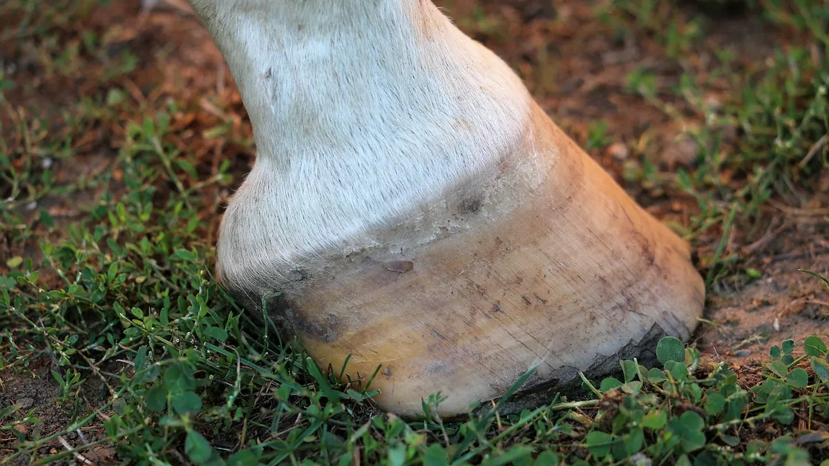
How to care for a horse with navicular disease comes down to four things you can control: correct hoof balance through skilled farriery, exercise that keeps the horse moving without repeated concussion, medication prescribed by your veterinarian, and regular monitoring so you catch changes early. Navicular syndrome is a progressive condition, so the goal is comfort and function for as long as possible, not a cure [1].
This article is educational and is not a substitute for veterinary diagnosis or treatment.
Quick Answer
- Work with a veterinarian and a farrier who have experience with palmar foot pain. Corrective farriery is the foundation of treatment [1].
- Keep your horse in consistent, low-impact exercise. Long periods of stall rest often make stiffness worse, so ask your veterinarian what level of work is appropriate.
- Give medications only as prescribed. Treatment commonly relies on corrective farriery, intrasynovial medication, and NSAIDs [1].
- Monitor stride length, stumbling, and willingness to work. These signs often change before obvious lameness appears [1].
- Expect a guarded to fair long-term outlook. The condition is progressive, so plan for periodic reassessment [1].
What Navicular Syndrome Actually Is
Navicular syndrome is a degenerative disease complex of the horse's foot [1]. The name is a little misleading because it does not describe one single injury. It can involve any of the structures in what veterinarians call the navicular apparatus: the navicular bone itself, the navicular bursa, the collateral sesamoid ligaments, the distal impar ligament, and the deep digital flexor tendon [1].
That last structure matters a lot. Current thinking holds that navicular syndrome is largely a biomechanical problem driven by chronic overload of the deep digital flexor tendon [2]. The tendon runs down the back of the leg, wraps around the navicular bone, and attaches to the coffin bone. Every stride loads that area. When the load is repeated thousands of times a day, the bone and soft tissues can break down.
The navicular bone sits inside the hoof between the deep digital flexor tendon and the middle and end phalanges [3]. Its position and shape affect how force travels through the joint. Research on joint stress shows that small differences in the bone's position and the tendon's moment arms change where and how hard the joint surface is loaded [3]. That helps explain why conformation and hoof balance matter so much in this disease.
One more thing owners should know: navicular syndrome is almost always bilateral [1]. Even if your horse looks lame in only one front leg, the other foot is usually involved too.
Signs Owners Notice First
The most common presentation is a bilateral forelimb lameness that ranges from mild to severe, with one leg usually worse than the other [1]. Because both feet hurt, many horses do not limp in an obvious way. Instead, they shorten their stride.
Watch for these signs:
- A choppy, shortened stride, especially at the trot
- Shuffling instead of reaching
- Stumbling or tripping, particularly on uneven ground
- Reluctance to go downhill or to turn tightly
- Lameness that improves with warm-up and gets worse with hard work
- Sensitivity when the heels or frog are pressed with hoof testers [4]
Horses with navicular bone fractures show similar signs. They may have mild to severe lameness, often resent hoof testers over the heels or frog, and typically improve after a palmar digital nerve block [4]. If your horse's lameness blocks out that way, your veterinarian will want imaging.
If you are sorting out which lameness signs matter and which are normal aging, Common Horse Health Problems Every Owner Should Know is a useful companion read.
Causes and Risk Factors
Certain breeds are predisposed to navicular syndrome [1]. Beyond breed, the biggest factors are biomechanical. Chronic overload of the deep digital flexor tendon is now considered the central driver of the disease process [2].
Conformation plays a role. Horses with upright pasterns, small feet relative to body size, or a long toe and low heel put more stress through the back of the foot. The geometry of the navicular bone itself matters too. The relative lengths of the tendon's moment arms and the bone's position influence the magnitude and location of joint force [3].
Age is another factor. Navicular syndrome typically shows up in mature, working horses instead of youngsters. Concussive injury from hard footing, repeated jumping on unforgiving ground, and long careers on hard surfaces all add up.
Fractures of the navicular bone are much less common than fractures of the coffin bone, but they do occur. They can happen after a single concussive injury or in horses that already have navicular bone degeneration [4].
How Navicular Syndrome Is Diagnosed
Diagnosis starts with a lameness exam. Your veterinarian will watch your horse move, flex the limbs, and use hoof testers. Nerve blocks help localize the pain to the back of the foot.
Imaging confirms the diagnosis. Radiographs are the standard first step and can show changes such as enlarged vascular channels along the distal border of the navicular bone [1]. Advanced imaging, including MRI, adds detail about soft tissue structures that radiographs cannot show well [5].
That soft tissue detail matters more than many owners realize. Horses with a clinical diagnosis of navicular syndrome often have pathology in the navicular bursa, deep digital flexor tendon, collateral sesamoid ligaments, and impar ligament, not just in the bone [6]. Radiographs alone can miss a lot of that.
Newer imaging research using micro-computed tomography has shown that changes in the bone's internal trabecular architecture occur in affected horses [2]. Standard radiography, scintigraphy, and ultrasound lack the sensitivity to detect these early microarchitectural changes [2]. This is one reason early diagnosis can be frustrating.
Your veterinarian may also want to rule out a bipartite or tripartite navicular bone. These are congenital abnormalities where the bone failed to fully fuse before birth, and they can look like fractures on radiographs [4].
Treatment Options for Navicular Disease
Treatment for navicular disease is palliative. There is no way to reverse the degeneration, so the plan focuses on reducing pain and slowing functional decline [1].
Corrective Farriery
Farriery is the cornerstone. The goal is to restore proper hoof balance, support the heels, and reduce strain on the deep digital flexor tendon. Your veterinarian and farrier should work together, because the right shoeing package depends on your horse's individual conformation and radiographs.
Shoeing changes are not one-size-fits-all. Some horses do well with a rolled toe and heel support. Others need a different approach. Reassess regularly, because what works at diagnosis may not work a year later.
Exercise Modification
Consistent, controlled exercise is usually better than stall rest. Turnout on firm, level ground keeps the foot loaded in a predictable way. Deep, muddy footing and hard, rocky ground are both problematic.
Ask your veterinarian to define your horse's work level. Many horses with navicular syndrome can keep working at a reduced intensity for a long time. Jumping on hard ground and fast work on unforgiving surfaces are usually the first things to go.
Medication
Treatment relies on corrective farriery, intrasynovial medication, and NSAIDs [1]. Intrasynovial medication means injecting medication directly into the navicular bursa or the coffin joint. Your veterinarian decides which structure to treat based on imaging and exam findings.
Bisphosphonates have been used to modify bone turnover in horses with navicular syndrome. In one field study, horses received an intramuscular dose of clodronic acid of 765 mg per horse, split across three injection sites [5]. Six of eleven horses showed clinical improvement, with a mean reduction of two lameness grades, along with increased velocity, stride length, and stride regularity [5]. That is a meaningful response in a subset of horses, not a universal fix.
Your veterinarian calculates any medication dose from your horse's weight and health status. Never adjust or repeat a prescription on your own.
Joint and Soft Tissue Support
Some owners ask about nutraceuticals. A 12-week study of natural eggshell membrane supplementation at 12 mg/kg orally in Warmblood horses with chronic lameness found rider-reported function improved by about half a grade at both walk and trot, with a small trend toward improvement in examiner-graded walk-trot scores [7]. Rest lameness did not change [7]. This is modest evidence in a small group, so treat it as a possible add-on instead of a primary treatment.
Surgery
Palmar digital neurectomy is sometimes used as a salvage procedure for horses that stop responding to medical management [6]. It is not a cure. It removes sensation, which means your horse cannot feel pain that would normally protect the foot from further damage.
Complications can be serious. Two reported cases developed navicular bone fracture with severe deep digital flexor tendinopathy and distal interphalangeal joint subluxation 12 and 19 weeks after bilateral palmar digital neurectomy [6]. Those cases highlight how important patient selection is, since soft tissue pathology often goes undiagnosed alongside radiographic changes [6]. Discuss the risks carefully with your veterinarian before considering this route.
Comparison of Management Approaches
| Approach | What it does | Owner role |
|---|---|---|
| Corrective farriery | Restores hoof balance and supports the heels | Schedule regular farrier visits and share radiographs |
| Exercise modification | Keeps controlled load on the foot without repeated concussion | Follow the veterinarian's work plan and manage footing |
| NSAIDs and intrasynovial medication | Reduces pain and joint inflammation | Give medications exactly as prescribed [1] |
| Bisphosphonates | Modifies bone turnover rate | Understand that response varies between horses [5] |
| Palmar digital neurectomy | Removes pain sensation as a salvage option | Weigh the risk of undiagnosed soft tissue damage [6] |
Recovery, Outlook, and Long-Term Management
The prognosis for navicular syndrome is guarded to fair [1]. Guarded means your horse may stay comfortable and useful for years with good management, or may decline despite your best efforts. Fair means many horses do continue working at some level.
The condition is progressive [1]. Plan on periodic reassessments instead of a single treatment plan that never changes. Your veterinarian may repeat lameness exams, adjust medications, or re-image the feet if your horse's comfort level shifts.
Monitoring at home is where you add the most value. Track these over time:
- Stride length at the trot on a consistent surface
- Number of stumbles per ride or turnout session
- Willingness to move forward
- Response to hoof testers if your farrier checks
- Attitude and appetite
Write things down. A horse that stumbles twice a week instead of twice a month is telling you something, even if the lameness grade has not changed.
Older horses with navicular syndrome often have other issues layered on top. Senior Horse Care Guide covers the broader picture for aging equines, and Horse Hoof Care and Lameness Basics helps you build a solid hoof care routine.
Prevention
You cannot prevent every case, since breed predisposition and bone geometry are fixed [1][3]. You can reduce the load your horse's feet absorb over a lifetime.
Keep hooves balanced with regular farrier care. Avoid chronic work on hard, concussive surfaces. Condition your horse gradually instead of pushing through fatigue. Maintain a healthy body weight, since extra pounds mean extra force through the foot with every stride.
If your horse has a known predisposition, ask your veterinarian about baseline radiographs. Early detection gives you more options.
Limitations and When to Contact a Veterinarian
This article cannot tell you what is happening inside your specific horse's feet. Individual cases need a veterinarian who can examine, image, and treat your horse directly.
Contact your veterinarian the same day if your horse:
- Becomes suddenly and severely lame
- Develops heat, swelling, or a pounding digital pulse in the foot
- Refuses to bear weight on one leg
- Shows a new, obvious head bob at the walk
Treat these as emergencies and call right away if your horse:
- Has a wound near the hoof or coronary band with deep structures exposed
- Shows signs of laminar pain in multiple feet, such as rocking back on the hind legs
- Cannot or will not move despite pain medication
Schedule a routine visit if you notice:
- A gradually shortening stride over weeks
- More frequent stumbling
- Reluctance to work downhill
- Any change in response to hoof testers
Frequently Asked Questions
Can a horse with navicular syndrome keep working?
Many can, with a modified program. Consistent low-impact exercise is usually better tolerated than stall rest, and your veterinarian can set a work level that matches your horse's comfort. Horses that stop responding to medical management may need a different plan.
Is navicular disease the same as navicular syndrome?
The terms are often used interchangeably, but syndrome is more accurate. It describes a degenerative disease complex that can involve the navicular bone, bursa, ligaments, and deep digital flexor tendon together. That is why two horses with the same label can look very different on imaging.
Does my horse need special shoes forever?
Most horses with this diagnosis benefit from ongoing corrective farriery. The specific shoeing package changes over time as the disease progresses and as your horse's comfort level shifts. Work with a farrier who will coordinate with your veterinarian.
Can I ride my navicular horse on trails?
Often yes, if the footing is firm and level and your veterinarian approves the workload. Deep mud, loose rock, and steep downhill grades tend to be the hardest on these horses. Start with short, easy rides and watch for increased stumbling.
Will my horse get better over time?
The underlying degeneration does not reverse. Some horses improve noticeably with treatment, especially in the first months after diagnosis, and then plateau. Others decline slowly. Regular reassessment helps you adjust the plan as things change.
Is surgery a cure for navicular syndrome?
No. Palmar digital neurectomy is a salvage procedure that removes pain sensation, not the disease. It carries real risks, including damage that goes unnoticed because the horse cannot feel it. Discuss the tradeoffs thoroughly with your veterinarian before deciding.
Related Articles
- Caring for a Horse in Winter
- Common Horse Health Problems Every Owner Should Know
- Senior Horse Care Guide
- Horse Hoof Care and Lameness Basics
- Foal Care Basics for New Owners
- Complete Horse Care Guide for New Owners
- Horse Hoof Abscess: How to Treat and Care for It
- Sore Feet in Horses: Causes, Care, and When to Call the Vet
- How to Help a Horse With Colic: Steps and Warning Signs
References
- Navicular Syndrome in Horses - Musculoskeletal System. Merck Veterinary Manual.
- Three-dimensional anatomical description of the microarchitecture of the distal sesamoid bone in healthy and navicular syndrome-affected horses by computed microtomography. Anatomical record (Hoboken, N.J. : 2007), 2026.
- Joint Stress Analysis of the Navicular Bone of the Horse and Its Implications for Navicular Disease. Bioengineering (Basel, Switzerland), 2024.
- Fractures of the Navicular Bone in Horses - Musculoskeletal System. Merck Veterinary Manual.
- Clinical efficacy of clodronic acid in horses diagnosed with navicular syndrome: A field study using objective and subjective lameness evaluation. Research in veterinary science, 2019.
- Navicular bone fracture and severe deep digital flexor tendinopathy after palmar digital neurectomy in two horses. Open veterinary journal, 2023.
- Natural eggshell membrane supplementation for chronic lameness in warmblood horses: a 12-week prospective before-after study. Frontiers in veterinary science, 2026.