IV Fluids: Types, Rates, and Vet Calculations
By Dr. Zubair Khalid, DVM, MS, PhD ·
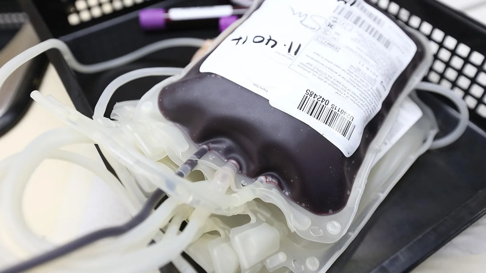
Intravenous fluids are one of the most common treatments a dog or cat receives in an emergency room, and they are also one of the easiest to get wrong. A fluid plan is not a single number typed into a pump. It is a short, repeatable calculation that adds three separate quantities together: the fluid the patient has already lost, the fluid the patient needs just to stay alive, and the fluid the patient keeps losing while you treat them. Get those three components right and the plan works. Ignore one of them and the patient either stays dehydrated or slides into volume overload.
This guide explains how veterinarians choose an intravenous fluid, how they build a rate from the three components, and how they reassess after every intervention. It covers the practical differences between 0.9% sodium chloride, lactated Ringer's solution, and balanced solutions such as Plasma-Lyte, including the calcium interaction that matters when blood products are running and the acid-base problem that follows large volumes of saline. It also covers the species difference that changes everything in cats, which tolerate volume loads poorly and tip into volume overload faster than dogs do.
The hands-on time for placing an intravenous catheter and starting fluids is usually 10 to 20 minutes in a cooperative patient. The elapsed time for the full resuscitation and stabilization process is measured in hours, because a fluid plan is rewritten at every reassessment point rather than set once and forgotten.
This article is educational and is not a substitute for veterinary diagnosis or treatment.
What Intravenous Fluid Therapy Achieves
Fluid therapy restores circulating volume, corrects dehydration, supports tissue perfusion, and gives the body a route to excrete metabolic waste and excess potassium. In shock, the immediate goal is to restore perfusion to vital organs. In dehydration without shock, the goal is slower replacement of a measured deficit. In ongoing illness such as vomiting, diarrhea, or urinary obstruction, the goal is to keep pace with losses the patient cannot replace by drinking.
The choice between crystalloid and colloid solutions, and between specific crystalloid formulations, is a decision about what the patient needs most: volume, sodium, potassium, calcium, a buffer, or a combination. Crystalloids distribute across the whole extracellular space, so a large fraction leaves the blood vessels within the first hour. Colloids stay in the vascular space longer. In a study of hemorrhagic shock resuscitation, only about 8% of infused water and less than 25% of infused saline remained in the intravascular compartment after one hour, while nearly the entire volume of an iso-oncotic colloid stayed intravascular over the same period [1].
That distribution difference explains why crystalloid resuscitation requires substantially more volume than colloid resuscitation to achieve the same hemodynamic endpoint. In a long-term canine shock model, keeping central hemodynamics at pre-shock levels required at least four times the volume of crystalloid compared with colloid [2]. This is not a reason to avoid crystalloids. It is a reason to understand that the number on the bag is not the number that stays in the vein.
The Three Components of a Fluid Plan
Every fluid rate is built from three parts. Write them down separately before you add them together.
1. The Deficit
The deficit is the fluid the patient has already lost before you met them. It comes from vomiting, diarrhea, inadequate intake, fever, third-space loss into the abdomen or gut wall, or blood loss. The deficit is estimated from clinical signs of dehydration, most often as a percentage of body weight.
A practical clinical estimate uses these ranges:
- Under 5% dehydration: not reliably detectable on physical examination.
- 5% to 6%: subtle loss of skin elasticity.
- 7% to 8%: obvious skin tenting, tacky mucous membranes.
- 9% to 10%: persistent skin tent, sunken eyes, dull corneas.
- Over 10%: moribund, often with concurrent shock.
The deficit in milliliters is body weight in kilograms multiplied by the dehydration percentage expressed as a decimal, then multiplied by 1000. For a 10 kg dog that is 8% dehydrated, the deficit is 10 × 0.08 × 1000, or 800 mL.
The deficit is usually replaced over 12 to 24 hours, not in one bolus, unless the patient is also in shock. Replacing a chronic deficit too quickly can overload the patient, especially if the heart or kidneys are compromised.
2. Maintenance
Maintenance replaces the water and electrolytes the patient loses through normal metabolism, urine, and insensible losses from the respiratory tract and skin. It is not the same as the deficit and it does not stop when the deficit is corrected.
A practical maintenance rate for dogs and cats is roughly 2 to 3 mL/kg/h. A 10 kg dog therefore needs about 20 to 30 mL/h just to maintain normal balance, before any deficit or ongoing loss is added.
Maintenance requirements are higher in young animals, in fever, and in hot environments. They are lower in patients with heart failure, kidney disease, or oliguria, where the body cannot excrete a normal fluid load.
3. Ongoing Losses
Ongoing losses are the fluid the patient continues to lose while you are treating them. Vomiting, diarrhea, suction, drain output, polyuria, and third-space sequestration all count. These losses are measured or estimated and replaced milliliter for milliliter, on top of maintenance and any remaining deficit.
A patient vomiting 200 mL three times a day is losing roughly 600 mL per day on top of maintenance. That number has to appear in the plan or the patient will remain dehydrated despite receiving fluids.
Building the Total Rate
The total hourly rate is the sum of the three components:
Total rate (mL/h) = maintenance + ongoing losses + (remaining deficit ÷ replacement hours)
Worked example. A 10 kg dog presents with 8% dehydration and has vomited about 300 mL over the past 12 hours. The veterinarian decides to replace the deficit over 24 hours and to add ongoing losses as they occur.
- Maintenance: 10 kg × 2 to 3 mL/kg/h = 20 to 30 mL/h.
- Deficit: 800 mL ÷ 24 h = about 33 mL/h.
- Ongoing losses: 300 mL ÷ 24 h = about 12.5 mL/h, adjusted as losses are measured.
Total: roughly 65 to 75 mL/h, reassessed every few hours.
This is why a fluid plan is never a single static number. The deficit shrinks as it is replaced. The ongoing loss changes with the patient's clinical course. The maintenance rate changes if the patient develops oliguria or heart failure. The plan is recalculated at each reassessment.
Shock Boluses and the Reassessment Rule
A patient in shock needs volume fast, and the tool for that is a bolus. In dogs and cats, a typical isotonic crystalloid shock bolus is given in aliquots of 10 to 20 mL/kg, with a full reassessment after each aliquot before deciding whether to give another.
A 10 kg dog therefore receives 100 to 200 mL per aliquot. A 4 kg cat receives 40 to 80 mL per aliquot. The aliquot is given over 10 to 15 minutes, or faster in a crashing patient, and then the veterinarian reassesses perfusion.
Reassessment means checking the same variables before and after each bolus:
- Heart rate and pulse quality.
- Mucous membrane color and capillary refill time.
- Blood pressure, ideally by Doppler or oscillometric measurement.
- Mentation and lactate if available.
- Respiratory rate and effort, and lung sounds, to detect early volume overload.
The reassessment rule is not optional. In a retrospective study of 35 hypotensive dogs treated in an emergency room, blood pressure rose significantly after fluid resuscitation, and 23 dogs normalized their blood pressure after bolus intravenous fluid therapy within the first hour [3]. That same study found that dogs who responded to a fluid bolus were significantly less likely to be euthanized than dogs who did not respond [3]. Response to a bolus is itself diagnostic information. A patient who does not improve after two appropriate aliquots is not simply "more dehydrated." They may be bleeding, may have a failing heart, may have a mechanical obstruction to filling, or may need blood products rather than crystalloid.
In dogs with pericardial effusion, a randomized trial compared a 10 mL/kg bolus of compound sodium lactate before pericardiocentesis against no bolus. The bolus did not produce adverse respiratory effects, and no dogs required supplemental oxygen by the four-hour mark [4]. That is reassuring for the specific scenario of tamponade, where clinicians often worry that a fluid bolus will worsen respiratory compromise. It does not mean every patient with respiratory signs should receive a bolus.
Comparing the Common Isotonic Crystalloids

.jpg)
The table below compares the isotonic crystalloids most often stocked in small animal practice. Values are approximate and reflect standard commercial formulations. Always read the label on the bag you are using.
| Fluid | Sodium (mEq/L) | Potassium (mEq/L) | Calcium (mEq/L) | Buffer | Notes |
|---|---|---|---|---|---|
| 0.9% Sodium chloride | 154 | 0 | 0 | None | No buffer. Large volumes cause hyperchloremic metabolic acidosis. |
| Lactated Ringer's solution | 130 | 4 | 2 to 3 | Lactate | Calcium content. Avoid or flush between lines when running with blood products. |
| Plasma-Lyte A | 140 | 5 | 0 | Acetate and gluconate | Balanced, calcium-free, no lactate. |
| Ringer's solution | 147 | 4 | 4 to 5 | None | Higher calcium than LRS, no buffer. |
| 5% Dextrose in water | 0 | 0 | 0 | None | Not a resuscitation fluid. Distributing water, not volume expander. |
Sodium
Sodium is the main determinant of extracellular fluid volume. All isotonic crystalloids are roughly similar in sodium content, which is why they all expand the extracellular space. The differences that matter clinically are in the other columns.
Potassium
Potassium content matters most in patients with hyperkalemia, such as a male cat with urethral obstruction. Lactated Ringer's and Plasma-Lyte both contain potassium in low concentrations. In a study of male cats with urethral obstruction and potassium above 7.5 mEq/L, all cats received intravenous fluids and calcium gluconate, and the median potassium fell from 9.1 mEq/L to 5.4 mEq/L over four hours [5]. Adding insulin and dextrose, terbutaline, or sodium bicarbonate did not produce a significantly different percentage reduction in potassium compared with fluids and calcium gluconate alone [5]. Fluids plus relief of the obstruction do most of the work.
Calcium
Calcium in lactated Ringer's solution is the reason it should not be run through the same line as blood products. Calcium chelates citrate anticoagulant and can promote clot formation in the line or in the bag. If a patient needs both, use a calcium-free fluid such as Plasma-Lyte A or 0.9% sodium chloride, or run the blood product through a separate catheter or flush the line thoroughly between products. A study comparing Plasma-Lyte A and lactated Ringer's solution in anesthetized Beagle dogs after hemorrhage found no significant differences in macrocirculation between the two, with both improving cardiac output after each 10 mL/kg bolus [6]. A companion study found that buccal microcirculatory variables were more reduced in the lactated Ringer's group than in the Plasma-Lyte A group at hypovolemia and after the second bolus [7]. That is a microcirculatory signal, not a mortality difference, but it supports the practical point that the calcium-free balanced solution is a reasonable default when blood products or microcirculatory concerns are in play.
Buffer
Lactate and acetate are buffers that the liver and other tissues convert to bicarbonate. They help correct the metabolic acidosis that often accompanies shock. The tradeoff is that lactate-containing fluids depend on hepatic metabolism, which may be impaired in a patient with severe liver dysfunction or profound hypoperfusion. Plasma-Lyte uses acetate and gluconate instead, which are metabolized more widely.
The Saline Problem
0.9% sodium chloride has no buffer and a supraphysiologic chloride concentration relative to plasma. Large volumes produce hyperchloremic metabolic acidosis. This is usually well tolerated in a patient receiving one or two boluses, but it becomes clinically relevant when saline is the only fluid a patient receives for days. A balanced solution is generally preferred when a patient needs sustained crystalloid therapy.
Crystalloid Versus Colloid
Colloids expand the vascular space with much less volume than crystalloids, and they stay there longer. The tradeoff is cost, coagulation effects, and the fact that colloid molecules can leak into the interstitium when capillary permeability is increased.
The evidence in veterinary patients is mixed and does not support a blanket preference for either class. In a canine hemorrhagic shock model, resuscitation with crystalloid, albumin, or hydroxyethyl starch produced similar pulmonary effects in the presence of moderate increases in capillary permeability [8]. A separate canine study of hemorrhagic pancreatitis found that balanced crystalloid resuscitation adequately restored plasma volume and supported tissue perfusion without detrimental effects on pulmonary pressures or oxygenation, with no apparent advantage to colloid [9]. Another long-term canine shock study found crystalloids and colloids equally effective at maintaining macrohemodynamics, though crystalloid required at least four times the volume [2].
Where colloids do show a signal is in specific endpoints. In dogs with gastric dilatation-volvulus, a study comparing a hemoglobin-based oxygen carrier against 6% hetastarch found that the hemoglobin-based product required significantly less colloid and crystalloid to reach resuscitation endpoints [10]. That is a study about a specific product, not a general endorsement.
Synthetic colloids have real coagulation effects. In healthy dogs and dogs with systemic inflammation, tetrastarch administration produced significantly increased activated partial thromboplastin time, decreased platelet count, and decreased von Willebrand factor antigen and collagen binding activity compared with saline [11]. In a thromboelastometry study, colloid treatment correlated with higher clot firmness in the extrinsic pathway and a shorter prothrombin time, with fibrinogen concentration significantly different between treatments [12]. These are laboratory findings, and their clinical relevance depends on the patient. A dog with normal coagulation receiving one dose of colloid is unlikely to bleed because of it. A dog with trauma, thrombocytopenia, or disseminated intravascular coagulation is a different story.
Species Differences: Cats Are Not Small Dogs
Cats tolerate volume loads poorly. They have a smaller blood volume relative to body weight, a lower tolerance for rapid volume expansion, and a well-documented tendency to develop volume overload with pulmonary edema and pleural effusion when fluid rates are too high or too fast.
The practical consequences are:
- Use smaller bolus aliquots. A 10 mL/kg aliquot in a cat is already a substantial volume. Many clinicians start at the lower end of the 10 to 20 mL/kg range and reassess.
- Reassess more often. Cats can develop respiratory signs from volume overload within hours of a fluid rate that would be unremarkable in a dog.
- Watch the respiratory rate and effort closely. An increasing respiratory rate in a cat receiving intravenous fluids is a volume overload warning until proven otherwise.
- Reduce maintenance rates in cats with heart disease. A cat with hypertrophic cardiomyopathy may need a maintenance rate at the low end of the range, or below it, with careful monitoring.
- Consider the fluid type. A balanced solution is generally preferred over saline for sustained therapy, for the same acid-base reasons as in dogs.
In a study of healthy dogs receiving resuscitative fluid therapy, crystalloid produced the most significant and prolonged increase in NT-proBNP concentration above baseline compared with colloid or hypertonic saline, though none of the protocols pushed NT-proBNP above the cutoff used to distinguish cardiac from noncardiac causes of respiratory signs [13]. That is a dog study, and it does not tell us what happens in a cat with heart disease, but it reinforces that fluid therapy has measurable cardiac effects even in healthy animals.
The Decision Path
The flowchart below shows the main decision path for a patient presenting with signs of hypoperfusion or dehydration.
flowchart TD
    A[Patient presents] --> B{Perfusion adequate}
    B -->|No| C[Shock bolus 10 to 20 mL per kg]
    C --> D[Reassess perfusion]
    D --> E{Improved}
    E -->|No| F[Repeat bolus and reassess]
    F --> E
    E -->|Yes| G[Calculate deficit maintenance and losses]
    B -->|Yes| G
    G --> H[Choose fluid type]
    H --> I[Start total rate]
    I --> J[Reassess every few hours]
    J --> K{On track}
    K -->|No| G
    K -->|Yes| L[Continue and taper]
Materials and Reagents
| Item | Working concentration or specification | Purpose |
|---|---|---|
| Isotonic crystalloid | 0.9% NaCl, LRS, or Plasma-Lyte A | Volume expansion and maintenance |
| Intravenous catheter | 20 to 24 gauge for most dogs and cats | Vascular access |
| Fluid administration set | Macro or micro drip, or infusion pump | Rate control |
| Infusion pump | Calibrated, with volume and rate alarms | Accurate delivery |
| Extension set and T-port | Luer lock | Drug and bolus access |
| Syringes | 1, 3, 5, 10, 20, 60 mL | Bolus delivery and flushing |
| Blood pressure monitor | Doppler or oscillometric | Reassessment |
| Blood glucose and lactate meter | Point of care | Reassessment |
| Blood product administration set | With filter | If blood products are needed |
| Calcium-free flush | 0.9% NaCl | Line flush before blood products |
Step-by-Step Procedure
- Assess perfusion and hydration. Check heart rate, pulse quality, mucous membrane color, capillary refill time, blood pressure, and skin turgor. This establishes the baseline against which every later reassessment is compared. Reason: without a baseline, you cannot tell whether a bolus worked.
- Place intravenous access. Use the largest catheter the vein will accept. Secure it well. Reason: a catheter that fails mid-resuscitation costs time you may not have.
- Decide whether the patient needs a bolus or a calculated rate. A patient with cold extremities, weak pulses, prolonged capillary refill, or a systolic blood pressure below 90 mm Hg needs a bolus. A patient who is dehydrated but perfusing needs a calculated rate. Reason: boluses treat shock, calculated rates treat dehydration, and the two are not interchangeable.
- Give the first bolus aliquot. Use 10 to 20 mL/kg of an isotonic crystalloid. Give it over 10 to 15 minutes, faster if the patient is crashing. Reason: aliquots allow reassessment between doses instead of committing the patient to a fixed volume.
- Reassess immediately after the aliquot. Repeat the same variables you measured at baseline. Reason: the response to the bolus is the single most useful piece of information you will get.
- Decide whether to repeat the bolus. If perfusion has improved, move to a calculated rate. If it has not, give another aliquot and reassess. If two or three aliquots produce no improvement, reconsider the diagnosis. Reason: a non-responsive patient may be bleeding, may have cardiac failure, or may have an obstructive process that fluids cannot fix.
- Calculate the total rate. Add maintenance, ongoing losses, and the remaining deficit divided by the replacement hours. Reason: this is the number that keeps the patient stable after the emergency is over.
- Choose the fluid type. Use a balanced solution for sustained therapy. Use a calcium-free fluid if blood products are running. Use saline when a specific indication exists. Reason: fluid choice affects acid-base status, electrolyte balance, and compatibility with other therapies.
- Start the infusion and set alarms. Use a pump for accuracy. Reason: gravity drip rates drift, and a drifting rate in a cat can mean volume overload.
- Reassess every few hours. Check perfusion, hydration, respiratory rate and effort, urine output, and electrolytes. Recalculate the plan. Reason: the plan that was correct at hour zero is rarely correct at hour six.
- Taper the rate as the patient improves. Reduce the deficit component as it is replaced. Reduce maintenance if the patient is eating and drinking. Reason: the goal is to stop intravenous fluids, not to continue them indefinitely.
- Document everything. Record the fluid type, rate, bolus volumes, reassessment findings, and urine output. Reason: the next clinician needs to know what has already been given.
Expected Results and How to Read Them
A patient responding appropriately to fluid therapy shows improving pulse quality, warming extremities, pinker mucous membranes, a faster capillary refill, improving mentation, and a rising blood pressure. In the retrospective study of hypotensive dogs, 23 of 35 normalized their blood pressure within the first hour of bolus therapy [3]. That is a realistic expectation for a fluid-responsive patient.
A patient who is not responding shows persistent tachycardia, weak pulses, pale or muddy mucous membranes, dull mentation, and a blood pressure that does not improve. This pattern should prompt a search for an alternative explanation: ongoing hemorrhage, cardiac disease, sepsis with vasodilation, or an obstructive process such as gastric dilatation-volvulus or pericardial effusion.
A patient developing volume overload shows an increasing respiratory rate and effort, new or worsening crackles on auscultation, serous nasal discharge, or a new cough. In cats, volume overload can present as tachypnea, dyspnea, or a new pleural effusion. These signs require the fluid rate to be reduced or stopped immediately and the patient assessed for diuretic therapy.
Troubleshooting
| Symptom | Likely cause | Fix |
|---|---|---|
| Blood pressure does not improve after two boluses | Ongoing hemorrhage, cardiac failure, or obstructive shock | Reassess for bleeding, perform point-of-care ultrasound, consider blood products or surgical intervention |
| Increasing respiratory rate during fluids | Volume overload or pulmonary edema | Stop or reduce fluids, auscultate, consider diuretics, provide oxygen |
| Potassium not falling in urethral obstruction | Inadequate fluid rate, ongoing obstruction, or severe hyperkalemia | Confirm unobstruction, reassess fluid rate, monitor ECG, follow serial potassium |
| Clotting in the line during blood product administration | Calcium-containing fluid running in the same line | Switch to a calcium-free fluid, use a separate line, flush thoroughly |
| Persistent metabolic acidosis | Large-volume saline administration | Switch to a balanced solution if ongoing therapy is needed |
| Cat becomes dyspneic on fluids | Volume overload, often with underlying heart disease | Stop fluids, assess for effusion or edema, treat accordingly |
| Patient remains dehydrated despite fluids | Ongoing losses not accounted for | Measure and replace losses, recalculate the plan |
| Catheter site swelling | Phlebitis or infiltration | Remove and replace the catheter at a new site |
Variations in Practice
Some clinicians use hypertonic saline for rapid small-volume resuscitation in dogs with head trauma or severe hypovolemia. It expands the vascular space with a small volume and draws water from the intracellular and interstitial compartments. It is not a replacement for isotonic crystalloid and is not appropriate for dehydration correction.
Some clinicians use a combination of crystalloid and colloid to reduce the total crystalloid volume needed. The evidence does not show a consistent advantage for this approach in terms of pulmonary outcomes [8][9], but it may reduce the total volume administered and the associated hemodilution.
Some clinicians use a "goal-directed" approach, where fluid is titrated to a specific endpoint such as lactate clearance, central venous oxygen saturation, or stroke volume variation. These endpoints require monitoring that is not available in every practice.
Some clinicians use a lower maintenance rate in cats, particularly cats with heart disease, and rely more heavily on reassessment. This is a reasonable adaptation of the general 2 to 3 mL/kg/h guideline.
Storage and Stability
Intravenous fluid bags should be stored at room temperature, protected from direct sunlight, and used before their expiration date. Once a bag is spiked, it should be used within the time frame specified by the manufacturer, typically 24 hours, because of the risk of bacterial contamination. Bags that have been used for a patient should not be reused for another patient.
Do not add drugs to a fluid bag unless the drug is specifically labeled for that use and the compatibility is known. Do not use a bag that is cloudy, discolored, or contains visible particles. Do not use a bag that has been frozen or exposed to extreme heat.
Blood products have their own storage requirements and should be handled according to the blood bank's protocols. Calcium-containing fluids should not be added to or run in the same line as blood products.
Limitations and When to Contact a Veterinarian
This article describes general principles. Every patient is different, and the fluid plan for a specific dog or cat depends on their weight, hydration status, perfusion status, electrolyte values, heart function, kidney function, and the disease process. A veterinarian who has examined the patient is the only person who can set a safe rate.
Contact a veterinarian immediately if your pet shows any of the following:
- Difficulty breathing, especially if it started or worsened after fluids were started.
- A respiratory rate that is increasing over hours.
- Collapse, weakness, or inability to stand.
- A heart rate that is very fast or very slow.
- Pale, white, or blue mucous membranes.
- Vomiting that continues despite treatment.
- No urine production for more than a few hours.
- Swelling at the catheter site, or a catheter that has come out.
- Any change in mentation, including confusion or unresponsiveness.
If your pet is receiving fluids at home under veterinary direction, follow the prescribed rate exactly. Do not increase the rate because your pet seems "more dehydrated," and do not stop the fluids without speaking to the veterinarian. Both changes can cause harm.
Frequently Asked Questions
What are IV fluids?
IV fluids are sterile solutions of water, electrolytes, and sometimes buffers or colloids that are delivered directly into a vein. They restore circulating volume, correct dehydration, and replace ongoing losses.
How do veterinarians calculate a fluid rate?
They add three components: maintenance at roughly 2 to 3 mL/kg/h for dogs and cats, the dehydration deficit divided over a replacement period, and measured ongoing losses. The total is recalculated at each reassessment.
What is a shock bolus?
A shock bolus is a rapid volume of fluid given to restore perfusion in a patient with shock. In dogs and cats, a typical isotonic crystalloid bolus is 10 to 20 mL/kg, given in aliquots with a full reassessment after each one.
Why can't lactated Ringer's solution run with blood products?
Lactated Ringer's solution contains calcium, which can chelate the citrate anticoagulant in blood products and promote clotting in the line. Use a calcium-free fluid or a separate line when blood products are running.
Why does 0.9% sodium chloride cause acidosis?
0.9% sodium chloride has no buffer and a chloride concentration higher than plasma. Large volumes produce hyperchloremic metabolic acidosis, which is why a balanced solution is generally preferred for sustained therapy.
Are cats more sensitive to IV fluids than dogs?
Yes. Cats tolerate volume loads poorly and are prone to volume overload, which can present as tachypnea, dyspnea, or pleural effusion. Smaller aliquots and more frequent reassessment are standard practice in cats.
Can I give IV fluids to my pet at home?
Only under direct veterinary direction, with the correct fluid, rate, and equipment. Giving too much or too little fluid can cause harm, and a cat can develop volume overload within hours of an incorrect rate.
What are the signs of volume overload?
Increasing respiratory rate and effort, new or worsening crackles on auscultation, coughing, and in cats, difficulty breathing or a new pleural effusion. Stop the fluids and contact a veterinarian immediately if these signs appear.
Related Articles
- Flow Rate and Water Exchange Calculations for Aquaculture Tanks
- Mastering NAVLE Pharmacology Calculations: Dosing and Fluid Therapy
- How to Calculate Bacterial Growth Rate and Doubling Time from a Growth Curve
- What Is a Vet Appointment? Types and How to Book
- How to Calculate the Specific Growth Rate of a Microorganism from Batch Culture Data
- Development and Validation of a Multiplex Real-Time RT-PCR Panel for Simultaneous Detection of Porcine Reproductive and Respiratory Syndrome Virus, Porcine Circovirus Type 2, and Swine Influenza A Virus in Oral Fluids
- Third Spacing Explained: Fluid Shifts in the Body
- Peritoneal Cavity Anatomy: Boundaries, Organs, and Fluids
- Fluid Mosaic Model: Definition and Key Components
Sources
- Colloid and crystalloid fluid resuscitation.
- Long-term observation following traumatic-hemorrhagic shock in the dog: a comparison of crystalloidal vs. colloidal fluids.
- Effectiveness of intravenous fluid resuscitation in the emergency room for treatment of hypotension in dogs: 35 cases (2000-2010).
- Intravenous Bolus Fluid Therapy Versus No Fluid Therapy Prior to Pericardiocentesis in Dogs: A Randomized Controlled Trial in 30 Dogs.
- Pharmacological therapy for hyperkalemia in feline urethral obstruction has no additional benefit over intravenous fluid and calcium gluconate therapy and prompt unobstruction.
- The influence of haemorrhage and fluid resuscitation with either Plasma-Lyte A or Ringer's Lactate in Beagle dogs under sevoflurane anaesthesia-part 1: Macrocirculation.
- The influence of haemorrhage and fluid resuscitation with either Plasma-Lyte A or Ringer's Lactate in Beagle dogs under sevoflurane anaesthesia-part 2: Buccal mucosal microcirculation.
- Pulmonary effects of crystalloid and colloid resuscitation from hemorrhagic shock in the presence of oleic acid-induced pulmonary capillary injury in the dog.
- Crystalloid versus colloid resuscitation in experimental hemorrhagic pancreatitis.
- Comparison of Hb-200 and 6% hetastarch 450/0.7 during initial fluid resuscitation of 20 dogs with gastric dilatation-volvulus.
- Effect of synthetic colloid administration on coagulation in healthy dogs and dogs with systemic inflammation.
- The effect of 3 resuscitative fluid therapies on hemostasis as measured by rotational thromboelastometry in dogs.
- Effect of three resuscitative fluid therapy strategies on NT-proBNP concentration in healthy dogs.