Trichobezoar in Pets: Hairball Blockage Guide
By Dr. Zubair Khalid, DVM, MS, PhD ·
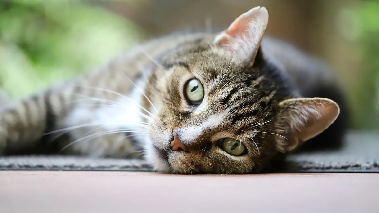
A trichobezoar is a compacted mass of ingested hair, sometimes mixed with food or other material, that collects in the stomach or intestine and can cause a physical blockage. Most small hair masses pass through or are vomited without harm, but a large or lodged trichobezoar can obstruct the stomach or bowel and may require surgery to remove.
Hairballs are so common in cats that owners often treat them as normal. In most cases a cat coughs up a tubular wad of hair and carries on. The problem is the hairball that does not come up and does not pass down. That mass sits in the stomach or jams in the small intestine, and the pet becomes genuinely ill. This guide explains how trichobezoars form, which animals are most at risk, what the signs look like, how veterinarians confirm the diagnosis, and when an obstruction crosses the line from a nuisance into a surgical emergency.
What Is a Trichobezoar?
A trichobezoar is a dense, felted ball of hair inside the gastrointestinal tract. The word combines tricho (hair) and bezoar (a mass trapped in the gut). Hair is essentially indigestible. The keratin that makes up hair resists the digestive enzymes that break down food, so swallowed hair tends to stay in the stomach rather than dissolve. Over time, strands mat together, trap mucus and food particles, and harden into a solid object.
The stomach is the usual location, but a trichobezoar can extend through the pylorus into the duodenum and beyond. When a hair mass trails out of the stomach and into the small intestine, the condition is called Rapunzel syndrome [1][2]. That variant is especially dangerous because the intestinal portion can act like a ball-and-socket obstruction, blocking the bowel even when the gastric portion is not fully plugging the stomach.
Trichobezoars are distinct from other bezoars. A phytobezoar is built from plant or fiber material, and a pharmacobezoar forms from medications or other ingested substances. The mechanism is similar, but the material differs. In veterinary patients, hair is the most common building block, which is why long-haired cats and rabbits dominate the clinical picture.
How a Trichobezoar Forms
Three ingredients drive trichobezoar formation: hair entering the gut, hair staying in the gut, and time. Any condition that increases grooming, increases hair swallowing, or slows the movement of material out of the stomach raises the risk.
Grooming Behavior and Overgrooming
Cats are the classic example. A cat spends a large part of its day grooming, and the rough papillae on its tongue catch loose hair and pull it into the mouth. Most of that hair is swallowed. In a healthy cat with normal gut motility, small amounts of hair move through or are vomited periodically.
Overgrooming changes the equation. Stress, skin disease, parasites, and allergies can all push a cat to groom far more than usual. The more hair goes in, the more likely it is to accumulate. The same logic applies to any animal that grooms itself or a companion.
Pica and Hair Ingestion
Pica is the deliberate eating of non-food material. Some animals chew and swallow hair, wool, or fabric. In the Canadian lynx case reported in the veterinary literature, fleas were identified as the likely cause of increased hair ingestion through grooming, and the trichobezoar recurred until flea control and a hairball laxative were added to the plan [3]. That case shows how an underlying skin or parasite problem can drive the behavior that creates the mass.
Why Hair Stays in the Stomach
The stomach is a holding area. Hair that reaches the stomach tends to swirl into a ball rather than pass smoothly through the pylorus. Once a small ball forms, it can grow as more hair arrives. The result is a mass that behaves like a foreign body. In dogs and cats, bezoars appear on imaging as intraluminal masses with mottled gas patterns and indistinct boundaries, and they tend to cause longer-lasting signs than sharp foreign bodies like bones or plastic [4].
Species That Cannot Vomit
Rabbits are a special case. They groom constantly, they swallow a great deal of hair, and they cannot vomit. A cat can bring a hairball up. A rabbit cannot. Hair that would be expelled in a cat has to travel all the way through the rabbit's digestive tract. When it does not, it contributes to a blockage, often alongside a diet too low in fiber or a slowdown in gut motility. This is one reason rabbits are so prone to hair-related gastrointestinal stasis and obstruction.
The flowchart below shows the main decision path from hair ingestion to treatment.
flowchart TD
    [Hair ingested] --> [Hair reaches stomach]
    [Hair reaches stomach] --> [Small mass forms]
    [Small mass forms] --> {Passes or vomited}
    {Passes or vomited} --> [No treatment needed]
    {Passes or vomited} --> [Mass grows]
    [Mass grows] --> {Obstruction develops}
    {Obstruction develops} --> [Vomiting and anorexia]
    [Vomiting and anorexia] --> [Imaging confirms mass]
    [Imaging confirms mass] --> {Small and stable}
    {Small and stable} --> [Medical management]
    {Small and stable} --> [Surgery needed]
Species at Risk
Trichobezoars are not evenly distributed across species. The animals most affected are those that groom heavily, have long coats, or cannot vomit.
Long-haired cats are the most familiar group. Cats with gastrointestinal obstruction from trichobezoars have been studied directly, and the histopathology around the obstruction site shows acute inflammation and mucosal erosion or ulceration, which means the mass is physically damaging the gut wall as it sits there [5]. Older cats in that study were more likely to have a concurrent diagnosis of alimentary lymphoma, so a hairball obstruction in a senior cat can be the event that uncovers a deeper problem [5].
Rabbits are highly prone. They groom continuously, ingest large amounts of hair, and have no ability to vomit. Their gastrointestinal tract is also sensitive to any slowdown in motility, so a hair mass that would be trivial in another species can become a life-threatening blockage.
Cattle develop trichobezoars as well, typically related to grooming and licking behavior, and these masses can obstruct the gut. They are a recognized cause of gastrointestinal obstruction in ruminants.
Dogs are affected less often, but they are not immune. Dogs that groom excessively, chew hair, or live with a heavy-shedding companion can swallow enough hair to form a mass. Foreign body obstruction studies in dogs show that bezoars behave differently from sharp objects: they cause longer clinical courses and more frequent transition zones on imaging [4].
Exotic and zoo species can also form trichobezoars. The Canadian lynx case is a documented example, with a pyloric obstruction removed by gastrotomy and enterotomy [3]. The underlying driver there was flea-related grooming.
Comparison Table by Species
| Species | Why at risk | Typical location | Vomiting possible | Key clinical concern |
|---|---|---|---|---|
| Long-haired cat | Heavy grooming, long coat, swallows hair | Stomach or small intestine | Yes | Obstruction, concurrent enteropathy or lymphoma in older cats [5] |
| Rabbit | Constant grooming, cannot vomit, motility-sensitive gut | Stomach or intestine | No | Rapid decline, stasis, surgical emergency |
| Dog | Grooming, pica, hair chewing | Stomach or intestine | Yes | Longer clinical course than sharp foreign bodies [4] |
| Cattle | Grooming and licking, rumen hair accumulation | Stomach or intestine | No (ruminant) | Obstruction, often chronic |
| Exotic felids | Grooming driven by parasites or skin disease | Pylorus or proximal duodenum | Yes | Recurrence unless the trigger is treated [3] |
Symptoms of a Trichobezoar
The signs of a trichobezoar depend on where the mass sits and how completely it blocks the gut. A small gastric hairball may cause intermittent vomiting and nothing more. A complete obstruction causes progressive, serious illness.
Vomiting
Vomiting is the most common sign in cats and dogs. Early on it may be occasional and may contain hair. As the mass grows or lodges, vomiting becomes more frequent. Bilious vomiting, meaning yellow or green fluid, suggests the obstruction is below the stomach and bile is backing up [6]. In the reported Rapunzel syndrome case, a patient initially managed for presumed pancreatitis deteriorated and developed bilious vomiting before imaging revealed trichobezoars in the stomach and duodenum [6].
Anorexia and Weight Loss
Animals with an obstruction stop eating. A gastric mass creates a feeling of fullness, so the pet may eat a little and then quit, or refuse food entirely. Weight loss follows. In a reported case of a giant gastric trichobezoar, the patient had a six-month history of abdominal pain, vomiting, weight loss, and intolerance to oral intake [1]. Rapid weight loss in a young animal with vague abdominal pain should always raise the question of a bezoar [7].
Palpable Mass
A large trichobezoar can sometimes be felt through the abdominal wall. In one adult case, palpation revealed a foreign mass occupying the epigastric area [8]. A palpable abdominal mass in a pet with vomiting and weight loss is a strong signal for imaging.
Ileus and Gut Motility Changes
Ileus means the normal muscular activity of the gut has slowed or stopped. A trichobezoar can cause ileus both by physically blocking the lumen and by irritating the bowel wall. Reduced gastrointestinal motility is a recognized finding in animals with gastrointestinal perforation, and it also appears with obstruction [9]. When motility drops, gas and fluid build up behind the blockage, and the pet's abdomen may become distended and painful.
Signs of a Surgical Emergency
Some signs mean the situation has moved beyond a simple blockage. Free air in the abdomen, a rigid or intensely painful belly, collapse, or a sudden deterioration point to perforation and peritonitis. In a reported fatal case, a teenager developed a massive trichobezoar that perforated the stomach and caused diffuse peritonitis, and she arrived in cardiopulmonary arrest [10]. A separate case described a perforated gastric ulcer secondary to a bezoar that required emergency surgery [11]. These outcomes are rare in pets, but they show why an untreated obstruction is dangerous.
How Veterinarians Diagnose a Trichobezoar
Diagnosis starts with a physical examination and a history. The veterinarian will ask about grooming habits, shedding, diet, and how long the signs have been present. Then imaging and, in some cases, endoscopy confirm the mass.
Radiography
Plain X-rays are often the first imaging step. They can show a mass effect, gas patterns, or dilated bowel loops. However, radiography is not reliable for bezoars. In one comparative study, only one of six available radiographs detected the bezoar [4]. That means a normal X-ray does not rule out a hairball. Radiographs are more useful for detecting free air, loss of serosal detail, or a suspected foreign body, all of which appeared in animals with gastrointestinal perforation [9].
Ultrasound
Ultrasound is more sensitive than plain radiography for bezoars. In the same comparative study, ultrasound showed acoustic shadowing in four of six bezoar cases [4]. A thickened, hyperechoic wall with acoustic shadowing in the stomach region has been described in a pediatric bezoar case that was later confirmed at surgery [12]. Ultrasound can also reveal free fluid, altered wall layering, reduced motility, and regional changes that suggest perforation [9].
Contrast Studies
A barium gastrointestinal study can outline a filling defect caused by a mass. In the Canadian lynx case, a barium study revealed a pyloric obstruction that had not been found on earlier diagnostics, and surgery confirmed a trichobezoar [3]. Contrast studies remain useful when ultrasound is inconclusive or when the mass is in a location that is hard to see.
Computed Tomography
CT is the most sensitive imaging option for bezoars. In dogs and cats, CT identifies bezoars as intraluminal masses with mottled gas patterns and indistinct boundaries, and it detects transition zones and bowel diameter ratios that predict obstruction severity [4]. CT is especially helpful when the clinical picture is confusing or when surgery is being planned.
Endoscopy
Endoscopy allows direct visualization of the stomach and upper intestine. A trichobezoar appears as a dark, hair-like mass, and the endoscopist can sometimes remove it or at least confirm the diagnosis. In a reported case, a foreign body resembling a kitchen scrubber was found partially obstructing the duodenum during gastroscopy [2]. Endoscopy is less useful for masses that have moved into the lower small intestine, which are beyond the reach of a standard scope.
Medical Management Versus Surgery
The decision to operate depends on the size and location of the mass, whether the pet is stable, and whether the gut is still moving.
When Medical Management May Work
A small gastric hairball that is not causing a complete obstruction may pass on its own or be managed with supportive care. Fluid therapy, anti-nausea medication, and sometimes a lubricant laxative can help move things along. In the lynx case, a hairball laxative was used three times a week after surgery to prevent recurrence [3]. Fiber can also play a role. A study in shorthaired cats found that beet pulp increased fecal production and reduced gastrointestinal transit time, though it did not change the amount of trichobezoar material excreted in feces [13]. Faster transit may help hair move through before it mats into a mass, but the evidence for fiber as a hairball cure is mixed.
Medical management is a reasonable trial only when the pet is stable, still passing stool, and not showing signs of complete obstruction. If the pet is vomiting everything, has a painful abdomen, or is deteriorating, medical management is not appropriate.
When Surgery Is Needed
Surgery is required when the trichobezoar is large, when it is causing a complete obstruction, when it has moved into the intestine, or when the pet is not responding to supportive care. The two main procedures are gastrotomy (opening the stomach) and enterotomy (opening the intestine).
Gastrotomy is used for gastric masses. In a reported adult case, laparoscopic gastrotomy with removal through a mini-laparotomy allowed discharge on the second postoperative day [8]. In a pediatric series, transumbilical single-incision laparoscopic-assisted removal achieved complete removal in all five patients without conversion to open surgery [14]. These minimally invasive approaches reduce recovery time, but they require the right equipment and an experienced surgeon.
Enterotomy is used when the mass has moved into the small intestine. In the lynx case, both a gastrotomy and a proximal duodenal enterotomy were needed to clear a pyloric obstruction [3]. Intestinal surgery carries more risk than gastric surgery because the bowel wall is thinner and the blood supply is more delicate.
Biopsy at the Time of Surgery
When a cat has a trichobezoar obstruction, taking biopsies from the gut wall is valuable. A study of 44 cats found that biopsies near the trichobezoar often showed neutrophilic inflammation and mucosal erosion or ulceration, while biopsies elsewhere showed lymphoplasmacytic or mixed inflammation, and some cats had alimentary small cell lymphoma [5]. Biopsies taken right at the obstruction site should be interpreted with caution because the mass itself causes acute inflammation, but biopsies from other areas can reveal chronic enteropathy or lymphoma that needs separate treatment [5].
Preventing Trichobezoars
Prevention focuses on reducing hair ingestion and keeping the gut moving.
Regular grooming removes loose hair before the pet swallows it. This is the single most useful step for long-haired cats and rabbits. Treating the underlying trigger matters too. In the lynx, monthly flea control and a hairball laxative prevented recurrence [3]. That case shows the principle: if parasites or skin disease drive grooming, controlling them reduces hair intake.
Dietary fiber may help some animals by speeding transit [13]. Fresh water and adequate hydration support normal gut motility. For rabbits, a high-fiber diet is the foundation of gut health, and any change in appetite or stool output needs prompt veterinary attention.
Some commercial hairball products are marketed for cats. Owners should discuss any supplement or laxative with their veterinarian before use, because the right choice depends on the individual animal and the underlying cause.
Limitations and When to Contact a Veterinarian
This article is educational and is not a substitute for veterinary diagnosis or treatment. Individual cases vary, and only a veterinarian who examines your pet can determine the right course of action.
Contact a veterinarian promptly if your pet has repeated vomiting, especially if it contains hair or bile, refuses food for more than a day, loses weight, has a painful or distended abdomen, or stops passing stool. These signs can indicate an obstruction that will not resolve on its own.
Seek emergency care immediately if your pet collapses, has a rigid or intensely painful belly, breathes rapidly, or shows sudden weakness. Free air in the abdomen, perforation, and peritonitis are life-threatening and require surgery without delay [9][10]. Rabbits that stop eating and stop producing stool need urgent care because their condition can deteriorate quickly.
Frequently Asked Questions
What is a trichobezoar?
A trichobezoar is a compacted mass of ingested hair, sometimes mixed with food or foreign material, that forms in the stomach or intestine and can cause a blockage.
Are hairballs in cats dangerous?
Most small hairballs are harmless and are either vomited or passed. A large hairball that lodges in the stomach or intestine can cause an obstruction that requires surgery.
Why are rabbits prone to trichobezoars?
Rabbits groom constantly, swallow a lot of hair, and cannot vomit, so hair has to travel through the entire digestive tract. Any slowdown in gut motility can turn a hair mass into a dangerous blockage.
Can dogs get trichobezoars?
Yes, though less often than cats and rabbits. Dogs that groom excessively, chew hair, or have pica can swallow enough hair to form a mass.
How do veterinarians diagnose a trichobezoar?
Diagnosis uses a combination of physical examination, radiography, ultrasound, contrast studies, CT, and endoscopy. Ultrasound and CT are more reliable than plain X-rays for detecting bezoars.
When does a trichobezoar require surgery?
Surgery is needed when the mass is large, causes a complete obstruction, moves into the intestine, or when the pet does not improve with supportive care. Gastrotomy and enterotomy are the main procedures.
Can a trichobezoar pass on its own?
A small gastric hairball may pass or be managed medically in a stable pet. A large or obstructive mass will not pass and needs surgical removal.
How can I prevent trichobezoars in my pet?
Regular grooming, controlling parasites and skin disease, adequate hydration, and a fiber-appropriate diet all help reduce hair accumulation. Discuss any hairball product with your veterinarian first.
Related Articles
- Dog Ate a Diaper: Blockage Risk and What to Do
- Dog Ate a Sponge: Signs of Blockage and What to Do
- Dog Hairball Remedy
- Cat Hairball Remedy Best
- Cat Hairball Remedy
- Cat Coughing Up Hairballs
Sources
- Giant gastric trichobezoar (Rapunzel syndrome) presenting as gastric outlet obstruction with imaging features suggestive of secondary superior mesenteric artery syndrome in a 27-year-old female: surgical management and multidisciplinary approach.
- Trichobezoar Causing Obstruction of a Percutaneous Endoscopic Gastrostomy (PEG) Feeding Tube: A Case Report.
- Pyloric trichobezoar in a Canadian lynx (Lynx canadensis).
- Computed Tomographic Features of Bezoars and Other Gastrointestinal Foreign Bodies in Dogs and Cats: A Comparative Analysis.
- Histopathologic diagnosis and patient characteristics in cats with small intestinal obstructions secondary to trichobezoars.
- Gastrointestinal trichobezoars presenting as abdominal pain and obstruction - Rapunzel syndrome.
- Trusting your gut: a hairy situation-gastric trichobezoar case report.
- Laparoscopic Removal of a Gastric Trichobezoar in Adulthood: A Case Report.
- Ultrasonographic findings in dogs and cats with gastrointestinal perforation.
- Fatal Gastric Perforation Caused by Undiagnosed Trichobezoar in an Adolescent: A Case Report.
- Pediatric case of trichobezoar leading to gastric perforation.
- Rare gastric bezoar in a school-aged child: a paediatric diagnostic conundrum.
- Beet pulp intake and hairball faecal excretion in mixed-breed shorthaired cats.
- Transumbilical single-incision laparoscopic-assisted removal of gastric trichobezoars in children: a technical refinement and feasibility study.