Perianal Glands in Dogs: Expression and Problems
By Dr. Zubair Khalid, DVM, MS, PhD ·

Perianal glands in dogs are not one structure. They are two separate systems that sit within a few centimeters of each other, cause overlapping signs, and get mixed up constantly in owner conversations and online advice. The first is the group of small hepatoid (sebaceous-type) glands embedded in the skin around the anus. The second is the paired anal sacs, which are glandular pouches with their own ducts, their own diseases, and their own surgical rules.
The word "expression" adds a second layer of confusion. For anal sacs, expression means physically emptying the sacs by pressure. For perianal gland tissue, expression means the activity of hormone receptors and proteins inside the cells, which is what determines whether a lump behaves as a benign adenoma or a carcinoma [1][2]. This article covers both meanings, both gland systems, and the evidence behind what veterinarians actually do about them.
This article is educational and is not a substitute for veterinary diagnosis or treatment.
The Short Answer and Triage Summary
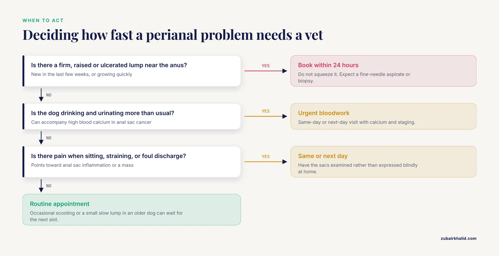
A swelling in the skin around the anus is a perianal gland problem until proven otherwise, and it needs a cytology or biopsy rather than squeezing. Repeated anal sac episodes, pain when sitting, or a changed stool shape point toward the anal sacs. A mass that is ulcerated, growing quickly, or accompanied by high blood calcium is a reason for urgent referral, not watchful waiting.
Seek veterinary care within 24 hours if you see any of the following:
- A firm, raised, or ulcerated lump beside or around the anus that was not there weeks ago.
- Repeated straining to defecate with little or no stool produced.
- A dog that cannot sit comfortably, cries when the tail base is touched, or guards the hind end.
- Blood, pus, or a foul discharge from the anal area.
- Any swelling accompanied by drinking more, urinating more, or unexplained weight loss, which can accompany elevated blood calcium in anal sac cancer [3][4].
- A previously removed anal sac tumor with a new swelling in the same area.
Routine (next available appointment) if you see:
- Occasional scooting or licking at the rear end without swelling or pain.
- A dog with known allergic skin disease and repeated anal sac flare-ups [5].
- A small, slow-growing skin lump near the anus in an older dog.
At a Glance: Two Gland Systems Owners Confuse
| Feature | Perianal (hepatoid) glands | Anal sacs |
|---|---|---|
| What they are | Modified sebaceous glands present in the skin of the perianal region [1] | Paired apocrine-type glandular sacs beside the anal canal [6] |
| Where the problem appears | A lump or thickening in the skin around the anus | Disease inside the sac or its duct, often without a visible lump |
| Main non-cancer problem | Hormonally driven hyperplasia [1] | Impaction and inflammation (sacculitis) [7][8][5] |
| Main cancer | Adenoma, epithelioma, carcinoma of hepatoid gland origin [1][9] | Apocrine gland anal sac adenocarcinoma (AGASACA) [3][10] |
| How "expression" applies | Receptor and protein expression inside tumor cells [1][2] | Manual emptying of the sacs by pressure |
| First diagnostic step | Fine-needle aspirate or biopsy of the mass [2][11][9] | Rectal examination, sac palpation, and cytology if a mass is felt |
| Definitive treatment for cancer | Wide or staged excision depending on grade, plus staging | Surgical excision, with radiation and systemic therapy in advanced cases [3][12][13] |
The practical value of this table is simple. A visible skin lump is not an anal sac problem, and a repeated anal sac problem is not a perianal gland tumor. Treating one as the other delays the correct diagnosis.
Anatomy and Physiology: What These Glands Do
Perianal (Hepatoid) Glands
Perianal gland tumors arise from modified sebaceous glands present in the skin of the perianal region, and the same tissue is referred to in the literature as hepatoid glands because the cells resemble liver cells under the microscope [1][11]. These glands sit in the skin, not inside the anal canal. That location matters, because it means a tumor here is a skin tumor that happens to be near the anus, and it is managed with skin and soft tissue surgical principles plus staging.
The hepatoid glands are hormone responsive. Androgen and estrogen receptors have been demonstrated both in normal perianal glands and in perianal gland tumors, and hormonal stimulation may drive hyperplasia of the glands or their progression toward neoplasia [1]. This is the biological reason perianal gland tumors are so closely tied to sex hormones, and it is the reason receptor testing has been investigated as a way to choose antihormonal treatment [1].
Anal Sacs
The anal sacs are paired structures beside the anal canal lined by apocrine-type glandular cells, the same cell type that gives rise to apocrine gland anal sac adenocarcinoma (AGASACA) [6]. They are anatomically separate from the hepatoid skin glands and behave completely differently. Anal sac disease covers three broad categories: impaction, inflammation (sacculitis), and neoplasia [7][8][5].
Anal sac disorders are common in primary care. A cohort of 104,212 dogs attending 110 UK practices found a one-year period prevalence of non-neoplastic anal sac disorders of 4.40 percent [8]. In a separate survey of 43,517 dogs, owner-reported anal sac impactions were among the most frequently reported gastrointestinal disorders at roughly 2 percent of the population [7]. These are not rare nuisance problems. They are a routine part of canine general practice.
The Two Meanings of "Expression"
Manual expression of the anal sacs is a physical procedure. A veterinarian or technician applies controlled pressure to empty the sacs when they are impacted or when material needs to be collected. It is a diagnostic and therapeutic maneuver, not a cure for every sac problem, and it does not address sacculitis, allergic skin disease, or cancer.
Cellular expression is a laboratory concept. Immunohistochemistry measures which proteins the tumor cells are producing. In perianal gland tumors, researchers have measured androgen and estrogen receptors [1], p63 [2], cyclooxygenase-2 [9], glycoconjugates [14], and immunoglobulin production by B cells and tumor cells [11]. These markers help pathologists classify tumors and, in some cases, estimate behavior.
Hormones and Perianal Gland Tissue
The hormone link in perianal gland tumors is well documented. In a study of 41 male dogs with perianal masses, histopathology identified 24 adenomas, 12 epitheliomas, and 5 carcinomas. Immunohistochemical staining showed mainly nuclear expression of both androgen and estrogen receptors in the neoplastic cells. Receptors were present in all three tumor types, but expression was highest in adenomas and epitheliomas and very weak in carcinomas [1].
That pattern has clinical implications. Strong receptor expression in the more benign end of the spectrum supports the long-standing observation that many perianal gland proliferations are hormone dependent. Weak expression in carcinomas suggests that the most aggressive tumors have partly escaped that dependency, which is one reason antihormonal therapy is not a universal answer. The authors concluded that determining sex hormone receptor expression could help build a diagnostic and therapeutic algorithm [1]. That is a research direction, not yet a standard of care.
For owners, the practical message is that hormone status matters in this tissue, and a veterinarian will factor in sex, neuter status, tumor type, and grade together rather than relying on any single variable.
Problems With Perianal Glands in Dogs
Hyperplasia
Hormonal stimulation can induce hyperplasia of the perianal glands [1]. Hyperplasia is an overgrowth of otherwise normal-appearing cells, and it can produce a palpable or visible swelling around the anus that mimics a tumor. Distinguishing hyperplasia from adenoma usually requires sampling, because the two can feel similar on examination.
Adenoma, Epithelioma, and Carcinoma
Canine perianal gland tumor is one of the most common skin neoplasms in dogs, yet its molecular characteristics remain poorly understood [2]. Hepatoid gland neoplasms are among the most common cutaneous tumors arising from perianal glands and are clinically characterized by rapid growth [9]. The three main categories are adenoma, epithelioma, and carcinoma, and the distinction is not academic. Adenomas are benign, epitheliomas occupy an intermediate position, and carcinomas are malignant with potential to invade and spread [1][2].
The molecular differences between these categories are becoming clearer:
- p63. In a study of 65 samples, p63 expression was significantly increased in perianal gland carcinomas compared with adenomas (P < 0.0001). The percentage of cells expressing p63 was higher in carcinomas, although staining intensity did not differ significantly between groups. The authors identified p63 as a candidate factor contributing to malignant transformation and progression [2].
- Cyclooxygenase-2. COX-2 expression was not detected in 60 percent of normal hepatoid glands and in 25 percent of hepatoid gland adenomas, while 75 percent of adenomas showed weak expression. Expression was also examined across epitheliomas and carcinomas, linking this inducible enzyme to tumorigenesis in this tissue [9].
- Glycoconjugates. Lectin-binding studies in normal and neoplastic hepatoid glands showed that most adenoma cells did not stain or stained only weakly with several lectins, while carcinoma basal cells lost specific binding for wheat germ agglutinin and showed heterogeneous concanavalin-A staining. These shifts track with progressive change in the tissue [14].
- Immune environment. Analysis of ten hepatoid adenomas, thirteen well-differentiated hepatoid carcinomas, and nine undifferentiated hepatoid carcinomas found that adenomatous structures had the greatest concentration of CD79a-positive, IgA-producing B cells. In malignant tumors, an inverse association appeared between CD79a expression and IgG, with a significant increase in tumor antigen-specific IgG. The authors suggested that IgG produced by neoplastic cells could contribute to tumor progression and might serve as a prognostic biomarker and therapeutic target [11].
What does this mean for an owner facing a diagnosis? It means the pathologist has more tools than a simple benign-versus-malignant label, and it means research is moving toward markers that predict behavior. It does not mean a COX-2 inhibitor or an immunoglobulin test is a standard treatment today.
Problems With Anal Sacs
Impaction
Impaction means the sacs are not emptying adequately and their contents accumulate. Owner-reported anal sac impaction was identified in about 2 percent of the 43,517 dogs surveyed in the Dog Aging Project, placing it alongside chronic diarrhea and foreign bodies as one of the most common gastrointestinal complaints [7]. Impaction is the mildest category of anal sac disease, and it can often be managed by emptying the sacs and identifying why they are not draining.
Anal Sacculitis
Sacculitis is inflammation of the anal sacs, and it tends to be a recurring problem rather than a single event. In a Swedish primary-care study of 190 dogs experiencing 290 episodes of sacculitis, the most common management (235 of 290 episodes, 81.0 percent) was flushing with or without infusion of an antimicrobial product into the sacs, with or without a nonsteroidal anti-inflammatory drug. This approach produced resolution in 213 of 235 episodes, or 90.6 percent. Median time to resolution was one week, with a range of one to sixteen weeks, and 70.7 percent of episodes resolved within one week [5].
Two findings from that study matter for long-term planning. First, 66.3 percent of dogs had only a single episode, which is reassuring. Second, dogs with concurrent cutaneous allergic disease were significantly more likely to have recurrent sacculitis than dogs with other comorbidities, and cutaneous allergic disease itself occurred more often in the sacculitis population than in the general practice population [5]. If your dog has recurrent anal sac inflammation and itchy skin, the skin disease and the sac disease are probably connected, and both need attention.
Anal Sac Adenocarcinoma
Apocrine gland anal sac adenocarcinoma is an aggressive malignancy with high metastatic potential [15]. Metastasis to intra-abdominal and pelvic lymph nodes occurs early in the disease course, and death is usually related to locoregional progression [10]. That combination, early spread and local progression as the main killer, explains why staging and local control dominate treatment planning.
Hypercalcemia of malignancy is a recognized complication. In two dogs with metastatic AGASACA and ionized calcium above the reference range, toceranib phosphate was used when owners declined surgery, and calcium response was monitored as part of case management [4]. In a larger surgical cohort, preoperative hypercalcemia was associated with reduced survival time after excision, at 7.5 months compared with the overall median [3]. Blood calcium is therefore not a routine add-on. It is a prognostic variable.
Signs Owners Notice and What They Mean
| What the owner sees | Likely category | What to do | |
|---|---|---|---|
| A visible lump in the skin beside the anus | Perianal (hepatoid) gland hyperplasia or tumor [1][9] | Book an exam and expect a fine-needle aspirate. Do not squeeze it. | |
| A dog that scoots, licks, or seems irritated at the tail base | Anal sac impaction or sacculitis [7][8] | Have the sacs examined rather than expressed blindly at home. | |
| Pain when sitting or when the tail base is touched | Anal sac inflammation, or a mass | Analgesics were prescribed in about 12 percent of anal sac disorder cases, so pain is a recognized feature [8] | Same-day or next-day appointment |
| Repeated episodes over months | Recurrent sacculitis, often with allergic skin disease [5] | Discuss the underlying skin disease, not just repeated emptying | |
| Straining to defecate | Anal sac disease or another colorectal problem | Examination, including rectal palpation | |
| Increased drinking and urination with a perianal problem | Possible hypercalcemia of malignancy [3][4] | Prompt bloodwork and staging |
Bloodwork matters here because hypercalcemia can produce vague signs that owners attribute to age or heat. If a dog with an anal sac mass is drinking more than usual, that detail should be raised at the appointment.
How Veterinarians Evaluate the Problem
Hands-On and Rectal Examination
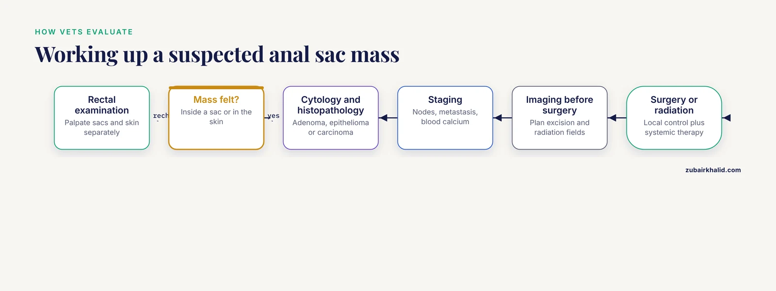
The physical examination starts with the skin around the anus. A hepatoid gland mass is usually palpable as a firm nodule in the skin, and its size, mobility, and relationship to the underlying tissue are recorded [1][9]. The anal sacs are assessed separately, because a mass inside a sac can be felt on rectal examination even when nothing is visible outside.
A thorough work-up for a presumed unilateral anal sac tumor should not assume the other side is normal. In 35 dogs that underwent bilateral anal sacculectomy for what appeared to be unilateral AGASACA, only 5 (14 percent) had histologically normal contralateral sacs. Non-neoplastic anal sac disease was found in 23 of 35 dogs (66 percent), and bilateral AGASACA was found in 7 of 35 (20 percent). None of the dogs with bilateral neoplasia had evidence of it before surgery, despite thorough work-up [16]. That last point is a caution against assuming the opposite side is clean without examining it.
Cytology, Histopathology, and Molecular Testing
Fine-needle aspiration is often the first step for a perianal skin mass. It can distinguish inflammatory lesions from round cell tumors from epithelial tumors, and it can be done in a conscious dog with minimal equipment. Definitive classification of hepatoid gland tumors relies on histopathology, with adenoma, epithelioma, and carcinoma separated on architectural and cellular criteria [1]. Where available, immunohistochemistry for markers such as p63 or COX-2 adds information about likely behavior [2][9].
Immune and glycoconjugate markers remain research tools. They are useful to understand where the science is going and are not part of a standard panel for every dog.
Staging for Anal Sac Tumors
Staging drives treatment. In AGASACA, the relevant questions are the size and invasiveness of the primary tumor, the status of the regional lymph nodes, and the presence of distant metastasis. Lymphovascular invasion on histopathology is an independent adverse factor, and mitotic count has also been associated with survival in surgical cohorts [3]. Preoperative hypercalcemia and the presence of metastatic disease both shortened survival in the same series [3].
Proliferation markers can refine prognosis. In a digital pathology study of 58 anal sac adenocarcinomas, a global Ki67 score of 26 or higher was associated with significantly shorter tumor-related survival (175 days) than a score below 26 (650 days). Anisokaryosis and the global Ki67 score were independent prognostic markers, while mitotic count did not affect survival in that analysis [15]. These are specialist pathology tools, but they can inform how aggressively a case is followed.
Imaging Before Surgery
Cross-sectional imaging is used to plan excision and to identify nodal disease. Advanced imaging also underpins radiation planning, since the organs at risk in this region include the colon, bladder, and spinal cord [10]. Stereotactic radiation has been used specifically for metastatic sacroiliac lymph nodes in dogs with AGASACA, with staging recorded in 3 stage IIIa, 14 stage IIIb, and 8 stage IV cases [13].
Evidence-Based Management
Flushing and Medical Therapy for Anal Sac Disease
For sacculitis, the practical first-line approach is mechanical and medical. Flushing the sacs, with or without instilling an antimicrobial product, and with or without an NSAID, resolved 90.6 percent of episodes in a large primary-care series, and median time to resolution was one week [5]. Most dogs that respond do so quickly, which means a dog that is not improving within a week or two needs re-evaluation rather than another round of the same treatment.
Antimicrobial use in anal sac disease is common but not universal. In the UK cohort, antimicrobials were prescribed in 480 of the recorded anal sac disorder cases (20.24 percent), and analgesics in 284 (11.97 percent). Anal sacculectomy was performed in under 1 percent of cases [8]. Those proportions are a useful reality check. Most anal sac problems are managed medically, and surgery is reserved for a small minority.
Recurrence is the main long-term issue. Because recurrent sacculitis is significantly associated with concurrent cutaneous allergic disease [5], a dog with repeat episodes deserves a skin work-up alongside sac management. Repeatedly expressing the sacs without addressing the underlying allergic dermatitis tends to produce a cycle rather than a cure.
Surgery for Anal Sac Disease
Surgical removal of the anal sacs is considered when medical management fails, when the sacs are chronically diseased, or when neoplasia is confirmed or suspected. Technique matters because the anatomy is unforgiving.
For non-neoplastic disease, a modified closed sacculectomy technique was evaluated in 50 dogs undergoing bilateral procedures. Intraoperative anal sac perforation occurred in 5 dogs (10 percent). Among 43 dogs with two-week follow-up, grade 1 complications occurred in 14 (32 percent), grade 2 in 2 dogs (5 percent), and grade 3B in 2 dogs (5 percent). At two weeks, 13 of the 14 dogs with grade 1 complications had resolved, both grade 2 complications had resolved, and both grade 3B complications had resolved after revision surgery [17]. In plain terms, minor complications are common, most settle quickly, and major complications are uncommon and usually fixable with a single revision.
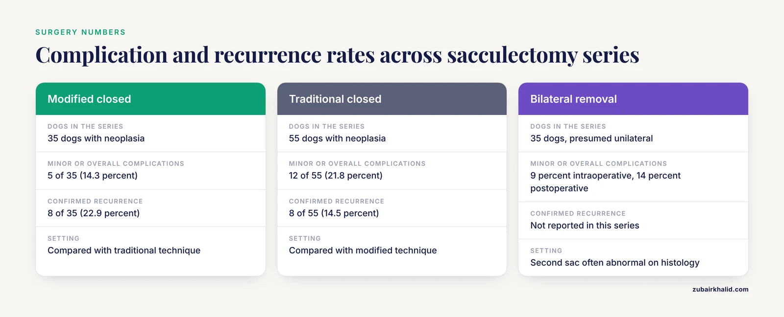
For anal sac neoplasia, a comparison of modified closed versus traditional closed sacculectomy in 90 dogs found minor postoperative complications in 5 of 35 modified-approach dogs (14.3 percent) and 12 of 55 traditional-approach dogs (21.8 percent). Confirmed tumor recurrence occurred in 8 of 35 modified-approach dogs (22.9 percent) and 8 of 55 traditional-approach dogs (14.5 percent), with suspected recurrence in a further 8.6 percent and 13.2 percent respectively [18]. A separate series of 47 dogs with AGASACA treated by modified closed sacculectomy reported postoperative complications in 15 dogs (31.9 percent), of which 14 were minor and 1 was major. Among 45 dogs followed for a median of 364 days, only 1 dog (2.2 percent) developed local recurrence, at 90 days [19].
The gap between those two recurrence figures (about 2 percent in one series and about 23 percent in another) shows why single-study numbers should not be treated as a guarantee. Follow-up duration, case mix, tumor stage, and whether adjunct therapy was used all differ. What is consistent is the principle: clean, complete removal of the sac and its duct, performed by a surgeon comfortable with the anatomy, gives the best local control.
Margin status is the most actionable surgical variable. In a cohort of 74 dogs undergoing AGASACA excision, 75.7 percent achieved an R0 (complete) margin. An R1 (incomplete) margin was associated with a markedly increased risk of local recurrence (odds ratio 23, p < 0.0001). Overall median survival time after surgery was 25 months. Survival was reduced in dogs with preoperative hypercalcemia (7.5 months), metastatic disease at presentation (17 months), and lymphovascular invasion on histopathology (21 months). Mitotic count and the absence of adjunct therapy also reduced survival time [3].
Removing both sacs when only one appears affected is a legitimate option, and the histology from the second side often surprises. In the bilateral sacculectomy series, 66 percent of presumed normal contralateral sacs had non-neoplastic disease and 20 percent harbored bilateral carcinoma [16]. Complications attributable to primary tumor removal occurred in 9 percent of dogs intraoperatively and 14 percent postoperatively, most often tumor capsule disruption and surgical site infection respectively [16]. Tumor capsule disruption is a specific reason not to manipulate a mass at home before surgery.
Perianal Gland Tumors: What the Evidence Supports
Treatment of a hepatoid gland tumor depends on the diagnosis. A benign adenoma may be monitored or excised depending on size, location, and whether it is causing discomfort. An epithelioma or carcinoma requires wider excision and staging.
Hormone receptor testing has been proposed as a way to guide antihormonal treatment, and receptor expression was highest in adenomas and epitheliomas and weak in carcinomas [1]. That is a rationale for considering hormonal manipulation in selected cases, not a validated protocol that replaces surgery for a malignant tumor. Any discussion of antihormonal therapy belongs with a veterinary oncologist who has the histopathology in hand.
p63 expression is a candidate marker for malignant transformation [2], and COX-2 expression has been documented in hepatoid neoplasms [9]. Neither is a treatment target with established clinical benefit in dogs at this time. Owners should be skeptical of any source that presents a COX-2 inhibitor as a proven cure for perianal gland cancer.
Anal Sac Adenocarcinoma: Combining Local and Systemic Treatment
Surgery is the treatment of choice for AGASACA when the tumor can be removed [10]. When surgery is not possible or is declined, radiation and systemic therapy become the main tools.
Palliative radiation has measurable benefit. A protocol of 20 Gy delivered in five consecutive 4 Gy fractions produced a median survival time of 384 days when used alone in 22 dogs, and 628 days when combined with additional therapies. Median time to progression was 337 days with radiation alone and 402 days with radiation plus additional treatment. Acute side effects were mild, with diarrhea in 61 percent of dogs, and one dog developed a grade III late effect 22 months after the first protocol following re-irradiation [20].
Dose escalation improves local control. A comparison of 10 fractions of 4.0 Gy (15 dogs) against 8 fractions of 3.8 Gy (11 dogs) found median overall survival of 1534 days versus 821 days, progression-free survival of 926 days versus 258 days, and local progression-free survival of 1554 days versus 351 days. The treatment response rate at three-month follow-up CT was higher in the dose-escalated group at 90.9 percent [12]. These are retrospective data from a single institution, so they guide protocol design rather than define a universal standard.
Stereotactic radiation is an option for macroscopic nodal disease. In 25 dogs receiving stereotactic body radiation therapy to metastatic sacroiliac lymph nodes, overall median survival time was 451 days and median event-free survival was 246 days. Stage did not significantly affect survival. Male sex, higher dose per fraction, and higher total dose were positive prognostic factors, and dogs receiving 6 to 7.5 Gy per fraction to a total of 30 to 37.5 Gy outperformed dogs on other protocols. Twelve dogs developed hind limb gait changes during the late radiation effects period. The best response was complete in 21 percent, partial in 58 percent, and stable in 17 percent at a median of 100 days, and resolution of hypercalcemia in 5 dogs was inconsistent and transient [13].
An exploratory study of stereotactic radiation in five dogs with advanced AGASACA found the protocol feasible and produced complete response in one dog, partial response in two, and stable disease in one on follow-up CT, with minimal acute side effects [10]. Small case numbers mean these results describe possibility, not probability.
Systemic options are evolving. Toceranib phosphate has been used in the gross disease setting for metastatic AGASACA with hypercalcemia of malignancy when surgery was declined [4]. Laboratory work with ten AGASACA organoid lines showed different sensitivity profiles among lineages for carboplatin, mitoxantrone, toceranib, and lapatinib, with carboplatin and lapatinib splitting into sensitive and resistant lines while mitoxantrone and toceranib showed generally high efficacy across all organoids [6]. Organoid testing is a research platform for precision medicine, not a service available in routine practice.
The realistic summary for owners is that AGASACA is treatable but not simple. Cure is possible when the tumor is small, completely excised, and not metastatic. Advanced disease is managed with combined local and systemic therapy with the goal of prolonging good-quality survival.
Unsafe Home Remedies and Common Mistakes
- Squeezing a lump near the anus. Tumor capsule disruption is a documented intraoperative complication during anal sac tumor removal [16]. Applying that kind of pressure at home on an uncharacterized mass risks the same problem plus infection.
- Routine home expression instead of diagnosis. Manual emptying can relieve an impacted sac, but it does nothing for sacculitis that needs flushing and medication [5] and nothing for a tumor. Repeated home expression can delay the appointment where the real problem is found.
- Assuming a perianal lump is a "blocked gland." Hepatoid gland tumors are common skin neoplasms in dogs [2], and they are not anal sacs. A lump in the skin around the anus needs sampling.
- Using human hemorrhoid creams or steroid ointments on a perianal mass. These do not treat hepatoid tumors or anal sac disease and can mask progression.
- Adding antibiotics from a previous prescription. Antimicrobials were prescribed in roughly one in five anal sac disorder cases in a large cohort [8], but the decision requires an examination, and recurrent cases usually need the underlying cause addressed rather than another course [5].
- Waiting on a fast-growing mass. Hepatoid gland neoplasms are clinically characterized by rapid growth [9], and delay can convert an easily excised tumor into a difficult one.
Prevention and Surveillance
There is no vaccine or diet that prevents perianal gland tumors, and owners should ignore products claiming to do so. What does help is structured surveillance.
- Know the breed risk for anal sac disease. Compared with crossbred dogs, Cavalier King Charles Spaniels, King Charles Spaniels, Cockapoos, Shih Tzus, Bichon Frises, and Cocker Spaniels had increased odds of non-neoplastic anal sac disorders, while Labrador Retrievers, Border Collies, Staffordshire Bull Terriers, Lurchers, German Shepherd Dogs, and Boxers had reduced odds. Brachycephalic dogs had 2.6 times the odds of dolichocephalic dogs [8].
- Treat the skin, not just the sacs. Recurrent sacculitis is significantly associated with cutaneous allergic disease [5].
- Check the rear end during grooming or routine handling. Small perianal masses are easier to treat than large ones.
- Ask about calcium and lymph nodes if a sac mass is found. Hypercalcemia and metastatic disease both carry prognostic weight [3].
- Keep follow-up appointments after surgery. Local recurrence after AGASACA excision is strongly linked to margin status, and recurrence risk does not disappear after the incision heals [3].
Prognosis
Prognosis in perianal gland disease spans an enormous range. A benign adenoma removed with clean margins has an excellent outlook. A carcinoma with weak hormone receptor expression and aggressive histologic features carries a guarded outlook [1][2].
For anal sac adenocarcinoma, the variables that consistently matter are stage at diagnosis, completeness of excision, the presence of hypercalcemia, lymphovascular invasion, and proliferation index [3][15]. Dogs with complete excision and no metastasis do substantially better than dogs with incomplete margins or distant spread. Radiation and systemic therapy extend survival in advanced disease, and dose-escalated protocols have produced long-term local control in selected cases [12][13][20].
A realistic conversation with an oncologist should cover what the goal is (cure versus control), what the treatment burden looks like, and what the dog's quality of life will be during treatment. Numbers from published cohorts inform that conversation but do not predict an individual dog.
Limitations and When to Contact a Veterinarian
This article summarizes published evidence and standard clinical reasoning. It cannot diagnose your dog, and it cannot tell you whether a specific lump is benign. Individual cases require a veterinarian who can examine the tissue, review the history, and interpret test results in context.
Contact a veterinarian promptly, and consider an emergency visit, in these situations:
- A perianal mass that is growing, ulcerated, bleeding, or causing pain.
- Straining to defecate that persists for more than 24 hours, especially with a tense abdomen or vomiting.
- Signs of severe pain at the tail base, inability to sit, or reluctance to walk.
- Fever, lethargy, or loss of appetite combined with a swollen anal area.
- Increased drinking and urination in a dog with a known or suspected anal sac mass.
- A patent anal sac that ruptures and drains foul material, which needs cleaning, antibiotics, and pain control rather than home squeezing.
- Any dog with recurrent anal sac episodes more than three or four times a year, which warrants investigation for allergic skin disease and anatomical factors.
If surgery is being considered, ask specifically about the planned technique, the surgeon's experience with anal sac anatomy, whether both sacs will be assessed, and what the follow-up schedule will be. Those questions change outcomes more than any home remedy.
Related Guides
- Anal Fistula in Dogs: Perianal Fistula Guide: Helps differentiate perianal masses from anal fistula disease
- How Much Does Dog Anal Gland Expression Cost in the UK?: Practical cost information for owners considering anal gland expression
- How to Treat Dog Diarrhea After Gland Expression at Home: Covers diarrhea after expression, a possible post-procedure concern
- Anal Gland Supplements for Dogs: Do Fiber Chews Actually Help?: Reviews whether fiber chews help anal gland disease
- Dog Anal Gland Abscess Home Treatment: Useful for abscess signs and what to do at home
- Dog Hemorrhoids or Anal Gland Trouble? What to Do: Helps distinguish hemorrhoids from anal gland problems
- Dog Anal Gland Infection Symptoms: Explains infection signs that may accompany anal gland disease
Frequently Asked Questions
Are perianal glands the same as anal sacs?
No. Perianal (hepatoid) glands are modified sebaceous glands in the skin around the anus, while anal sacs are separate paired glandular pouches beside the anal canal. They cause different diseases and are treated differently.
Should I express my dog's anal glands at home?
Only if your veterinarian has specifically shown you how and asked you to do it. Repeated home expression does not treat sacculitis or a tumor, and squeezing an uncharacterized lump near the anus risks rupturing it.
Do perianal gland tumors mean my dog needs to be neutered?
Not automatically, and neutering is not a treatment for a tumor that is already present. Hormone receptors do influence this tissue, so discuss neuter timing and status with your veterinarian alongside the tumor's histologic type and grade.
Are perianal gland tumors cancer?
Some are. Adenomas are benign, epitheliomas are intermediate, and carcinomas are malignant. A pathologist distinguishes them on tissue examination, and the distinction drives treatment.
What causes anal sac infection?
Anal sacculitis usually reflects poor drainage plus inflammation, and it is significantly more common in dogs that also have allergic skin disease. Breed and skull shape also affect risk.
How long does anal sac treatment take to work?
Most episodes that respond to flushing and medication resolve within about a week. A dog that is not improving in that window needs re-evaluation rather than repeat treatment.
Can anal sac cancer be cured?
Yes, in some cases. Cure is most likely when the tumor is small, completely excised, and has not spread. Advanced disease is managed rather than cured, with surgery, radiation, and systemic therapy combined.
When should I worry about a lump near my dog's anus?
Worry when it is new, firm, growing, ulcerated, or painful, and when your dog is also drinking and urinating more than usual. Any of those findings justifies an appointment within days, not months.
Sources
- Androgen and Estrogen Receptor Expression in Different Types of Perianal Gland Tumors in Male Dogs.
- Increased p63 Expression in Canine Perianal Gland Tumours.
- Investigation of the "R" tumor margin classification and prognostic factors in apocrine gland anal sac adenocarcinoma of dogs.
- Toceranib phosphate for treatment of hypercalcaemia of malignancy in two dogs with metastatic anal sac apocrine gland adenocarcinoma.
- A retrospective study of treatment, outcome, recurrence and concurrent diseases in 190 dogs with anal sacculitis.
- Establishment of an experimental model of canine apocrine gland anal sac adenocarcinoma organoid culture using a three-dimensional culture method.
- Cross-sectional survey of 43,517 dogs in the Dog Aging Project identifies owner-reported lifetime prevalence and characteristics of gastrointestinal disease.
- Non-neoplastic anal sac disorders in UK dogs: Epidemiology and management aspects of a research-neglected syndrome.
- First study on the immunohistochemical expression of cyclooxygenase-2 and clinicopathological association in canine hepatoid gland neoplasms.
- Stereotactic radiotherapy for advanced canine anal sac adenocarcinoma: an exploratory study.
- B-cells and non-B-cells immunoglobulins expression in canine perianal gland tumours: a preliminary study.
- A dose-escalated radiation protocol of 10 × 4.0 Gy provides long-term local control in canine apocrine gland anal sac adenocarcinoma.
- Retrospective Study Evaluating the Outcome and Efficacy of Stereotactic Body Radiation Therapy for the Treatment of Metastatic Abdominal Lymph Nodes in Dogs With Apocrine Gland Anal Sac Adenocarcinoma.
- Glycoconjugate expression in normal hepatoid glands and hepatoid gland adenomas and carcinomas in dogs.
- Proliferation scores in canine anal sac adenocarcinomas: Ki67 global score is superior to Ki67 hotspot indices and mitotic count for prognosis.
- Histopathological diagnosis and surgical complications following bilateral anal sacculectomy for the treatment of unilateral canine apocrine gland anal sac adenocarcinoma: 35 cases (2019-2023).
- Modified closed sacculectomy in 50 dogs with non-neoplastic anal sac disease.
- Long-term outcomes associated with a modified versus traditional closed anal sacculectomy for treatment of canine anal sac neoplasia.
- Outcomes of dogs with apocrine gland anal sac adenocarcinoma treated via modified closed anal sacculectomy (2015-2022).
- Radiotherapy for Canine Apocrine Gland Anal Sac Adenocarcinoma: Survival Outcomes and Side Effects of a Palliative Treatment Protocol of 20 Gy in Five Consecutive Fractions.