
This article is educational and is not a substitute for veterinary diagnosis or treatment.
Dog Ear Yeast Infection: Signs, Diagnosis, Treatment Principles, and Recurrence Prevention
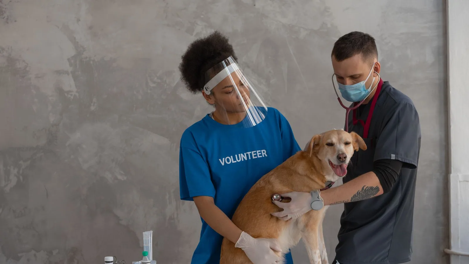
A dog ear yeast infection, clinically termed Malassezia otitis externa, is one of the most common dermatologic conditions seen in small animal practice. The condition occurs when the commensal yeast Malassezia pachydermatis overgrows within the external ear canal, leading to inflammation, pruritus, and a characteristic odour. While many owners attempt home remedies or over-the-counter drops, effective management requires a proper veterinary diagnosis, cytological confirmation, and treatment of the underlying cause. This article provides a definitive, evidence-based review of canine Malassezia otitis, covering anatomy, diagnostic principles, treatment options, and strategies for preventing recurrence.
At a Glance: Clinical Decision Table for Suspected Yeast Otitis
| Clinical Feature | Typical Yeast Otitis | Possible Bacterial Otitis | Emergency Red Flags |
|---|---|---|---|
| Discharge appearance | Dark brown, waxy, crumbly | Yellow-green, purulent, bloody | Blood, pus, or necrotic tissue |
| Odour | Musty, sweet, "yeasty" | Foul, rancid, or sweet-purulent | Foul with systemic signs |
| Pruritus | Moderate to severe head shaking, ear scratching | Variable, often painful | Head tilt, circling, nystagmus |
| Pain on ear handling | Mild to moderate | Moderate to severe | Severe pain, inability to open mouth |
| Cytology (stained) | Yeast organisms (budding) | Cocci, rods, or both | Neutrophils with intracellular bacteria |
| Tympanic membrane | Intact (usually) | May be intact or ruptured | Ruptured (requires non-ototoxic therapy) |
If your dog shows head tilt, circling, loss of balance, or severe pain, seek emergency veterinary care immediately.
Understanding Ear Anatomy and Why Yeast Thrives
The canine external ear canal is L-shaped, consisting of a vertical canal leading to a horizontal canal that ends at the tympanic membrane (eardrum). This anatomy predisposes dogs to otitis because the vertical orientation traps debris, moisture, and exudate. The horizontal canal is narrow and poorly ventilated, creating a warm, humid environment ideal for microbial overgrowth [3].
The ear canal is lined by stratified squamous epithelium that produces cerumen, a waxy secretion containing lipids, proteins, and antimicrobial peptides. In healthy ears, the microbiota is balanced, with low numbers of bacteria and yeast. Malassezia pachydermatis is a lipophilic yeast that normally resides on canine skin and in the ear canal. It attaches to cornified epithelial cells via lipid-mediated adhesion, not direct binding, which explains why excessive cerumen production promotes yeast colonisation [9].
When the ear canal environment is altered by inflammation, moisture, or cerumen accumulation, Malassezia can proliferate rapidly. The yeast does not invade living tissue; instead, it triggers inflammation through metabolic byproducts and cell wall components that activate the host immune response. This inflammation further damages the epithelial barrier, perpetuating a cycle of infection and inflammation.
Causes and Risk Factors for Malassezia Otitis
Primary Causes
Malassezia otitis is almost always secondary to an underlying primary disease that disrupts the ear canal environment. The most common primary causes include:
- Allergic skin disease: Atopic dermatitis, food allergy, and contact allergy are the most frequent triggers. Allergic inflammation alters cerumen composition and impairs the skin barrier, allowing yeast to overgrow [6].
- Endocrine disorders: Hypothyroidism, hyperadrenocorticism (Cushing's disease), and sex hormone imbalances can alter skin lipid production and immune function.
- Anatomic abnormalities: Pendulous pinnae (e.g., Cocker Spaniels, Basset Hounds), stenotic ear canals, and excessive hair within the canal impair ventilation and cleaning.
- Moisture: Swimming, bathing, or living in humid climates can macerate the ear canal epithelium.
- Foreign bodies: Grass awns, foxtails, or plant material can cause inflammation and secondary infection.
- Iatrogenic causes: Overcleaning with harsh products, use of cotton swabs that push debris deeper, or inappropriate topical medications.
Breed Predispositions
Certain breeds are overrepresented for otitis externa. Golden Retrievers and West Highland White Terriers show significantly higher risk [3]. Dogs with pendulous pinnae are also more likely to develop otitis compared to those with erect ears [3]. Breeds with stenotic ear canals, such as Shar-Peis and Bulldogs, are also predisposed.
Perpetuating Factors
Once otitis is established, perpetuating factors prevent resolution. These include biofilm formation, antimicrobial resistance, and chronic inflammatory changes such as hyperplasia and fibrosis of the ear canal lining [5][15].
Clinical Signs: What Owners Notice
The Classic Signs
The hallmark of a yeast infection in a dog ear is a dark brown, waxy discharge that resembles coffee grounds or chocolate pudding. The ear canal may appear red and swollen. Dogs typically show:
- Head shaking (often vigorous)
- Ear scratching (using hind paws)
- Rubbing the ear along furniture or carpet
- A musty or sweet odour (the characteristic dog ear infection smell)
- Holding the affected ear down
- Redness and swelling of the pinna
The Yeast Odour
The dog ear infection smell associated with Malassezia is often described as musty, sweet, or "yeasty," similar to bread dough or corn chips. This odour is distinct from the foul, rancid smell of bacterial otitis caused by Pseudomonas or Proteus species. Owners may notice the smell before visible discharge appears.
Pain vs. Pruritus
Yeast otitis is primarily pruritic (itchy) rather than painful. Dogs with Malassezia otitis often scratch and shake their heads vigorously. Pain on palpation of the ear base suggests deeper infection, middle ear involvement, or concurrent bacterial infection.
Why Diagnosis Matters Before Medication
Many owners purchase over-the-counter ear drops or use home remedies before seeing a veterinarian. This approach is risky for several reasons:
- Incorrect diagnosis: Clinical signs of yeast, bacterial, and mixed infections overlap. Using an antifungal product when bacteria are present will not resolve the infection.
- Tympanic membrane rupture: If the eardrum is ruptured, many topical medications are ototoxic and can cause permanent hearing loss, vestibular dysfunction, or facial nerve paralysis.
- Delayed treatment of primary disease: Treating the infection without addressing the underlying allergy or endocrine disorder guarantees recurrence.
- Antimicrobial resistance: Inappropriate or incomplete treatment can select for resistant organisms [10][20].
Veterinary Examination and Diagnostic Approach
Otoscopic Examination
A thorough otoscopic examination is essential to evaluate the ear canal, assess the tympanic membrane, and identify foreign bodies or masses. In painful dogs, sedation may be required. The otoscope allows visualisation of:
- Discharge colour and consistency
- Erythema and swelling
- Ulceration or hyperplasia
- Tympanic membrane integrity
- Presence of foreign bodies
Cytology: The Cornerstone of Diagnosis
Cytological examination of otic exudate is the most important diagnostic test for otitis externa. It is quick, inexpensive, and provides immediate information about the type and number of microorganisms present [2][8].
Sampling technique: A sterile cotton-tipped swab is inserted into the vertical ear canal and rotated gently. The swab is then rolled onto a glass slide in a thin, even line. Alternatively, a cytobrush can be used and shows comparable results to swabs [19]. For deep horizontal canal sampling, a rubber tube aspiration method may yield more inflammatory cells [8].
Staining: The slide is heat-fixed (though heat-fixing does not systematically affect yeast counts [4]) and stained with a Romanowsky-type stain such as Diff-Quik. This stain allows visualisation of yeast, bacteria, and inflammatory cells. Diff-Quik shows high agreement with culture results for yeast detection [2].
Interpretation: Under oil immersion (1000x magnification), Malassezia yeast appear as oval, "peanut-shaped" or "footprint-shaped" organisms that reproduce by budding. A finding of 5 or more yeast per high-power field is considered clinically significant [6]. The presence of budding yeast indicates active proliferation.
Culture and Sensitivity
Fungal culture is not routinely necessary for Malassezia otitis because the yeast is almost always M. pachydermatis and responds predictably to standard antifungals. However, culture may be indicated when:
- Cytology shows yeast but treatment fails
- Unusual species are suspected
- Antifungal resistance is suspected (increasingly reported with miconazole [10])
Bacterial culture and sensitivity are indicated when rods are seen on cytology or when bacterial otitis fails to respond to empirical therapy.
Imaging
Radiography, computed tomography (CT), or magnetic resonance imaging (MRI) may be indicated for chronic or recurrent otitis to evaluate for otitis media, polyps, or masses.
Treatment Principles for Malassezia Otitis
Treatment of Malassezia otitis follows a stepwise approach: ear cleaning, topical medication, systemic therapy when indicated, and management of the primary cause.
Ear Cleaning
Effective ear cleaning is essential to remove debris, exudate, and biofilm that impede medication penetration. Cerumenolytic ear cleaners dissolve wax and debris. A 2025 study demonstrated that a cerumenolytic ear cleaner significantly reduced pruritus scores, clinical scores, and cytological scores in dogs with Malassezia-associated otitis, with fungal culture becoming negative in all ears after 14 days [1].
Cleaning technique: The ear canal is filled with cleaner, the base of the ear is massaged for 30-60 seconds, and the dog is allowed to shake out the debris. This is repeated until the discharge clears. In severe cases, professional cleaning under sedation may be necessary.
Cleaning frequency: During active infection, cleaning may be performed daily to twice weekly depending on secretion volume [1]. Once the infection resolves, cleaning frequency can be reduced.
Topical Antifungal Therapy
Topical therapy is the mainstay for Malassezia otitis. Common antifungal agents include:
- Miconazole: An azole antifungal that inhibits ergosterol synthesis. Available in combination products with antibiotics and corticosteroids.
- Clotrimazole: Another azole, often combined with marbofloxacin and dexamethasone.
- Terbinafine: An allylamine that inhibits squalene epoxidase. Available in combination with florfenicol and betamethasone.
- Posaconazole: A newer triazole with broad antifungal activity. A single-dose product containing gentamicin, posaconazole, and mometasone furoate showed 89.5% treatment success at day 28 [14].
- Nystatin: A polyene antifungal that binds to ergosterol. Less commonly used due to availability of more effective agents.
Combination products: Many commercial otic products contain an antifungal, an antibiotic, and a corticosteroid. This combination addresses mixed infections and reduces inflammation. The corticosteroid component is critical for breaking the itch-inflammation cycle.
Dosing frequency: Traditional products are applied once or twice daily for 7-14 days. Newer formulations allow for less frequent dosing. A single-dose product applied by the veterinarian on day 0 showed non-inferior efficacy to a twice-daily product applied for 7 days [14]. A gel product applied twice with a one-week interval also showed comparable efficacy to daily therapy [7][12].
Systemic Antifungal Therapy
Systemic antifungals are reserved for cases where topical therapy is impossible (e.g., stenotic canals, painful ears, uncooperative dogs) or when otitis media is present. Options include:
- Ketoconazole: 5-10 mg/kg orally once daily with food. Requires monitoring for hepatotoxicity.
- Itraconazole: 5 mg/kg orally once daily. Better safety profile than ketoconazole.
- Fluconazole: Less effective against Malassezia and not recommended as first-line.
- Terbinafine: 30-40 mg/kg orally once daily. Good activity against Malassezia.
Corticosteroids
Topical or systemic corticosteroids are important adjuncts to reduce inflammation, pain, and pruritus. They also decrease cerumen production and oedema, improving ventilation of the ear canal. Most commercial otic products contain a corticosteroid (e.g., betamethasone, dexamethasone, mometasone).
Novel and Adjunctive Therapies
N-acetylcysteine (NAC): NAC has antifungal activity against Malassezia and can disrupt biofilms. In vitro, NAC showed synergistic effects with terbinafine against some isolates [16]. NAC-containing ear cleaners may be beneficial as adjunctive therapy.
Antimicrobial peptides: Lactoferricin, a peptide derived from lactoferrin, showed in vitro activity against Malassezia at 20% solution [18]. Further studies are needed to establish clinical efficacy.
Probiotics: Probiotic ear drops containing Lactiplantibacillus plantarum and Lacticaseibacillus rhamnosus can engraft in the ear canal and inhibit Malassezia growth in vitro [13]. This approach may have a role in preventing recurrence.
Farnesol: This natural sesquiterpene has antibiofilm activity against Malassezia at concentrations above 1.6 µM/mL [17]. Not yet available in commercial otic products.
Pomegranate extract: A product containing prednisolone plus pomegranate extract showed clinical efficacy comparable to conventional antimicrobial therapy in nonpurulent otitis, with favourable effects on the otic microbiome [11].
Physicochemical Properties of Otic Products
The efficacy of topical otic products depends on their physicochemical properties. A study comparing nine commercial products found that pH, viscosity, spreadability, and bioadhesion vary significantly [5]. Products with higher viscosity and bioadhesion may require less frequent application but may not spread as evenly. Aqueous-based products with pH near 6.26 (similar to healthy ear canal) may be better tolerated.
Unsafe Cleaning Practices and Ruptured Eardrum Concerns
What Not to Do
- Do not use cotton swabs (Q-tips) in the ear canal. They push debris deeper, can damage the delicate epithelium, and may rupture the tympanic membrane.
- Do not use hydrogen peroxide. It is irritating to inflamed tissue and can delay healing.
- Do not use vinegar or alcohol solutions. They sting, cause pain, and can damage the ear canal lining.
- Do not use essential oils undiluted. Tea tree oil, for example, can be toxic to dogs.
- Do not use human ear drops. Many contain ingredients that are ototoxic in dogs.
The Ruptured Eardrum Problem
The tympanic membrane separates the external ear canal from the middle ear. When it is ruptured, topical medications can enter the middle ear and cause:
- Ototoxicity: Aminoglycosides (gentamicin, neomycin) and some antiseptics (chlorhexidine) can damage the cochlea and vestibular apparatus, causing hearing loss and balance disorders.
- Facial nerve paralysis: The facial nerve runs through the middle ear; toxic medications can cause temporary or permanent paralysis.
- Vestibular syndrome: Head tilt, nystagmus, circling, and ataxia.
How to know if the eardrum is intact: Only a veterinarian can determine this through otoscopic examination. In painful or uncooperative dogs, sedation may be needed. Never administer ear drops without a veterinary examination that confirms an intact tympanic membrane.
Safe Cleaning Solutions
Veterinary-approved ear cleaners contain:
- Cerumenolytics: Carbamide peroxide, squalene, or dioctyl sodium sulfosuccinate to dissolve wax.
- Antimicrobials: Chlorhexidine (at safe concentrations), tris-EDTA, or polyhexamethylene biguanide (PHMB).
- Drying agents: Isopropyl alcohol (in low concentrations) or boric acid.
- Anti-inflammatories: Some contain hydrocortisone or other corticosteroids.
Products containing PHMB-EDTA show antibiofilm activity against Malassezia and other pathogens [15]. Tris-EDTA disrupts bacterial cell walls and enhances antimicrobial penetration.
Recurrence Prevention: Addressing the Root Cause
Malassezia otitis recurs in most dogs unless the primary cause is identified and managed. The key to prevention is controlling the underlying disease.
Allergy Management
Allergic skin disease is the most common primary cause. Management strategies include:
- Allergen avoidance: For food allergy, a strict elimination diet followed by challenge testing.
- Allergen-specific immunotherapy (ASIT): Allergy shots or sublingual drops for atopic dermatitis.
- Antipruritic therapy: Oclacitinib (Apoquel), lokivetmab (Cytopoint), or corticosteroids for flare control.
- Topical therapy: Regular use of antifungal or antiseptic ear cleaners to maintain microbial balance.
Endocrine Management
For hypothyroidism, levothyroxine replacement therapy. For hyperadrenocorticism, medical management with trilostane or mitotane, or surgical removal of adrenal or pituitary tumours.
Anatomic Correction
For dogs with stenotic ear canals or excessive hair, surgical options include lateral ear resection or total ear canal ablation (TECA) for end-stage disease.
Maintenance Ear Cleaning
Regular ear cleaning with a veterinary-approved cleaner can prevent wax accumulation and microbial overgrowth. Frequency depends on the individual dog, ranging from once weekly to once monthly. A 2025 study showed that a cerumenolytic cleaner used as needed based on secretion score effectively reduced yeast counts and improved clinical scores [1].
Environmental Control
- Keep ears dry after swimming or bathing.
- Use cotton balls (not swabs) to gently dry the outer ear.
- Avoid excessive bathing, which can increase ear moisture.
- In humid climates, consider using a drying ear cleaner after water exposure.
Dietary Supplements
Omega-3 fatty acids (fish oil) may reduce inflammation and improve skin barrier function. Probiotics, including topical ear probiotics, may help maintain a healthy otic microbiome [13].
Prognosis
With appropriate diagnosis and treatment, most cases of Malassezia otitis resolve within 7-14 days. However, recurrence is common if the primary cause is not addressed. Dogs with chronic allergic disease may require lifelong maintenance therapy. End-stage otitis with fibrosis and stenosis has a guarded prognosis and may require surgical intervention.
Emergency Red Flags
Seek immediate veterinary care if your dog shows:
- Head tilt, circling, or loss of balance
- Nystagmus (rapid eye movements)
- Facial droop or inability to blink
- Severe pain when opening the mouth
- Blood or pus draining from the ear
- Sudden hearing loss
- Lethargy, fever, or loss of appetite
These signs may indicate otitis media or interna, which requires systemic therapy and possibly surgical intervention.
Clinical Reasoning: Differentiating Yeast from Bacterial and Mixed Infections
The clinical decision table provided earlier offers a useful framework, but real-world cases often present with overlapping features. A dog with Malassezia otitis may develop secondary bacterial infection, creating a mixed infection that complicates both diagnosis and treatment. Understanding the clinical reasoning behind differentiation is essential for owners preparing for a veterinary visit.
Cytological Clues Beyond Yeast Count
While the presence of five or more yeast per high-power field is considered clinically significant, the context of other cytological findings matters equally. Neutrophils indicate active inflammation, and their presence in large numbers suggests a more severe inflammatory response. When neutrophils contain intracellular bacteria, this confirms bacterial infection requiring antibiotic therapy. Eosinophils may suggest allergic or parasitic causes. The ratio of yeast to bacteria, the morphology of bacteria (cocci versus rods), and the presence of inflammatory cells all guide treatment decisions.
Rods on cytology are particularly concerning because they often indicate Gram-negative bacteria such as Pseudomonas aeruginosa or Proteus species, which can be resistant to many commonly used topical antibiotics. When rods are identified, bacterial culture and sensitivity testing become important to guide appropriate antibiotic selection.
The Limitations of Owner Observation
Owners may notice the characteristic dog ear infection smell and dark discharge, but these signs alone cannot distinguish between yeast, bacterial, or mixed infections. The musty, yeasty odour of Malassezia can change as secondary bacteria proliferate. A dog that initially had a sweet, yeasty smell may develop a foul odour over several days, indicating bacterial overgrowth. This progression underscores why early veterinary intervention is important rather than waiting to see if the infection resolves on its own.
When Clinical Signs Disagree with Cytology
Occasionally, a dog presents with classic signs of yeast otitis but cytology shows few organisms. This discrepancy may occur due to recent cleaning, topical medication use, or sampling error. In such cases, the veterinarian may recommend repeating cytology after withholding cleaning for 24 to 48 hours or sampling from deeper within the horizontal canal. Alternatively, the inflammation may be driven by allergic disease alone without significant microbial overgrowth, in which case anti-inflammatory therapy takes priority over antimicrobial treatment.
Diagnostic Workflow: What Happens During a Veterinary Visit
Understanding the diagnostic process helps owners prepare for their appointment and know what to expect.
History Taking
The veterinarian will ask about the duration of signs, previous ear infections, response to past treatments, and any underlying conditions such as allergies, hypothyroidism, or Cushing's disease. Questions about bathing frequency, swimming habits, diet, and environmental changes help identify triggers. A thorough history often reveals patterns, such as seasonal recurrence suggesting atopic dermatitis or year-round signs pointing to food allergy.
Otoscopic Examination Technique
The veterinarian uses an otoscope with a speculum of appropriate size to visualise the ear canal. In dogs with severe pain or swelling, sedation may be necessary to perform a complete examination without causing further trauma. The otoscope allows assessment of the vertical and horizontal canal, evaluation of discharge character, and visualisation of the tympanic membrane.
A normal tympanic membrane appears as a translucent, pearly grey membrane with visible landmarks. Signs of rupture include absence of the membrane, visible fluid or debris in the middle ear, or a dark, bulging appearance suggesting middle ear effusion. The veterinarian may also assess the integrity of the tympanic membrane by gently applying pressure and observing for movement.
Cytology Collection and Interpretation
The veterinarian collects a sample using a sterile cotton-tipped swab inserted into the vertical canal. The swab is rolled onto a glass slide, heat-fixed, and stained with Diff-Quik or a similar Romanowsky stain. Under the microscope, the veterinarian examines the slide at low power (100x) to assess overall cellularity and distribution, then switches to oil immersion (1000x) for detailed identification of organisms.
Malassezia yeast appear as oval, peanut-shaped, or footprint-shaped organisms approximately 3 to 8 micrometres in size. Budding forms indicate active replication. The veterinarian counts yeast per high-power field and notes the presence of bacteria, inflammatory cells, and any other findings such as fungal hyphae or foreign material.
When Additional Testing Is Needed
If cytology shows rods, if the infection fails to respond to initial therapy, or if the dog has recurrent infections, bacterial culture and sensitivity testing may be recommended. This involves sending a swab to a laboratory where bacteria are grown and tested against various antibiotics to determine the most effective treatment.
For dogs with chronic or recurrent otitis, imaging such as radiography or CT may be indicated to evaluate for otitis media, polyps, or masses. These advanced diagnostics are typically reserved for cases that do not resolve with standard therapy.
Owner Observation and Preparation for a Veterinary Visit
Owners play a crucial role in the diagnostic process by providing accurate observations and preparing their dog for examination.
What to Observe and Record
Before the veterinary visit, owners should note:
- Duration of signs and when they first noticed the problem
- Any previous ear infections and their treatment
- Current medications or home remedies used
- Changes in behaviour such as head shaking, scratching, or rubbing
- Appearance and odour of ear discharge
- Any signs of pain, such as yelping when the ear is touched
- Recent activities such as swimming, bathing, or grooming
- Dietary history and any recent diet changes
- Other skin problems such as itching, hair loss, or hot spots
Preparing the Dog for Examination
Owners should avoid cleaning the ears for 24 to 48 hours before the veterinary visit unless directed otherwise. Cleaning removes the discharge needed for cytology and can make diagnosis more difficult. If the ears are extremely dirty, the veterinarian may clean them during the examination.
Bringing a recent food label or list of medications can be helpful. For dogs with suspected allergies, keeping a symptom diary that correlates flare-ups with environmental changes or dietary exposures provides valuable information.
Questions to Ask the Veterinarian
Owners should feel empowered to ask questions during the visit:
- What did cytology show, and what does that mean?
- Is the eardrum intact?
- What is the treatment plan, and how long should it take to see improvement?
- What are the potential side effects of the prescribed medications?
- How should I clean the ears, and how often?
- What underlying causes might be contributing to this infection?
- What follow-up is needed?
Prevention: A Comprehensive Approach
Preventing recurrence requires addressing the underlying cause while maintaining ear health through routine care.
Identifying and Managing Primary Causes
Allergic skin disease is the most common primary cause of recurrent Malassezia otitis. For dogs with atopic dermatitis, allergen-specific immunotherapy (allergy shots or sublingual drops) can reduce the frequency and severity of flare-ups. Antipruritic medications such as oclacitinib or lokivetmab help control itch and reduce the inflammation that predisposes to yeast overgrowth.
Food allergy requires a strict elimination diet followed by challenge testing to identify offending ingredients. Once identified, lifelong avoidance of those ingredients prevents recurrence.
Endocrine disorders such as hypothyroidism require lifelong medication with levothyroxine, with regular monitoring of thyroid hormone levels. Cushing's disease may be managed medically with trilostane or mitotane, or surgically in some cases.
Maintenance Ear Cleaning Protocols
Regular ear cleaning with a veterinary-approved cleaner helps maintain a healthy ear environment. The frequency depends on the individual dog's needs. Dogs with a history of recurrent infections may benefit from cleaning once or twice weekly, while those with minimal issues may only need cleaning after swimming or bathing.
The cerumenolytic cleaner studied by Briand et al. (2025) showed that cleaning as needed based on secretion score effectively reduced yeast counts and improved clinical scores [1]. This approach allows owners to adjust cleaning frequency based on the amount of wax produced.
Environmental Modifications
Keeping ears dry is essential for prevention. After swimming or bathing, owners should gently dry the outer ear with a soft towel. Cotton balls placed in the ear canal during bathing can prevent water entry, but they must be removed afterward. Drying ear cleaners containing isopropyl alcohol or boric acid can help evaporate residual moisture.
In humid climates, using a drying ear cleaner after water exposure may be beneficial. Dogs that swim frequently may need more frequent ear cleaning and monitoring.
Dietary Considerations
Omega-3 fatty acids from fish oil supplements may reduce inflammation and improve skin barrier function, potentially reducing the frequency of ear infections. While evidence is limited, many veterinarians recommend omega-3 supplementation as part of a comprehensive allergy management plan.
Probiotics, including topical ear probiotics containing Lactiplantibacillus plantarum and Lacticaseibacillus rhamnosus, may help maintain a healthy otic microbiome and prevent yeast overgrowth [13]. These products are becoming more available and may have a role in prevention.
Regular Veterinary Check-ups
Dogs with a history of ear infections should have regular veterinary examinations, typically every six to twelve months. These visits allow early detection of problems before they become severe. The veterinarian can perform otoscopic examination and cytology if needed, and adjust the prevention plan based on the dog's current status.
Prognosis and Long-Term Outlook
The prognosis for Malassezia otitis depends on the underlying cause and the owner's commitment to prevention.
Acute Infections
Most acute Malassezia otitis cases resolve within 7 to 14 days of appropriate treatment. With proper ear cleaning and topical antifungal therapy, clinical signs improve rapidly. Owners should see reduced discharge, less odour, and decreased head shaking within a few days.
Recurrent Infections
Dogs with allergic skin disease often experience recurrent ear infections throughout their lives. However, with good allergy management and maintenance ear cleaning, the frequency and severity of infections can be significantly reduced. Some dogs may only need treatment once or twice yearly, while others require more frequent intervention.
Chronic and End-Stage Disease
Chronic otitis that is not adequately managed can lead to irreversible changes in the ear canal. Hyperplasia (thickening) and fibrosis (scarring) narrow the canal, creating a cycle of poor ventilation, moisture retention, and recurrent infection. In severe cases, the ear canal may become stenotic (narrowed) to the point where topical medications cannot penetrate.
End-stage otitis may require surgical intervention such as lateral ear resection or total ear canal ablation (TECA). These procedures remove the diseased tissue and provide drainage, but they also alter the ear's anatomy and may affect hearing. Surgery is typically reserved for cases that have failed medical management and where the dog's quality of life is significantly impaired.
Special-Population Considerations
Certain groups of dogs require special consideration in the diagnosis and management of Malassezia otitis.
Puppies
Puppies can develop ear infections, but the underlying causes differ from those in adult dogs. Allergies may not manifest until after six months of age, so infections in very young puppies may be related to anatomic factors, moisture, or primary skin conditions such as demodicosis. Treatment should be cautious, using medications safe for young animals.
Senior Dogs
Older dogs are more likely to have underlying endocrine disorders such as hypothyroidism or Cushing's disease. These conditions should be considered in any senior dog with recurrent ear infections. Additionally, senior dogs may have other health problems that affect treatment choices, such as liver or kidney disease that limits the use of systemic medications.
Dogs with Preexisting Conditions
Dogs with diabetes mellitus are at increased risk for infections, including otitis. Their immune function may be compromised, and they may heal more slowly. Careful glucose monitoring is important during treatment, as stress from infection can affect blood sugar levels.
Dogs with immune-mediated diseases or those receiving immunosuppressive medications may be more susceptible to yeast overgrowth. Treatment should address both the infection and the underlying condition.
Brachycephalic Breeds
Breeds with flat faces and narrow ear canals, such as Bulldogs, Pugs, and French Bulldogs, are predisposed to otitis due to poor ventilation and moisture retention. These dogs often require more frequent ear cleaning and may need surgical intervention if stenosis develops.
Dogs with Pendulous Ears
Breeds with heavy, drooping ears such as Cocker Spaniels, Basset Hounds, and Labrador Retrievers have reduced air circulation in the ear canal. This creates a warm, humid environment ideal for yeast growth. Regular ear cleaning and keeping the ears dry are particularly important for these breeds.
Owner Education: Recognizing Early Signs
Teaching owners to recognise early signs of ear infection can lead to earlier intervention and better outcomes.
Early Warning Signs
- Increased head shaking, especially after sleeping or eating
- Scratching at the ears more than usual
- Rubbing the head along furniture or carpet
- A change in ear odour, even before visible discharge
- Redness or swelling of the ear flap
- Increased wax production visible at the ear opening
When to Seek Veterinary Care
Owners should seek veterinary care if they notice any of these signs, especially if the dog has a history of ear infections. Early treatment is simpler, less expensive, and more effective than waiting until the infection is severe.
The Danger of Delayed Treatment
Delaying treatment allows inflammation to damage the ear canal lining, creating a cycle of chronic disease. The longer an infection persists, the more likely it is to become chronic and difficult to treat. In severe cases, delayed treatment can lead to tympanic membrane rupture, middle ear infection, and permanent hearing loss.
The Role of the Veterinary Team
Successful management of Malassezia otitis requires collaboration between the veterinarian, veterinary technicians, and the owner.
Veterinary Technicians
Veterinary technicians play a crucial role in ear cleaning, cytology collection, and owner education. They can demonstrate proper ear cleaning technique, explain medication administration, and answer questions about prevention.
Follow-up Care
Follow-up visits are important to confirm that the infection has resolved. The veterinarian may repeat cytology to ensure yeast counts have returned to normal. If the infection persists, additional diagnostics or treatment adjustments may be needed.
Long-term Monitoring
Dogs with recurrent infections benefit from regular monitoring, even when they appear healthy. Periodic ear examinations and cytology can detect early changes before clinical signs develop, allowing for proactive management.
Frequently Asked Questions
1. Can I treat my dog's ear yeast infection at home without seeing a vet?
No. Home treatment is not recommended because you cannot confirm the diagnosis, rule out a ruptured eardrum, or identify the underlying cause without a veterinary examination. Using the wrong medication can worsen the infection or cause permanent damage.
2. What does a yeast infection in a dog ear smell like?
The smell is typically musty, sweet, or yeasty, similar to bread dough or corn chips. This is distinct from the foul, rancid odour of bacterial otitis.
3. How is a dog ear yeast infection diagnosed?
Diagnosis is made through otoscopic examination and cytology. A swab of ear discharge is stained and examined under a microscope for yeast organisms. This is the gold standard for diagnosis [2][8].
4. What is the best treatment for Malassezia otitis in dogs?
The best treatment involves thorough ear cleaning followed by a topical antifungal medication (e.g., miconazole, clotrimazole, terbinafine, or posaconazole) often combined with a corticosteroid. Treatment should continue for 7-14 days or as directed by your veterinarian.
5. Why does my dog keep getting ear yeast infections?
Recurrent infections usually indicate an underlying primary cause such as allergies (atopic dermatitis or food allergy), endocrine disorders (hypothyroidism), or anatomic abnormalities. Treating the infection without addressing the primary cause guarantees recurrence.
6. Can a dog ear yeast infection cause hearing loss?
Yes, if left untreated. Chronic inflammation and infection can damage the ear canal and tympanic membrane, leading to conductive hearing loss. In severe cases, infection can spread to the middle and inner ear, causing permanent hearing loss.
7. Is it safe to use apple cider vinegar for my dog's ear infection?
No. Apple cider vinegar is acidic and can irritate inflamed ear tissue. It may also sting and cause pain. If the eardrum is ruptured, it can enter the middle ear and cause serious damage.
8. How can I prevent my dog from getting ear yeast infections?
Prevention focuses on managing the underlying cause (e.g., allergy treatment, thyroid medication), regular ear cleaning with a veterinary-approved cleaner, keeping ears dry, and avoiding irritants. Regular veterinary check-ups are essential.
Related Veterinary Guides
- Canine Atopic Dermatitis: Diagnosis and Management
- Food Allergy in Dogs: Elimination Diet Protocols
- Otitis Media in Dogs: When Infection Spreads Beyond the Ear Canal
- Hypothyroidism in Dogs: Clinical Signs and Treatment
- Ear Cleaning in Dogs: Step-by-Step Guide for Owners
References
[1] Briand A, Bensignor E, Dropsy H et al. Effect of a ceruminolytic ear cleaner on clinical, microbiological and ear canal microbiome evolution in canine erythemato-ceruminous otitis externa associated with proliferation of Malassezia yeasts. Vet Dermatol. 2025. https://pubmed.ncbi.nlm.nih.gov/40364797/
[2] Bouassiba C, Osthold W, Mueller RS. [Comparison of four different staining methods for ear cytology of dogs with otitis externa]. Tierarztl Prax Ausg K Kleintiere Heimtiere. 2013. https://pubmed.ncbi.nlm.nih.gov/23403782/
[3] Lehner G, Louis CS, Mueller RS. Reproducibility of ear cytology in dogs with otitis externa. Vet Rec. 2010. https://pubmed.ncbi.nlm.nih.gov/20605955/
[4] Griffin JS, Scott DW, Erb HN. Malassezia otitis externa in the dog: the effect of heat-fixing otic exudate for cytological analysis. J Vet Med A Physiol Pathol Clin Med. 2007. https://pubmed.ncbi.nlm.nih.gov/17877584/
[5] Song Y, Abdella S, Afinjuomo F et al. Physicochemical properties of otic products for Canine Otitis Externa: comparative analysis of marketed products. BMC Vet Res. 2023. https://pubmed.ncbi.nlm.nih.gov/36759841/
[6] Bradley CW, Lee FF, Rankin SC et al. The otic microbiota and mycobiota in a referral population of dogs in eastern USA with otitis externa. Vet Dermatol. 2020. https://pubmed.ncbi.nlm.nih.gov/31960536/
[7] King SB, Doucette KP, Seewald W et al. A randomized, controlled, single-blinded, multicenter evaluation of the efficacy and safety of a once weekly two dose otic gel containing florfenicol, terbinafine and betamethasone administered for the treatment of canine otitis externa. BMC Vet Res. 2018. https://pubmed.ncbi.nlm.nih.gov/30305092/
[8] Choi N, Edginton HD, Griffin CE et al. Comparison of two ear cytological collection techniques in dogs with otitis externa. Vet Dermatol. 2018. https://pubmed.ncbi.nlm.nih.gov/30109742/
[9] Masuda A, Sukegawa T, Tani H et al. Attachment of Malassezia pachydermatis to the ear dermal cells in canine otitis externa. J Vet Med Sci. 2001. https://pubmed.ncbi.nlm.nih.gov/11459014/
[10] Belcher CM, Souza CP, McGuire E et al. Phylogenetic and Structural Analysis of Miconazole Susceptibility in Malassezia pachydermatis Isolates From Dogs With Otitis Externa. Vet Dermatol. 2026. https://pubmed.ncbi.nlm.nih.gov/41852310/
[11] Puigdemont A, D'Andreano S, Ramió-Lluch L et al. Effect of an anti-inflammatory pomegranate otic treatment on the clinical evolution and microbiota profile of dogs with otitis externa. Vet Dermatol. 2021. https://pubmed.ncbi.nlm.nih.gov/33399257/
[12] Bouassiba C, Mueller RS. [In vivo-efficacy of an ear medication using gelatin powder as a vehicle for the therapy of canine otitis externa]. Tierarztl Prax Ausg K Kleintiere Heimtiere. 2017. https://pubmed.ncbi.nlm.nih.gov/28594048/
[13] Salichs M, Beasley S, Gagroo H et al. Probiotic Engraftment and Suppression of Canine Otitis Externa Pathogens by Probiotic Ear Drops. Vet Dermatol. 2026. https://pubmed.ncbi.nlm.nih.gov/42104600/
[14] Heuer L, Wilhelm C, Roy O et al. Clinical safety and efficacy of a single-dose gentamicin, posaconazole and mometasone furoate otic suspension for treatment of canine otitis externa. Vet Rec. 2024. https://pubmed.ncbi.nlm.nih.gov/38462781/
[15] Savaliya BF, Kim S, Veltman T et al. Comparison of the in vitro antibiofilm activities of otic cleansers against canine otitis externa pathogens. Vet Dermatol. 2025. https://pubmed.ncbi.nlm.nih.gov/39976169/
[16] Jeon M, Bae S. In vitro effects of N-acetylcysteine in combination with antifungal agents against Malassezia pachydermatis isolated from canine otitis externa. Vet Med Sci. 2024. https://pubmed.ncbi.nlm.nih.gov/39042563/
[17] Olabode IR, Sachivkina N, Karamyan A et al. In Vitro Activity of Farnesol against Malassezia pachydermatis Isolates from Otitis Externa Cases in Dogs. Animals (Basel). 2023. https://pubmed.ncbi.nlm.nih.gov/37048514/
[18] Corona A, Vercelli A, Bruni N et al. In vitro activity of lactoferricin solution against Malassezia pachydermatis from otitis externa in dogs and cats. Vet Dermatol. 2021. https://pubmed.ncbi.nlm.nih.gov/34009660/
[19] Atanasio NR, Ledur GR, Filho DM et al. Use of a Cytobrush for Sampling the Ear Canal of Dogs With Otitis Externa. Vet Dermatol. 2026. https://pubmed.ncbi.nlm.nih.gov/41208598/
[20] Popa I, Iancu I, Gligor A et al. Prevalence and Antimicrobial Resistance Profiles of E. coli, P. mirabilis, and E. cloacae Complex Isolated from Dogs with Otitis Externa. Antibiotics (Basel). 2026. https://pubmed.ncbi.nlm.nih.gov/42041306/
[21] Merck Veterinary Manual: Otitis Externa in Animals. https://www.merckvetmanual.com/ear-disorders/otitis-externa/otitis-externa-in-animals
[22] Merck Veterinary Manual: Ear Infections and Otitis Externa in Dogs. https://www.merckvetmanual.com/dog-owners/ear-disorders-of-dogs/ear-infections-and-otitis-externa-in-dogs