
Dog Cancer Symptoms: Changes That Need a Veterinary Examination
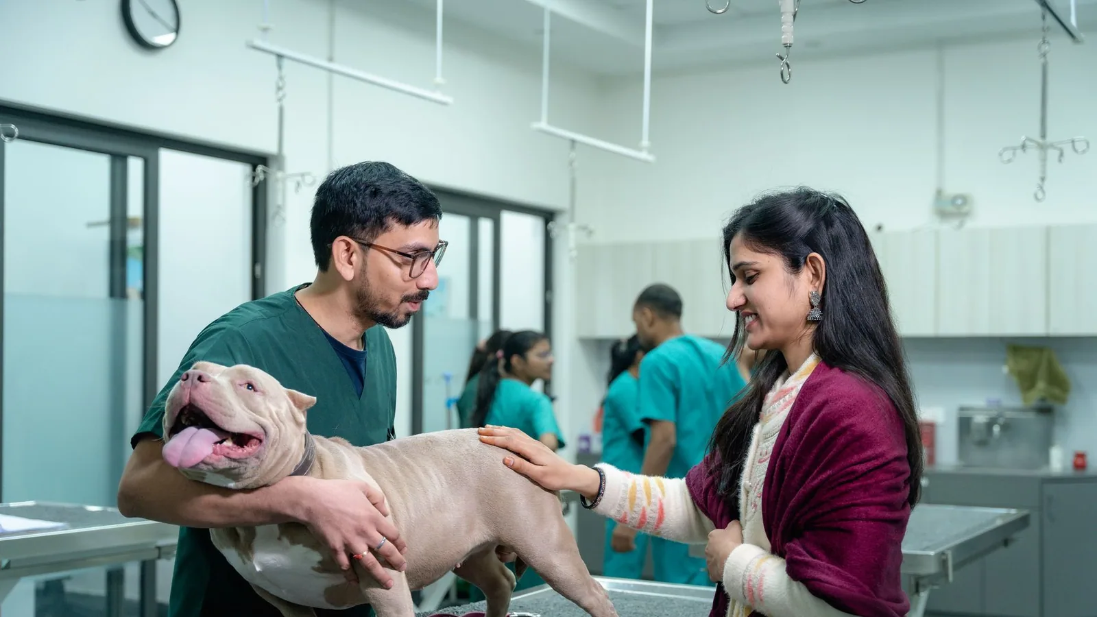
Cancer in dogs is a leading cause of morbidity and mortality, particularly in older animals. For the veterinary clinician and the concerned pet owner alike, the ability to distinguish benign age-related changes from early signs of cancer in dogs is critical. This article provides a definitive, evidence-based guide to the clinical presentation of canine neoplasia, focusing on the specific changes that warrant immediate diagnostic investigation.
The challenge in veterinary oncology is that many canine cancer warning signs are subtle and easily attributed to other conditions. A lump may be dismissed as a harmless lipoma, or lethargy may be blamed on arthritis. However, early detection directly correlates with improved treatment outcomes. This guide synthesises current research and clinical consensus to help you identify when a change in your dog's health requires a thorough veterinary examination.
At a Glance: When to Seek Veterinary Examination
| Clinical Sign | Possible Cancer Association | Urgency |
|---|---|---|
| New or growing lump | Mast cell tumour, sarcoma, lipoma (benign vs malignant) | Immediate if growing rapidly |
| Non-healing wound | Squamous cell carcinoma, soft tissue sarcoma | Within 1-2 weeks |
| Unexplained weight loss | Lymphoma, gastrointestinal carcinoma | Within 1 week |
| Lethargy or exercise intolerance | Haemangiosarcoma, leukaemia | Within 1 week |
| Changes in appetite or swallowing | Oral melanoma, oesophageal carcinoma | Immediate if dysphagia present |
| Lameness or bone swelling | Osteosarcoma (most common primary bone tumour) | Immediate |
| Abnormal bleeding or discharge | Transitional cell carcinoma, haemangiosarcoma | Immediate |
| Neurological signs (seizures, ataxia) | Meningioma, glioma, metastatic disease | Emergency |
Understanding the Clinical Presentation of Canine Cancer
Cancer is not a single disease but a collection of over 100 distinct neoplastic conditions, each with its own biological behaviour. The clinical signs reflect the tumour's location, size, growth rate, and metastatic potential. A thorough understanding of these presentations allows the clinician to prioritise differential diagnoses and select appropriate diagnostic tests.
The Spectrum of Clinical Signs
The presentation of neoplasia in dogs ranges from an incidental finding on physical examination to an acute, life-threatening emergency. The most common dog cancer symptoms include:
- Palpable Masses: The most frequently reported sign. While many subcutaneous masses are benign lipomas, any new, growing, or changing lump requires cytological evaluation. The classic teaching that a lipoma is "soft and moveable" is not absolute; some lipomas (e.g., infiltrative lipomas) can be firm and adherent, while some sarcomas can feel deceptively benign.
- Non-Healing Wounds or Sores: A wound that fails to heal within 2-3 weeks of appropriate care should raise suspicion for neoplasia, particularly squamous cell carcinoma or soft tissue sarcoma. These lesions may bleed intermittently and are often painful.
- Unexplained Weight Loss (Cachexia): Cancer cachexia is a multifactorial syndrome involving metabolic alterations, reduced appetite, and increased energy expenditure. It is a hallmark of advanced disease, particularly lymphoma, leukaemia, and gastrointestinal carcinomas.
- Lethargy and Exercise Intolerance: Non-specific but significant. Dogs with haemangiosarcoma, for example, may present with acute collapse due to tumour rupture and haemorrhage, but more commonly exhibit a gradual decline in energy.
- Gastrointestinal Signs: Vomiting, diarrhoea (or diarrhoea), and changes in appetite can indicate gastric carcinoma, intestinal lymphoma, or pancreatic neoplasia. Haematochezia or melena may suggest a bleeding tumour.
- Lameness and Bone Swelling: Persistent, progressive lameness, especially in a large-breed dog, is a classic presentation of osteosarcoma. The pain is often severe and may not respond to standard anti-inflammatory therapy. Swelling may be palpable over the distal radius, proximal humerus, or tibia.
- Respiratory Signs: Cough, dyspnoea, or tachypnoea can indicate primary lung tumours or, more commonly, pulmonary metastases from distant primary sites.
- Neurological Deficits: Seizures, ataxia, head tilt, circling, or behavioural changes may be the first signs of a primary brain tumour such as meningioma or glioma, or of metastatic disease affecting the central nervous system.
Lumps and Masses: The Most Common Warning Sign
The discovery of a lump is the single most common reason owners seek veterinary attention for suspected cancer. However, not all lumps are malignant. The key is to differentiate between benign and malignant lesions through systematic evaluation.
Benign vs Malignant: A Clinical Framework
The clinical characteristics of a mass can provide initial clues, but cytology or histopathology is required for definitive diagnosis.
Benign Masses (e.g., Lipoma, Adenoma)
- Slow growing or static in size
- Well-defined borders, often freely moveable over underlying tissue
- Soft to firm consistency
- Non-painful on palpation
- No ulceration or discharge
Malignant Masses (e.g., Mast Cell Tumour, Sarcoma)
- Rapid growth over weeks to months
- Irregular, poorly defined borders; may be fixed to underlying muscle or bone
- Firm to hard consistency
- May be painful or pruritic (especially mast cell tumours)
- Ulceration, necrosis, or serosanguinous discharge
- Satellite nodules or regional lymphadenopathy
The Diagnostic Approach to a Palpable Mass
The standard of care for any new mass is fine-needle aspiration (FNA) and cytology. This minimally invasive procedure can provide a rapid, preliminary diagnosis. For example, a mast cell tumour will show characteristic granules on Diff-Quik staining, while a lipoma will reveal clusters of adipocytes.
If cytology is non-diagnostic or suggests a malignant process, a biopsy (incisional or excisional) with histopathology is indicated. Histopathology provides a definitive diagnosis, tumour grade, and assessment of surgical margins.
Important Consideration: A negative cytology does not rule out malignancy. Some tumours, particularly sarcomas, are poorly exfoliative and may yield a false-negative result. If clinical suspicion remains high, a biopsy is warranted.
The Significance of Lymph Node Enlargement
Lymphadenopathy is a critical finding in the staging of canine cancer. Enlarged lymph nodes may indicate local metastasis (e.g., from a mammary or oral tumour) or systemic disease (e.g., lymphoma).
Regional vs Generalised Lymphadenopathy
- Regional Lymphadenopathy: Enlargement of a single lymph node or chain draining a known tumour site. This is a common finding in dogs with sinonasal cancer, where the mandibular or retropharyngeal lymph nodes may be affected. A recent study evaluating dogs with sinonasal carcinomas and sarcomas found that physical examination alone had a sensitivity of 71.4% and specificity of 64% for detecting lymph node metastasis [1]. This underscores the need for cytological evaluation even when nodes feel palpably normal.
- Generalised Lymphadenopathy: Enlargement of multiple peripheral lymph nodes (mandibular, prescapular, popliteal) is highly suggestive of multicentric lymphoma. The nodes are typically firm, non-painful, and freely moveable.
Diagnostic Sampling
Cytology of an enlarged lymph node is a straightforward, high-yield procedure. In the study of sinonasal cancer, the combination of palpation and computed tomography (CT) achieved 100% sensitivity and 59.5% specificity for detecting metastasis [1]. Critically, a palpably and tomographically normal lymph node had a 100% negative predictive value, meaning that a normal-appearing node can confidently rule out cytologically detectable metastasis [1]. This finding is clinically useful for treatment planning.
Systemic Signs: When Cancer Affects the Whole Body
Beyond localised lumps, cancer often manifests through systemic effects. These signs of cancer in dogs can be vague and are easily mistaken for other chronic diseases.
Unexplained Weight Loss and Cachexia
Cancer cachexia is a distinct syndrome from simple starvation. It involves the loss of lean body mass (muscle) despite adequate caloric intake. It is driven by pro-inflammatory cytokines and tumour-derived factors. A dog that is eating well but still losing weight should be evaluated for neoplasia, particularly lymphoma or gastrointestinal carcinoma.
Lethargy and Exercise Intolerance
Chronic, progressive lethargy is a common but non-specific sign. In the context of cancer, it may be due to anaemia (e.g., from chronic blood loss due to a gastrointestinal tumour), metabolic derangements, or the tumour's energy demands.
Fever of Unknown Origin
Persistent or intermittent pyrexia that does not respond to antibiotics can be a paraneoplastic syndrome associated with lymphoma, leukaemia, or other neoplasms. The fever is caused by cytokines released by the tumour or the host's immune response.
Specific Cancer Types and Their Warning Signs
Different cancers have unique clinical presentations. Recognising these patterns can expedite diagnosis and treatment.
Mammary Cancer
Canine mammary cancer (CMC) is the most common neoplasm in intact female dogs [2]. It is a hormone-sensitive tumour with a multifactorial origin, sharing several risk factors with human breast cancer [2].
Warning Signs:
- Palpable masses in the mammary chain (inguinal or thoracic)
- Single or multiple nodules
- Firm, irregular, or fixed to underlying tissue
- Ulceration or discharge from the nipple
- Enlargement of the ipsilateral inguinal lymph node
Diagnostic Advances: Recent research has focused on non-invasive biomarkers. A study evaluating serum TP53 autoantibodies found that these antibodies were present in 60% of dogs with mammary tumours and absent in healthy controls, demonstrating high diagnostic accuracy (AUC 0.9587) [5]. This suggests that a simple blood test could aid in early detection. Furthermore, microRNA (miRNA) profiling has identified specific patterns, such as the downregulation of cfa-miR-133a in both adenomas and carcinomas, which may serve as a biomarker for early malignant transformation [6].
Oral Tumours
Oral tumours are common in dogs and can be aggressive. They include melanoma, squamous cell carcinoma, fibrosarcoma, and osteosarcoma.
Warning Signs:
- Halitosis (bad breath)
- Excessive drooling (ptyalism) with or without blood
- Difficulty eating or dropping food (dysphagia)
- Visible mass in the mouth
- Loose teeth without periodontal disease
- Facial swelling
Oral Osteosarcoma: A rare but aggressive subtype is telangiectatic osteosarcoma, which consists of blood-filled spaces and can be difficult to differentiate from haemangiosarcoma [4]. These tumours present as rapidly growing, ulcerated, and occasionally haemorrhagic masses involving the maxilla, mandible, or gingiva [4]. Diagnosis relies on histopathology and immunohistochemistry, with RUNX2 positivity and CD31 negativity confirming osteosarcoma [4].
Bone Tumours
Osteosarcoma is the most common primary bone tumour in dogs, accounting for over 85% of skeletal malignancies. It is highly aggressive and metastatic.
Warning Signs:
- Progressive, non-weight-bearing lameness
- Swelling at the metaphysis of long bones (distal radius, proximal humerus, distal femur, proximal tibia)
- Pain on palpation
- Pathological fracture (rare at presentation)
Diagnosis: Radiographs typically show a "sunburst" pattern of periosteal reaction, cortical lysis, and Codman's triangle. However, biopsy is required for definitive diagnosis. Immunohistochemistry for the transcription factor osterix (Osx) has shown high sensitivity (92.5%) and specificity (95%) for diagnosing canine osteosarcoma, and it can help differentiate OSA from other bone tumours like chondrosarcoma [19].
Brain Tumours
Meningioma is the most common primary brain tumour in dogs [18]. Other types include glioma and choroid plexus tumours.
Warning Signs:
- Seizures (especially in older dogs with no prior history)
- Ataxia (incoordination)
- Head pressing or circling
- Behavioural changes (aggression, disorientation)
- Visual deficits
- Head tilt or nystagmus
Prognosis: Surgical resection with or without radiation therapy can provide long-term control. A study of 21 dogs with intracranial meningioma reported a median survival time of 845 days [18]. Grade 1 tumours and intact male status were associated with favourable outcomes [18].
Urothelial Carcinoma (Bladder Cancer)
Transitional cell carcinoma (TCC) is the most common bladder tumour in dogs. It is locally invasive and has a moderate metastatic rate.
Warning Signs:
- Stranguria (straining to urinate)
- Pollakiuria (frequent urination of small volumes)
- Haematuria (blood in urine)
- Urinary tract infections that are difficult to treat
Treatment Advances: For unresectable tumours, transarterial chemoembolization (TACE) using quick-soluble gelatin microparticles has shown promise. In a prospective study, TACE resulted in a median tumour volume reduction of 76.41% and a median survival time of 569 days [13].
Peritoneal Mesothelioma
This is a rare and difficult-to-diagnose cancer that arises from the mesothelial lining of the abdominal cavity.
Warning Signs:
- Recurrent aseptic peritonitis (abdominal fluid accumulation without infection)
- Abdominal distension
- Weight loss
- Lethargy
Diagnostic Challenge: A case report highlighted a dog with recurrent aseptic peritonitis that was diagnosed with peritoneal mesothelioma only post-mortem [11]. The authors emphasise the need for high-quality biopsy samples early in the disease course to improve the chance of definitive diagnosis [11].
Diagnostic Tools: From Physical Exam to Advanced Imaging
The diagnostic workup for suspected cancer in dogs has evolved significantly. The goal is to confirm the diagnosis, determine the tumour type, and stage the disease (assess local invasion and distant metastasis).
The Role of Physical Examination
A thorough physical examination remains the cornerstone of cancer detection. This includes:
- Palpation of all lymph nodes (mandibular, prescapular, axillary, inguinal, popliteal)
- Abdominal palpation for organomegaly or masses
- Rectal examination for prostatic or pelvic masses
- Oral examination under sedation if necessary
- Skin and subcutaneous tissue palpation for masses
Cytology and Histopathology
- Fine-Needle Aspiration (FNA): Quick, minimally invasive, and often diagnostic for round cell tumours (lymphoma, mast cell tumour, histiocytoma) and some carcinomas.
- Biopsy: Required for definitive diagnosis of most solid tumours. Incisional biopsy (wedge or core) is preferred for large masses. Excisional biopsy (complete removal) is both diagnostic and therapeutic for small, accessible masses.
Advanced Imaging
- Computed Tomography (CT): Essential for staging many cancers. CT provides detailed anatomical information, assesses local invasion, and detects pulmonary metastases. For sinonasal cancer, CT has a sensitivity of 100% for detecting lymph node metastasis, though its specificity is lower at 46.3% [1].
- Magnetic Resonance Imaging (MRI): The gold standard for imaging the brain and spinal cord. It provides superior soft tissue contrast, making it ideal for evaluating intracranial masses like meningioma or colloid cysts [7].
- Ultrasound: Useful for evaluating abdominal organs (liver, spleen, kidneys, bladder) and guiding FNA or biopsy.
Liquid Biopsy: The Future of Early Detection
Liquid biopsy, which analyses circulating tumour DNA (ctDNA) or other biomarkers in the blood, is an emerging tool in veterinary oncology. A major obstacle has been the low abundance of ctDNA in early-stage disease. However, a recent study using a sequential CRISPR-EspCas9 enrichment strategy achieved a detection limit of 0.001% mutant allele frequency for the PIK3CA H1047R mutation, a significant improvement over standard next-generation sequencing [8]. This technology has the potential to revolutionise early cancer detection in dogs.
Additionally, NMR-based metabolomics combined with machine learning has shown high discriminative performance for identifying dogs with cancer (ROC AUC 0.911) and cardiovascular disease (ROC AUC 0.924) [15]. This approach analyses the serum metabolomic profile, offering a non-invasive screening tool.
Clinical Red Flags: When to Seek Immediate Veterinary Attention
While many cancers progress slowly, certain presentations constitute emergencies. These "red flags" require immediate veterinary evaluation:
- Acute Collapse or Weakness: May indicate haemorrhage from a ruptured haemangiosarcoma (spleen, heart) or a bleeding gastrointestinal tumour.
- Respiratory Distress: Sudden onset of difficulty breathing could be due to a pulmonary embolism, pleural effusion (from mesothelioma or metastatic disease), or a large airway tumour.
- Seizures: New-onset seizures in an older dog are a classic sign of a structural brain lesion, most commonly a brain tumour.
- Acute Abdomen: A tense, painful abdomen with pale mucous membranes suggests hemoabdomen (blood in the abdomen), often from a ruptured splenic mass.
- Inability to Urinate: Complete urethral obstruction from a bladder or urethral tumour is a life-threatening emergency.
- Pathological Fracture: A bone weakened by a tumour (e.g., osteosarcoma) can fracture with minimal trauma.
Prevention and Risk Factor Modification
While not all cancers are preventable, certain strategies can reduce risk.
- Early Spaying: Spaying before the first heat cycle dramatically reduces the risk of mammary cancer. The risk increases with each subsequent oestrus cycle. However, spaying is not without trade-offs; it may increase the risk of other conditions like urinary incontinence and some orthopaedic diseases.
- Castration: Castration eliminates the risk of testicular cancer and reduces the risk of prostatic disease. However, cryptorchidism (retained testicle) is a significant risk factor for testicular cancer, and the retained testicle should be surgically removed [3].
- Weight Management: Obesity is a known risk factor for several cancers, including mammary tumours. Maintaining a lean body condition score is recommended.
- Regular Veterinary Check-ups: Annual or semi-annual examinations allow for early detection of lumps, lymphadenopathy, and other abnormalities.
Prognosis and Quality of Life
The prognosis for a dog with cancer depends on several factors: tumour type, grade, stage, location, and the dog's overall health. Some cancers, like low-grade mast cell tumours or well-differentiated thyroid carcinomas, can be cured with surgery alone. Others, like osteosarcoma or haemangiosarcoma, have a guarded prognosis despite aggressive therapy.
Quality of Life (QoL) Assessment: For dogs undergoing cancer treatment, regular QoL assessment is essential. Tools like the HHHHHMM (Hurt, Hunger, Hydration, Hygiene, Happiness, Mobility, More good days than bad) scale help owners and veterinarians make informed decisions about continuing or discontinuing treatment.
The Clinical Reasoning Behind Diagnostic Decision-Making
When a dog presents with potential dog cancer symptoms, the veterinarian must navigate a complex differential diagnosis list. The clinical reasoning process is not merely about identifying a mass or abnormality; it is about integrating the signalment, history, physical examination findings, and initial diagnostic results to formulate a prioritised list of possibilities. This cognitive framework is essential for avoiding diagnostic delays and ensuring appropriate testing.
Signalment as a Diagnostic Clue
The age, breed, and sex of the dog provide critical context for interpreting clinical signs. For example, a young dog with a rapidly growing subcutaneous mass is more likely to have a histiocytoma (a benign, self-limiting tumour) than a mast cell tumour, whereas the same presentation in a 10-year-old Golden Retriever raises a high index of suspicion for malignancy. Similarly, osteosarcoma has a strong predilection for large and giant breed dogs, with the distal radius, proximal humerus, and distal femur being the most common sites. A small breed dog with lameness and bone swelling is more likely to have a different primary bone tumour, such as chondrosarcoma or synovial cell sarcoma, or even a metastatic lesion.
Breed predispositions extend beyond bone tumours. Boxers and Pugs are overrepresented for mast cell tumours, while Scottish Terriers have a markedly increased risk of transitional cell carcinoma of the bladder. Understanding these breed-specific risks allows the clinician to maintain a higher index of suspicion for certain neoplasms when evaluating canine cancer warning signs in predisposed breeds.
The Importance of Growth Rate Assessment
One of the most clinically useful pieces of information an owner can provide is the rate at which a lump or clinical sign has progressed. A mass that has been present and unchanged for two years is far less concerning than one that has doubled in size over two weeks. The veterinarian should specifically ask: "When did you first notice this lump?" and "Has it changed in size, shape, or texture since you first found it?"
Rapid growth is a hallmark of aggressive malignancies. High-grade mast cell tumours, sarcomas, and some carcinomas can exhibit exponential growth over a short period. Conversely, slow growth over months to years is more consistent with benign neoplasms like lipomas or well-differentiated soft tissue sarcomas. However, it is important to note that some malignant tumours, such as low-grade mast cell tumours, can grow slowly and be mistaken for benign lesions. This underscores the principle that any new or changing lump warrants cytological evaluation, regardless of its perceived growth rate.
The Diagnostic Value of Pain Assessment
Pain is a significant but often overlooked sign of cancer in dogs. While many benign masses are non-painful, certain malignancies are inherently painful due to their location, invasiveness, or paraneoplastic effects. Bone tumours, particularly osteosarcoma, cause severe, progressive pain that often does not respond adequately to non-steroidal anti-inflammatory drugs. Dogs with oral tumours may exhibit pain when eating or during oral examination. Mast cell tumours can be pruritic and painful due to the release of histamine and other vasoactive amines.
The absence of pain does not rule out cancer. Many internal tumours, such as lymphoma or splenic haemangiosarcoma, may be painless until they reach an advanced stage or cause a complication like organ rupture. Therefore, pain assessment should be integrated with other clinical findings rather than used as a standalone criterion for malignancy.
Preparing for the Veterinary Visit: What Owners Should Observe and Document
Owners play a crucial role in the early detection of dog cancer symptoms. However, the information they provide is only as useful as its accuracy and completeness. Veterinarians can empower owners by providing clear guidance on what to observe and document before the appointment.
Key Observations for Owners
Owners should be encouraged to monitor and record the following:
- Lump Characteristics: Note the location, size (using a ruler or a familiar object for scale), shape, colour, and texture. Document any changes over time, including growth, ulceration, bleeding, or discharge.
- Behavioural Changes: Record any alterations in energy level, appetite, thirst, urination, defecation, and sleep patterns. Specific behaviours like excessive licking of a particular area, head shaking, or scratching may indicate discomfort or a mass.
- Gastrointestinal Signs: Note the frequency and character of vomiting or diarrhoea, the presence of blood, and any changes in appetite (increased, decreased, or picky eating).
- Lameness and Mobility: Describe the onset (sudden vs gradual), which limb is affected, and whether the lameness is worse after exercise or rest. Note any swelling or heat over a joint or bone.
- Respiratory Signs: Record any coughing, sneezing, nasal discharge, or difficulty breathing. Note the timing (e.g., at night, after exercise) and any associated sounds (e.g., wheezing, honking).
- Neurological Signs: Document any seizures, including their duration, frequency, and any post-ictal behaviour. Note any changes in coordination, balance, head tilt, circling, or vision.
The Value of a Symptom Diary
A written or digital symptom diary can be invaluable for tracking subtle changes over time. Owners should record the date, time, and specific observations. This diary can help the veterinarian identify patterns, assess the progression of disease, and monitor response to treatment. For example, a dog with a brain tumour may have intermittent seizures that are easily forgotten or minimised by the owner. A diary provides an objective record that can guide treatment decisions.
What to Bring to the Appointment
Owners should be advised to bring the following to the veterinary visit:
- Any previous medical records, including vaccination history, laboratory results, and imaging reports.
- A list of all current medications, including supplements and over-the-counter products.
- A list of any known allergies or adverse reactions to medications.
- The symptom diary, if one has been kept.
- Photographs or videos of any concerning lumps, wounds, or behavioural episodes.
- A sample of urine or faeces, if requested by the veterinarian.
The Diagnostic Workup: A Step-by-Step Owner's Guide
Understanding the diagnostic process can help owners feel more informed and engaged in their dog's care. The workup for suspected cancer typically follows a logical sequence, though the specific tests depend on the clinical presentation.
Step 1: Physical Examination and History
The veterinarian will perform a thorough physical examination, paying close attention to the area of concern. They will palpate all lymph nodes, assess the abdomen for organomegaly or masses, and perform an oral examination if indicated. The history provided by the owner is critical for guiding the next steps.
Step 2: Fine-Needle Aspiration and Cytology
For any palpable mass or enlarged lymph node, fine-needle aspiration (FNA) is typically the first diagnostic test. This procedure involves inserting a small needle into the mass and aspirating cells. The cells are then placed on a slide, stained, and examined under a microscope. FNA is quick, minimally invasive, and often provides a preliminary diagnosis. For example, a mast cell tumour will show characteristic purple granules within the cells, while a lipoma will reveal clusters of fat cells.
Step 3: Blood and Urine Tests
Complete blood count (CBC), serum biochemistry profile, and urinalysis are essential for assessing the dog's overall health and identifying any paraneoplastic syndromes. For example, anaemia may indicate chronic blood loss from a gastrointestinal tumour, while hypercalcaemia can be a paraneoplastic effect of lymphoma or anal sac adenocarcinoma. Urinalysis can detect blood, infection, or abnormal cells that may suggest bladder cancer.
Step 4: Imaging
Depending on the suspected tumour type and location, imaging may include radiographs (X-rays), ultrasound, computed tomography (CT), or magnetic resonance imaging (MRI). Radiographs are useful for evaluating bone tumours and detecting pulmonary metastases. Ultrasound is ideal for examining abdominal organs and guiding FNA or biopsy. CT and MRI provide detailed anatomical information and are essential for surgical planning and radiation therapy.
Step 5: Biopsy and Histopathology
If cytology is non-diagnostic or suggests a malignant process, a biopsy is required for definitive diagnosis. A biopsy involves removing a small piece of tissue for histopathological examination. This can be performed using a needle (core biopsy), an endoscopic instrument, or during surgery. Histopathology provides a definitive diagnosis, tumour grade, and assessment of surgical margins.
Step 6: Staging
Once a cancer diagnosis is confirmed, staging is performed to determine the extent of the disease. Staging typically includes imaging of the chest and abdomen to detect metastases, as well as evaluation of regional lymph nodes. The stage of the disease is a critical factor in determining prognosis and treatment options.
The Role of the Owner in Monitoring for Recurrence
After a dog has been treated for cancer, vigilant monitoring is essential for detecting recurrence or metastasis. Owners should be educated on what to look for and how to perform regular at-home examinations.
At-Home Examination Protocol
Owners should be encouraged to perform a monthly "head-to-tail" examination of their dog. This includes:
- Skin and Coat: Run hands over the entire body, feeling for any new lumps, bumps, or changes in existing scars or surgical sites. Look for any areas of hair loss, redness, or ulceration.
- Lymph Nodes: Gently palpate the mandibular (under the jaw), prescapular (in front of the shoulder), axillary (in the armpit), inguinal (in the groin), and popliteal (behind the knee) lymph nodes. Note any enlargement, firmness, or asymmetry.
- Oral Cavity: Lift the lips and examine the gums, tongue, and roof of the mouth for any masses, ulcers, or discolouration.
- Abdomen: Gently palpate the abdomen for any masses or distension.
- Mammary Glands: In female dogs, palpate the mammary chain for any nodules or thickening.
- Behaviour and Appetite: Monitor for any changes in energy level, appetite, thirst, urination, or defecation.
When to Seek Re-Evaluation
Owners should be advised to seek veterinary re-evaluation if they detect any new lump, a change in an existing lump, unexplained weight loss, lethargy, loss of appetite, or any other concerning sign. Early detection of recurrence can significantly impact treatment options and outcomes.
Special Considerations for Senior Dogs
Senior dogs are at the highest risk for cancer, but they are also more likely to have concurrent age-related conditions that can complicate diagnosis and treatment. The clinical signs of cancer can be easily mistaken for "old age" changes, leading to diagnostic delays.
Differentiating Cancer from Age-Related Changes
Common age-related changes in senior dogs include arthritis, cognitive dysfunction, dental disease, and organ dysfunction (e.g., kidney or liver disease). The challenge is that many dog cancer symptoms overlap with these conditions. For example, lethargy and decreased activity can be due to arthritis, but they can also be early signs of lymphoma or haemangiosarcoma. Weight loss can be due to dental disease or chronic kidney disease, but it can also indicate gastrointestinal carcinoma.
The key differentiator is the rate and pattern of change. Age-related changes tend to be gradual and progressive over months to years. Cancer-related changes are often more rapid and may be accompanied by other signs such as a palpable mass, lymphadenopathy, or paraneoplastic syndromes. Any senior dog with a sudden or rapid decline in health should be evaluated for cancer.
The Importance of Wellness Screening
Regular wellness examinations, including blood work and urinalysis, are essential for senior dogs. These tests can detect early signs of disease, including anaemia, elevated liver enzymes, or abnormal kidney values, that may prompt further investigation for cancer. Annual or semi-annual screening is recommended for all senior dogs, with more frequent monitoring for breeds predisposed to specific cancers.
The Role of Nutrition and Lifestyle in Cancer Prevention
While the primary focus of this article is on detection, it is important to acknowledge the role of nutrition and lifestyle in cancer prevention. A growing body of evidence suggests that dietary factors and body condition can influence cancer risk.
Weight Management
Obesity is a well-established risk factor for several cancers in dogs, including mammary tumours and transitional cell carcinoma. Adipose tissue produces hormones and inflammatory cytokines that can promote tumour growth. Maintaining a lean body condition score throughout life is one of the most effective strategies for reducing cancer risk.
Dietary Considerations
While no single diet can prevent cancer, certain dietary patterns may be beneficial. A diet rich in antioxidants, omega-3 fatty acids, and fibre may help support the immune system and reduce inflammation. Conversely, diets high in processed carbohydrates and unhealthy fats may promote inflammation and oxidative stress. Owners should consult with their veterinarian to select a high-quality, balanced diet appropriate for their dog's age, breed, and health status.
Environmental Factors
Exposure to certain environmental toxins, such as second-hand smoke, pesticides, and herbicides, has been linked to an increased risk of cancer in dogs. Minimising exposure to these substances may help reduce cancer risk.
The Importance of a Multidisciplinary Approach
The management of canine cancer often requires a multidisciplinary team, including the primary care veterinarian, a veterinary oncologist, a surgeon, a radiologist, and a pathologist. Owners should be informed about the availability of specialty care and the potential benefits of referral.
When to Refer to a Specialist
Referral to a veterinary oncologist is indicated for complex cases, such as those requiring chemotherapy, radiation therapy, or advanced surgical techniques. Oncologists have specialised training in cancer diagnosis, staging, and treatment, and they can offer the most up-to-date treatment options. Referral should also be considered when the primary care veterinarian is unsure of the diagnosis or when the owner desires a second opinion.
The Role of Clinical Trials
Clinical trials offer access to novel therapies that may not be otherwise available. Owners should be informed about ongoing clinical trials at veterinary teaching hospitals and specialty centres. Participation in a clinical trial can provide access to cutting-edge treatments and contribute to the advancement of veterinary oncology.
The Emotional Impact of a Cancer Diagnosis
A cancer diagnosis in a beloved pet is emotionally devastating for owners. Veterinarians play a crucial role in providing support, empathy, and clear communication throughout the diagnostic and treatment process.
Communicating the Diagnosis
The way a cancer diagnosis is communicated can significantly impact the owner's ability to cope and make informed decisions. The veterinarian should provide the diagnosis in a clear, compassionate manner, using language that the owner can understand. They should explain the treatment options, prognosis, and potential side effects in a balanced and realistic way.
Supporting the Owner's Decision-Making
Owners may feel overwhelmed by the information and the need to make difficult decisions. The veterinarian should provide ample time for questions and discussion, and they should offer resources such as support groups, online forums, and educational materials. The goal is to empower the owner to make the best decision for their dog and their family.
Quality of Life Considerations
Throughout the treatment process, the focus should remain on the dog's quality of life. Regular assessments using validated quality-of-life scales can help guide treatment decisions and ensure that the dog is not suffering. When treatment is no longer effective or the dog's quality of life is unacceptable, humane euthanasia should be presented as a compassionate option.
Frequently Asked Questions
1. What are the most common early signs of cancer in dogs? The most common early signs include a new lump or bump that is growing, a non-healing wound, unexplained weight loss, and a gradual decline in energy or activity level.
2. How can I tell if a lump on my dog is cancerous? You cannot reliably tell by feel alone. Any new, growing, or changing lump should be evaluated by a veterinarian via fine-needle aspiration (FNA) and cytology. Benign lumps are often slow-growing and moveable, but malignant lumps can feel similar.
3. Is weight loss always a sign of cancer in dogs? No, but it is a significant warning sign. Weight loss can also be caused by chronic kidney disease, diabetes, or gastrointestinal disorders. However, unexplained weight loss, especially when the dog is still eating, warrants a thorough cancer workup.
4. Can a dog have cancer without showing any symptoms? Yes, some cancers, particularly slow-growing ones or those in internal organs, can be asymptomatic in early stages. This is why regular veterinary check-ups and wellness screening are important, especially for senior dogs.
5. What should I do if I find a lump on my dog? Schedule a veterinary appointment as soon as possible. Do not attempt to squeeze or drain the lump. Keep a record of the lump's size and any changes you observe. Your veterinarian will likely perform an FNA to obtain a sample for cytology.
6. Are some dog breeds more prone to cancer? Yes. Golden Retrievers, Boxers, Rottweilers, Bernese Mountain Dogs, and Scottish Terriers are among the breeds with a higher incidence of specific cancers. However, any dog can develop cancer.
7. Can dog cancer be cured? Yes, many cancers can be cured if detected early and treated appropriately. For example, low-grade mast cell tumours, well-differentiated thyroid carcinomas, and some soft tissue sarcomas can be cured with complete surgical excision.
8. Is chemotherapy as harsh for dogs as it is for humans? No. Dogs generally tolerate chemotherapy much better than humans. They experience fewer side effects, such as nausea and hair loss, due to differences in drug metabolism and dosing protocols. The goal of veterinary chemotherapy is to maintain a good quality of life while controlling the cancer.
Related Veterinary Guides
- Understanding Canine Lymphoma: Diagnosis and Treatment Options
- Mast Cell Tumours in Dogs: A Complete Clinical Guide
- Osteosarcoma in Dogs: From Diagnosis to Palliative Care
- Veterinary Oncology: Principles of Cancer Staging
- Senior Dog Health: Common Age-Related Diseases
Disclaimer: This article is educational and is not a substitute for veterinary diagnosis or treatment. If you suspect your dog has cancer, please consult a licensed veterinarian immediately.
References
[1] Simon SS, Horne-Reid BX, Love KR et al. Palpation and Computed Tomography as Surrogates for Lymph Node Cytology in Dogs With Sinonasal Cancer. J Am Anim Hosp Assoc. 2026. https://pubmed.ncbi.nlm.nih.gov/42379555/
[2] DO Rosário Pinheiro D, DE Andrade DM, DE Oliveira NCL et al. The role of epigenetic alterations in canine mammary cancer. J Reprod Dev. 2026. https://pubmed.ncbi.nlm.nih.gov/42309737/
[3] Nowacka-Woszuk J, Kajdasz A, Stachowiak M et al. Dysregulation of miRNA expression and 3'UTR length variation in target mRNAs in the testes of dogs with cryptorchidism. Sci Rep. 2026. https://pubmed.ncbi.nlm.nih.gov/42409905/
[4] Rissi DR, Mendes RE. Histological and immunohistochemical features of oral telangiectatic osteosarcomas in dogs and their differentiation from oral haemangiosarcomas. J Comp Pathol. 2026. https://pubmed.ncbi.nlm.nih.gov/42462441/
[5] Kumar P, Saxena S, Shrivastava S et al. Serum TP53 autoantibodies as a non-invasive diagnostic biomarker for canine mammary tumours: comparative insights from human and experimental rat model. Mol Biol Rep. 2026. https://pubmed.ncbi.nlm.nih.gov/42446626/
[6] Benevenuto LGD, Lacerda ZA, de Moraes RS et al. Biphasic tissue expression of cfa-miR-409-3p and cfa-miR-4270 during malignant transformation in canine mammary tumors: an exploratory study. Front Vet Sci. 2026. https://pubmed.ncbi.nlm.nih.gov/42416301/
[7] Jo H, Park KH, Shin D. Intracranial colloid cyst in the cerebellopontine angle of a dog: A novel case report and surgical outcome. Open Vet J. 2026. https://pubmed.ncbi.nlm.nih.gov/42375941/
[8] Hong S, Park CS, Been KW et al. Sequential CRISPR-EspCas9-Mediated Wild-Type Depletion Enhances the Detection Sensitivity of Rare Mutations for Canine Liquid Biopsy Application. Biosensors (Basel). 2026. https://pubmed.ncbi.nlm.nih.gov/42345886/
[9] Murthy VD, White LA, Rossmeisl J et al. Clinical characteristics, advanced imaging findings, and long-term outcomes in 50 dogs with calvarial multilobular tumor of bone treated with craniectomy. J Vet Intern Med. 2026. https://pubmed.ncbi.nlm.nih.gov/42315194/
[10] Vardanega MJ, Longo T, Aceino A et al. Use of the dual tyrosine kinase inhibitor lapatinib for the treatment of vulvovaginal squamous cell carcinoma in a dog. J Vet Intern Med. 2026. https://pubmed.ncbi.nlm.nih.gov/42315193/
[11] Anstead KD, Schneider SM, Vezzi NK. Recurrent aseptic peritonitis in a dog diagnosed with peritoneal mesothelioma post-mortem. Can Vet J. 2026. https://pubmed.ncbi.nlm.nih.gov/42266345/
[12] Chaudhari P, Bendale K, Bhere S et al. Sclerotherapy for Aneurysmal Bone Cyst in Canine. Vet Med Sci. 2026. https://pubmed.ncbi.nlm.nih.gov/42237883/
[13] Jeon S, Lee G, Kim JK et al. Leverage of quick-soluble gelatin microparticles in transarterial chemoembolization of canine urothelial carcinomas. Vet Q. 2026. https://pubmed.ncbi.nlm.nih.gov/42216429/
[14] Jacobs M, Valdivia N, Varas M et al. Cryptocarya alba and Laureliopsis philippiana Essential Oil-Loaded Hydrogels with Antibacterial Activity Against Staphylococcus pseudintermedius: Potential Topical Candidates for Canine Pyoderma. Vet Sci. 2026. https://pubmed.ncbi.nlm.nih.gov/42357742/
[15] Finotello R, Teoh ST, Nili H et al. Application of NMR-based metabolomics and machine learning for non-invasive disease screening in dogs. Front Vet Sci. 2026. https://pubmed.ncbi.nlm.nih.gov/42453966/
[16] Kruckman-Gatesy CR, LaRue SM, Griffin LR et al. Clinically Relevant Motion of Canine Thoracic Tumors Is Seen Between Inspiratory and Expiratory Breath Holds on Computed Tomography Performed for Radiation Treatment Planning. Vet Radiol Ultrasound. 2026. https://pubmed.ncbi.nlm.nih.gov/42444534/
[17] Bilhalva LC, Biedak NR, Granger J et al. Ectopic Splenic Tissue Mimicking Malignant Neoplasia in a Dog. Vet Radiol Ultrasound. 2026. https://pubmed.ncbi.nlm.nih.gov/42433008/
[18] Uno A, Nozue Y, Iwasaki R et al. Surgical Outcomes of Canine Intracranial Meningioma With or Without Radiation. J Am Anim Hosp Assoc. 2026. https://pubmed.ncbi.nlm.nih.gov/42379561/
[19] Ings K, Plog S, Jackson M et al. Immunohistochemical detection of the transcription factor osterix in canine and feline osteosarcoma. Vet Pathol. 2026. https://pubmed.ncbi.nlm.nih.gov/42376931/
[20] Ling XC, Pakhawala J, Phipps W. Perirenal lipoma in a 14-year-old spayed female dog: A rare diagnosis. Can Vet J. 2026. https://pubmed.ncbi.nlm.nih.gov/42368691/