Avian Anesthesia: Monitoring and Troubleshooting in Pet Birds
By Dr. Zubair Khalid, DVM, MS, PhD ·

Key Takeaways
- Respiratory monitoring is paramount: Due to their unique unidirectional airflow and air sac system, birds exhibit rapid changes in anesthetic depth. Respiratory rate and pattern are the most sensitive early indicators, with apnea exceeding 15-30 seconds in small birds necessitating immediate intervention. Capnography is valuable for trend monitoring, but avian respiratory anatomy requires careful interpretation of end-tidal CO2 values.
- Cardiovascular monitoring presents challenges: High heart rates in small psittacines (exceeding 400 bpm) can render standard pulse oximetry unreliable. Doppler ultrasound for systolic blood pressure and auscultation via esophageal stethoscope (for birds >200g) are more dependable methods for assessing cardiovascular status and detecting bradycardia, which signifies significant perfusion compromise in birds.
- Thermoregulation is a critical intervention: Birds have a higher body temperature and surface area to volume ratio, leading to rapid heat loss during anesthesia. Active warming with forced warm air, circulating water blankets, and warmed fluids is essential from induction through recovery to prevent hypothermia, which depresses metabolic rate and prolongs recovery.
- Preanesthetic assessment and risk stratification are vital: A thorough physical examination, including body condition scoring and respiratory auscultation, is mandatory to identify high-risk patients, particularly those with underlying respiratory disease. Fasting protocols must be species-specific to ensure crop emptying and reduce aspiration risk without inducing hypoglycemia.
- Anesthetic depth assessment relies on a composite approach: While palpebral and pedal withdrawal reflexes are useful, the respiratory pattern is the most reliable indicator of surgical plane. Changes in respiration from regular to shallow and irregular, or the onset of apnea, signal excessively deep anesthesia and require immediate adjustment of anesthetic delivery.
- Common complications require prompt recognition and intervention: Hypothermia, apnea, hypotension, and prolonged recovery are frequent issues. Troubleshooting involves reducing anesthetic depth, ensuring airway patency, providing active warming, and administering fluids as needed, with a systematic approach mirroring mammalian protocols but adapted for avian physiology.
Anesthetizing pet birds presents a distinct set of challenges that separate avian anesthesia from routine small animal practice. The small body mass, high metabolic rate, and unique respiratory anatomy of birds demand a monitoring approach that is both more intensive and more adaptable than that used for dogs and cats. This article addresses the practicing veterinarian who needs a practical framework for monitoring avian patients under anesthesia and for troubleshooting the complications that arise when physiologic reserve is exhausted. It focuses on the physiological principles that underpin monitoring choices, the specific parameters that are most informative in birds, and the common failure modes encountered during avian anesthetic events.
The clinical question at the core of this reference is straightforward: how does the anesthetist recognize and correct deterioration in a bird before it becomes irreversible? Birds mask signs of cardiovascular and respiratory compromise until late in the decompensation sequence. A monitoring protocol designed for a 20 kg dog cannot simply be scaled down for a 100 g parrot. The differences are qualitative, also quantitative, and they affect every stage from premedication through recovery.
At a Glance
| Parameter or Decision | Key Point |
|---|---|
| Preanesthetic assessment | Physical examination and body condition scoring are mandatory, birds with respiratory disease are high risk |
| Fasting protocol | Crop should be empty to reduce regurgitation and aspiration risk, duration varies with species and body size |
| Monitoring priority | Respiratory rate and depth are the most sensitive early indicators of anesthetic depth in birds |
| Capnography | End-tidal CO2 is valuable but requires careful interpretation due to avian respiratory anatomy |
| Heart rate | Auscultation and Doppler flow are more reliable than pulse oximetry in small patients |
| Temperature | Birds lose heat rapidly, active warming is required throughout the procedure |
| Anesthetic depth assessment | Palpebral reflex, withdrawal reflex, and respiratory pattern are primary indicators |
| Common complications | Hypothermia, apnea, hypotension, and prolonged recovery are the most frequent problems |
Physiologic Foundations of Avian Anesthesia
Respiratory Anatomy and Its Monitoring Implications
The avian respiratory system is fundamentally different from the mammalian system. Birds possess rigid lungs with unidirectional airflow driven by a system of air sacs that extend into the coelomic cavity and even into the bones. This arrangement means that gas exchange is continuous and highly efficient, but it also creates specific vulnerabilities. The air sacs are thin-walled and poorly vascularized, and they do not participate directly in gas exchange. However, they are in direct communication with the lungs, which means that any compromise to the air sac system, whether from disease, positioning, or iatrogenic injury, can rapidly impair ventilation.
The unidirectional flow pattern also affects how anesthetic gases are delivered and eliminated. Birds achieve a more rapid induction and recovery with inhalant anesthetics than mammals of similar size because of the efficiency of the avian respiratory system. The same efficiency means that changes in delivered anesthetic concentration produce rapid changes in depth, and the margin for error is correspondingly narrow. Capnography in birds requires an understanding that the waveform morphology differs from mammals, and the numeric values may not correlate directly with arterial CO2 in the same way. The anesthetist must therefore use capnography as a trend monitor instead of as an absolute measure of ventilation.
Cardiovascular Physiology and the Limits of Monitoring
The avian heart is proportionally larger than the mammalian heart, and cardiac output per unit body mass is higher. Heart rates in small psittacines can exceed 400 beats per minute, which creates technical challenges for monitoring equipment designed for mammalian patients. Standard pulse oximeters and electrocardiogram units may fail to acquire a signal at these rates, and the anesthetist must be prepared to rely on alternative methods.
Birds have a relatively fixed stroke volume compared with mammals, and they increase cardiac output primarily through heart rate changes. This means that bradycardia in a bird represents a more significant compromise to tissue perfusion than it would in a dog or cat. The baroreceptor response is present but less robust, and birds may not mount the same compensatory tachycardia in response to hypotension that mammals exhibit. Blood pressure measurement in birds is technically demanding. Doppler ultrasound over the ulnar or metatarsal artery provides a systolic reading that is useful for trend monitoring, but oscillometric devices are frequently unreliable in small patients.
Thermoregulation and Metabolic Demand
Birds have a higher body temperature than mammals, typically 39 to 42 degrees Celsius depending on species, and a higher surface area to volume ratio. This combination produces rapid heat loss during anesthesia, particularly in small species. Hypothermia is also a recovery problem. It depresses anesthetic requirements, slows drug metabolism, impairs coagulation, and prolongs recovery. The anesthetist must treat temperature management as an active intervention from the moment the bird is anesthetized, not as a recovery phase consideration.
The high metabolic rate of birds also means that oxygen consumption is substantial. Apnea in a bird leads to hypoxia more quickly than in a mammal of comparable size. This is compounded by the fact that many pet birds presented for procedures have underlying respiratory disease that may not be apparent on physical examination. The MSD Veterinary Manual notes that respiratory disease is among the most common reasons for anesthetic complications in avian patients, and a thorough preanesthetic assessment should include auscultation of the respiratory tract and evaluation of the choanal slit.
Preanesthetic Assessment and Risk Stratification
Identifying the High-Risk Patient
The preanesthetic examination in birds serves a dual purpose. It identifies abnormalities that may affect anesthetic safety, and it establishes baseline values against which intraoperative changes can be compared. Body condition scoring is essential, as both cachectic and obese birds have reduced anesthetic safety margins. Auscultation of the heart and lungs is technically challenging in birds due to their small size and rapid heart rate, but it should still be attempted. The presence of abnormal respiratory sounds, nasal discharge, or tail bobbing indicates significant respiratory compromise and should prompt a discussion with the owner about the risk-benefit ratio of the procedure.
The AAHA anesthesia and monitoring guidelines emphasize the importance of a structured preanesthetic assessment and individualized anesthetic planning for all patients. This principle applies with particular force to birds, where the margin for error is small. A bird that is dyspneic at rest should not be anesthetized for elective procedures. For emergency procedures in such patients, the anesthetist should plan for rapid induction, minimal handling, and immediate post-induction assessment of the airway.
Fasting and Crop Management
The decision to fast a bird before anesthesia requires judgment. Birds have a high metabolic rate and limited glycogen stores, and prolonged fasting can produce hypoglycemia and hypothermia. However, a full crop increases the risk of regurgitation and aspiration during induction and recovery. The crop should be evaluated before anesthesia, and birds with a full crop should either be fasted until the crop empties or have the procedure postponed. The duration of fasting varies with species and body size, and current formularies should be consulted for species-specific recommendations. Small birds with rapid gastrointestinal transit may require only a short fast, while larger species may need longer. The anesthetist should also consider that hand-fed chicks and birds with delayed crop emptying may require special management.
Anesthetic Depth Assessment
Reflexes and Their Limitations
Assessment of anesthetic depth in birds relies on a combination of reflexes and physiologic parameters. The palpebral reflex, elicited by gentle stimulation of the medial canthus, is a useful indicator of depth in most species. It is lost at a surgical plane of anesthesia and returns as the bird lightens. The pedal withdrawal reflex, elicited by pinching a toe, is also useful, but it may persist at planes of anesthesia that are otherwise adequate for surgery. The corneal reflex should be preserved throughout anesthesia, and its loss indicates excessively deep anesthesia.
The respiratory pattern is arguably the most valuable indicator of depth in birds. At a surgical plane, respiration is regular and rhythmic. As depth increases, respiration becomes shallow and irregular, and apnea may occur. The anesthetist must be alert to changes in respiratory pattern and respond immediately, as the progression from light to dangerously deep anesthesia can occur rapidly in birds. The WSAVA pain management guidelines remind practitioners that adequate analgesia is a component of anesthetic safety, and a bird that is too lightly anesthetized may respond to surgical stimulation with movement or an increase in heart rate.
Monitoring Parameters and Normal Ranges
The small body mass of most pet birds compresses the time available to recognize and correct physiologic deterioration. Monitoring must therefore be continuous, integrated, and interpreted against species-specific baselines obtained during the preanesthetic examination.
Respiratory Rate and Pattern
Resting respiratory rates in psittacines range from 20 to 60 breaths per minute depending on species and body mass, with smaller species breathing faster. During anesthesia, the target respiratory rate is typically 10 to 20 breaths per minute for a medium-sized parrot under isoflurane, but the rate alone is less informative than the pattern. Observe for paradoxic breathing, in which the keel moves inward during inspiration, because this indicates increased work of breathing or upper airway obstruction. Apnea for more than 15 to 30 seconds in a small bird warrants immediate intervention, not continued observation.
Capnography is valuable when available, but mainstream and sidestream adapters add dead space and resistance that can be significant in birds under 300 g. The avian respiratory cycle includes a brief end-inspiratory pause, and the capnogram waveform differs from the mammalian square wave. End-tidal carbon dioxide values in birds often read lower than arterial values because of the cross-current gas exchange system, so interpret the trend instead of the absolute number.
Heart Rate and Rhythm
Normal anesthetized heart rates vary widely: a budgerigar may run 300 to 400 beats per minute, while an African grey parrot sits closer to 120 to 180. The most useful reference is the patient's own preanesthetic rate. Doppler ultrasound provides an audible signal and detects rate and rhythm changes, but it cannot measure blood pressure. An esophageal stethoscope is practical in birds over 200 g and allows continuous auscultation without interfering with the surgical field.
Bradycardia in birds is frequently vagally mediated and can follow traction on the crop, tracheal intubation, or ocular pressure. It may also signal excessive anesthetic depth. Tachycardia more often reflects pain, hypovolemia, or hypercapnia. Arrhythmias in birds are poorly characterized compared with mammals, and the clinical significance of isolated premature complexes is uncertain. A sudden change in rhythm accompanied by hypotension warrants immediate reduction in vaporizer setting and assessment of perfusion.
Blood Pressure
Direct arterial blood pressure monitoring is technically challenging in birds because of small vessel calibre and the risk of hematoma formation. Indirect methods, particularly Doppler ultrasound with a cuff placed on the distal tibiotarsus or ulna, are more practical. Cuff width should approximate 30 to 40 percent of limb circumference. Systolic pressures of 90 to 150 mmHg are generally acceptable in anesthetized psittacines, though published reference intervals vary by species and method.
Oscillometric devices perform poorly in small birds because the signal amplitude is low and motion artifact is common. If an oscillometric device is the only option, use it only in birds over 500 g and interpret readings with caution. The Doppler signal quality itself is a useful perfusion indicator: a strong, crisp pulse with each heartbeat supports adequate cardiac output even when the numeric pressure is borderline.
Temperature
Birds lose heat rapidly because of their high surface area to volume ratio and elevated metabolic rate. Core temperature should be measured continuously with an esophageal or cloacal probe. Normal avian body temperature is 39 to 41 degrees Celsius, higher than in mammals. Hypothermia below 37 degrees Celsius slows anesthetic recovery, prolongs drug metabolism, and increases mortality risk. Active warming with forced warm air blankets, circulating water blankets, and warmed fluids should begin immediately after induction and continue through recovery.
Table 1. Monitoring Parameters in Anesthetized Pet Birds
| Parameter | Method | Target Range | Primary Abnormality Detected |
|---|---|---|---|
| Respiratory rate | Visual, capnography | 10 to 20 breaths/min (medium psittacine) | Anesthetic depth, airway obstruction |
| Heart rate | Doppler, ECG, esophageal stethoscope | Species-dependent, compare to baseline | Depth, vagal stimulation, hypovolemia |
| Systolic blood pressure | Doppler ultrasound | 90 to 150 mmHg | Hypovolemia, excessive depth |
| End-tidal CO2 | Capnography | Trend monitoring, often lower than arterial | Hypoventilation, equipment failure |
| Temperature | Esophageal or cloacal probe | 39 to 41 degrees Celsius | Hypothermia, hyperthermia |
| Mucous membrane color | Direct visualization | Pink to pale pink | Hypoxia, anemia, poor perfusion |
| Capillary refill time | Direct visualization | Less than 2 seconds | Dehydration, low cardiac output |
Anesthetic Circuit and Equipment Considerations
The choice of anesthetic circuit depends on patient size. Non-rebreathing circuits such as the Bain or modified Ayres T-piece are appropriate for birds under 3 to 5 kg because they minimize resistance and dead space. Rebreathing circuits with a soda lime canister are acceptable for larger birds but add significant dead space that small patients cannot overcome. Fresh gas flow rates for non-rebreathing circuits should be two to three times the minute ventilation to prevent rebreathing of carbon dioxide.
Endotracheal tube selection is critical. Birds have complete tracheal rings, so cuffed tubes can cause pressure necrosis and tracheal stenosis if overinflated. Use an uncuffed tube or a cuffed tube with the cuff deflated, selecting the largest tube that passes without resistance. The tube length must be measured carefully because the syrinx sits at the thoracic inlet and a tube advanced too far will enter a primary bronchus, producing unilateral ventilation.
The avian glottis is located at the base of the tongue and is easily visualized with a laryngoscope or otoscope cone. Intubation should be performed with the bird in sternal recumbency and the neck extended. Apnea during intubation attempts is common, and the bird should be preoxygenated for 30 to 60 seconds beforehand.
Troubleshooting Common Intraoperative Complications
Apnea
Apnea in an anesthetized bird demands immediate assessment of depth, airway patency, and equipment function. Reduce the vaporizer setting, confirm that the endotracheal tube is not obstructed or kinked, and verify that the breathing circuit delivers fresh gas. Manual ventilation at a rate of 10 to 15 breaths per minute with a peak inspiratory pressure below 15 cm H2O is appropriate. Prolonged apnea that does not respond to lightening of anesthesia may reflect hypothermia, hypercapnia, or a preexisting respiratory condition identified during the preanesthetic examination.
Hypotension
Hypotension, defined as a Doppler systolic pressure below 90 mmHg, most often results from excessive anesthetic depth. Reduce the vaporizer setting and reassess within 60 seconds. If pressure remains low, consider hypovolemia from blood loss or dehydration and administer warmed crystalloid fluids at a rate appropriate for the patient's estimated deficit. Vasopressor support is rarely needed in birds if depth is corrected promptly. The AAHA anesthesia and monitoring guidelines for dogs and cats describe a systematic approach to hypotension that translates well to avian patients, though the thresholds differ.
Hypothermia
Hypothermia develops faster in birds than in any mammalian patient of comparable size. Prevention is more effective than treatment. Warm the anesthetic circuit gases, use a heat pad under the patient, cover exposed body surfaces, and warm all administered fluids. Once core temperature drops below 37 degrees Celsius, recovery times lengthen and the risk of bradycardia and hypotension increases. Rewarming should be gradual to avoid peripheral vasodilation and distributive shock.
Cardiac Arrest
Cardiac arrest in birds is often preceded by a period of progressive bradycardia and hypotension that was either unrecognised or attributed to depth alone. When arrest occurs, cease anesthetic delivery, begin manual ventilation with 100 percent oxygen, and initiate closed-chest compressions at a rate of 100 to 200 per minute with the bird in dorsal or lateral recumbency. The avian heart sits more cranially than in mammals, so compress the cranial coelom just caudal to the thoracic inlet. Epinephrine and atropine may be administered, but current formulary references should be consulted for doses and routes in the specific species.
Documentation and Communication
Anesthetic records for birds should include the same core elements as for mammals: preanesthetic baseline values, induction and maintenance drug doses, vaporizer settings, fluid rates, monitoring parameters at five-minute intervals, and any complications with their interventions. The record should note the monitoring method used for each parameter because a Doppler systolic pressure is not equivalent to an invasive arterial measurement.
The MSD Veterinary Manual provides species-specific reference values that can be used to contextualise monitoring findings, though the clinician should always compare against the individual patient's baseline. When complications occur, document the sequence of events, the interventions attempted, and the response. This record supports both clinical decision-making during recovery and retrospective review of anesthetic outcomes.
Communication with the owner should include a realistic discussion of anesthetic risk before the procedure, particularly for birds with identified comorbidities. The AVMA practice resources offer guidance on informed consent and professional communication that applies to avian anesthesia as much as to other disciplines.
Recognized Complications and Early Detection
The avian patient deteriorates faster than mammalian counterparts because of high metabolic rate, small functional residual capacity, and limited cardiovascular reserve. Early detection depends on trend recognition instead of isolated threshold values.
Hypoventilation with progressive hypercapnia develops insidiously. Capnography in birds frequently underestimates arterial CO2 because of dead space in the mask or tracheal tube, parallel ventilation-perfusion mismatch, and the cross-current gas exchange pattern of avian lungs. A rising end-tidal CO2 trend with stable respiratory rate warrants immediate assessment of circuit function, airway patency, and anesthetic depth. When capnography is unavailable, observe thoracic and abdominal excursion directly. Birds rely on both costal and abdominal components, loss of either suggests diaphragmatic or air sac compromise.
Arrhythmias occur more commonly than recognized. The avian heart is sensitive to hypoxemia, hypercapnia, and catecholamine release during inadequate anesthetic depth. Premature ventricular complexes and bradyarrhythmias often precede hypotension. Auscultation every 5 minutes with an esophageal stethoscope or Doppler flow detection provides continuous auditory feedback. A sudden change in Doppler signal quality, from crisp to muffled or irregular, demands immediate evaluation before blood pressure falls further.
Hypoxemia may present without visible cyanosis because avian hemoglobin has high oxygen affinity and mucous membranes are often pigmented. Pulse oximetry readings below 90% at sea level warrant intervention, but probe placement on the ulnar artery, basilic vein, or distal tibiotarsus can produce motion artifact. Verify the waveform quality before acting on numeric values. When SpO2 is unreliable, assess mucous membrane color, capillary refill time, and heart rate trends together.
Hypothermia progresses rapidly in birds weighing under 500 g. Core temperature below 38°C impairs drug metabolism, prolongs recovery, and predisposes to arrhythmias. Continuous temperature monitoring with a cloacal or esophageal probe is mandatory. Forced-air warming devices designed for mammals may overwhelm small patients, use them on low settings with a thermal barrier between device and patient.
Common Errors and Corrective Actions
Less experienced clinicians frequently misjudge anesthetic depth in birds because avian reflexes differ from mammals. The corneal reflex is often absent at surgical planes, while the pedal withdrawal reflex may persist longer than expected. Relying on a single reflex instead of the composite picture leads to either overdosing or inadequate analgesia. The corrective action is to assess heart rate response to surgical stimulus, respiratory pattern, and muscle tone together.
Another recurring error is failure to adjust circuit dead space. Birds have small tidal volumes, and a mask or adapter that adds 3 to 5 mL of dead space can double the rebreathing fraction. Use the smallest possible mask, or intubate when the procedure exceeds 10 minutes. Verify that the endotracheal tube cuff, when present, is inflated only to the point of seal, overinflation compresses tracheal rings and can cause postoperative tracheal stenosis.
Intermittent positive pressure ventilation is often withheld because clinicians fear barotrauma. The greater risk is prolonged apnea with progressive hypercapnia. When spontaneous ventilation ceases, ventilate manually at 10 to 15 breaths per minute with peak inspiratory pressure below 15 cm H2O. Observe thoracic expansion visually and adjust pressure downward if the coelom distends excessively.
Limitations of Current Evidence
The evidence base for avian anesthesia monitoring relies heavily on small case series, extrapolation from poultry studies, and expert opinion. Controlled trials comparing monitoring modalities in pet birds are scarce. The MSD Veterinary Manual and the AAHA anesthesia and monitoring guidelines provide frameworks developed primarily for dogs and cats, their transfer to avian patients requires judgment. Normal reference ranges for heart rate, blood pressure, and temperature vary by species, body mass, and restraint method, and published values often derive from single institutions.
Expert opinion differs on the utility of invasive blood pressure monitoring in birds. Some authorities advocate arterial catheterization for procedures exceeding 30 minutes or in critically ill patients, while others consider the technical difficulty and risk of vessel damage disproportionate to benefit. Doppler ultrasound provides a systolic estimate but cannot detect diastolic pressure or mean arterial pressure reliably. Oscillometric devices frequently fail in birds because of small vessel caliber and high heart rates.
Referral and Escalation Criteria
Referral to a specialist or board-certified practitioner is warranted when the procedure exceeds the clinician's experience, when the patient has significant cardiopulmonary disease, or when intraoperative complications require interventions beyond the available equipment. A bird that requires vasopressor support, prolonged mechanical ventilation, or continuous invasive monitoring is better managed at a facility with dedicated avian anesthesia capability.
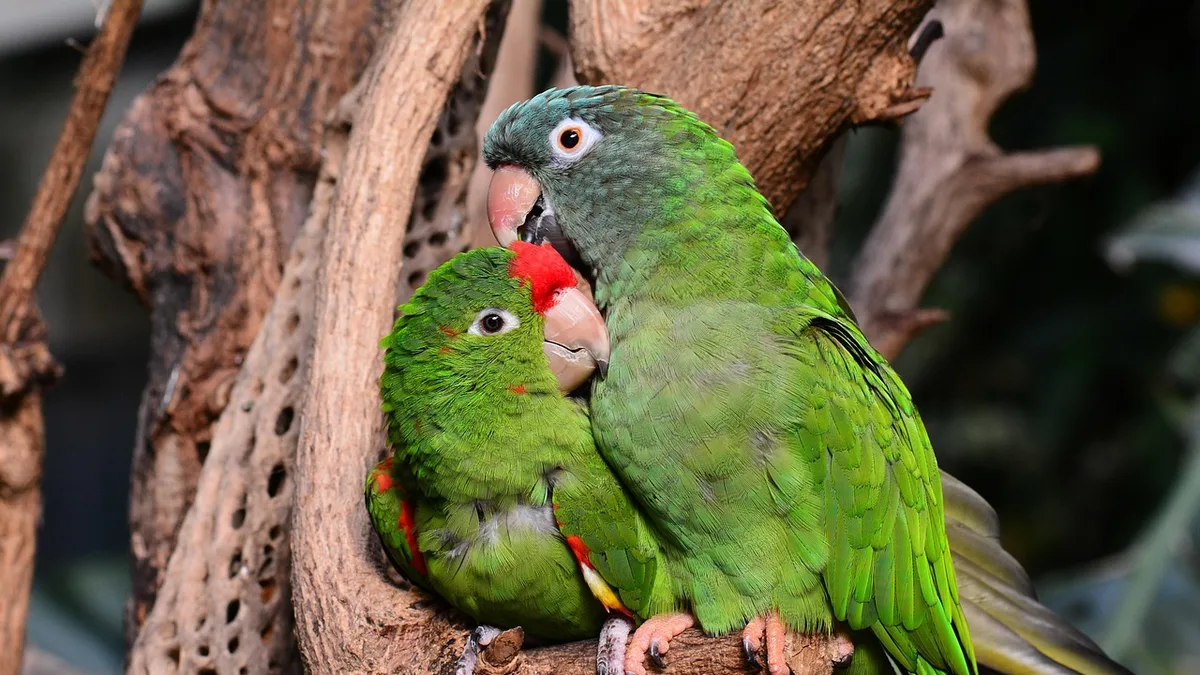 Feather condition and posture are the earliest external indicators of illness. Photo: 1931526 via Pixabay.
Laboratory involvement is indicated when preanesthetic assessment reveals abnormalities that could alter drug selection or fluid therapy. Point-of-care glucose, hematocrit, and total solids are reasonable minimum data for sick birds. Blood gas analysis during anesthesia is valuable when capnography and pulse oximetry conflict or when ventilation is being adjusted for a compromised patient.
Regulatory reporting obligations vary by jurisdiction. The WOAH terrestrial animal health standards address reportable diseases and trade-related health requirements, while the AVMA practice resources provide guidance on professional conduct and adverse event reporting. Anesthetic deaths in birds are not generally reportable, but suspected adverse drug reactions should be documented and reported through the appropriate pharmacovigilance system.
Troubleshooting Table
| Observation | Likely Cause | Discriminating Check |
|---|---|---|
| Rising ETCO2, stable rate | Hypoventilation or dead space | Compare ETCO2 to arterial blood gas if available, check circuit connections and mask fit |
| Sudden bradycardia | Hypoxemia, deep plane, vagal reflex | Assess depth, verify oxygen flow, check Doppler signal quality |
| Falling SpO2 with good waveform | Hypoxemia, shunt, equipment error | Check probe site, verify inspired oxygen, auscultate lungs |
| Progressive hypothermia | Heat loss exceeding input | Verify warming device function, check probe placement |
| Weak Doppler signal | Hypotension, probe displacement | Reposition probe, check blood pressure directly |
| Prolonged recovery | Hypothermia, drug accumulation, hepatic impairment | Measure temperature, review drug doses and intervals |
Frequently Asked Questions
What monitoring adaptations are acceptable when Doppler and capnography are unavailable?
When advanced monitors are absent, the physical examination becomes the primary monitoring tool. Pulse quality via Doppler or direct palpation of the basilic or metatarsal artery provides heart rate and rhythm information, while mucous membrane color and capillary refill time offer crude perfusion estimates. Capnography loss is partially compensated by observing the anesthetic reservoir bag excursion and thoracic wall movement, though these methods cannot detect hypoventilation as early as capnography. The AAHA anesthesia and monitoring guidelines emphasize that monitoring frequency and documentation become more critical when technology is limited. Increase assessment intervals to every 2 to 3 minutes, record all findings, and shorten anesthetic duration to reduce cumulative risk.
How should monitoring be adjusted for a budgerigar or cockatiel weighing under 50 grams?
Small psittacines tolerate minimal monitoring equipment weight and have higher metabolic rates that accelerate drug clearance and hypothermia onset. Use the lightest available Doppler probe and place it on the distal tibiotarsus instead of the wing to preserve limb mobility. Temperature probes sized for small patients are essential because heat loss is rapid and profound. Respiratory rate in these patients may exceed 60 breaths per minute, making visual counting difficult, use a magnifying lens or observe the keel's rise and fall. The MSD Veterinary Manual notes that small body mass magnifies the effects of anesthetic circuit dead space, so use non-rebreathing circuits exclusively. Shorten monitoring intervals to every minute and prioritize temperature and respiratory assessment over blood pressure measurement when equipment constraints force a choice.
What constitutes an acceptable recovery period, and when should intervention begin?
Most pet birds should show coordinated head movement within 5 to 10 minutes after volatile agent discontinuation and achieve sternal recumbency within 15 to 20 minutes. Prolonged recovery beyond 30 minutes warrants investigation for hypothermia, hypoglycemia, or residual drug effect. During recovery, maintain supplemental oxygen and active warming until the bird is perching or gripping normally. The WSAVA Global Pain Council guidelines remind clinicians that pain itself can delay recovery, so reassess analgesic adequacy in a bird that remains depressed. If recovery stalls, re-evaluate depth, check temperature, and consider whether the patient was hypotensive intraoperatively. Do not discharge a bird that cannot maintain sternal posture or thermoregulate independently.
How do I explain an anesthetic complication to a client without causing undue alarm?
Use precise, factual language that acknowledges the event without speculation. State what occurred, what was done, and what the current status is. For example, describe that the bird's breathing slowed during anesthesia, that oxygen support was increased, and that the bird is now recovering normally. The AVMA practice resources emphasize transparent communication about adverse events as part of professional standards. Offer the client a follow-up call within 24 hours and document the conversation in the medical record. Avoid assigning blame to equipment or staff in front of the client. If the complication resulted in death, provide a clear timeline and offer necropsy as an option, explaining what information it may provide.
What documentation is required for avian anesthesia beyond the standard anesthetic record?
Record the same parameters as for mammalian patients, but add species, body weight in grams, and preanesthetic crop status. Document the specific anesthetic circuit used, fresh gas flow rate, and any equipment adaptations such as a modified face mask. Include a body condition score and an assessment of pectoral muscle mass, as these affect drug distribution and recovery. The AAHA anesthesia and monitoring guidelines recommend recording monitoring intervals and any corrective actions taken. For avian patients, also note ambient room temperature and the warming method used, since hypothermia is a common contributor to prolonged recovery. Photographs of unusual equipment setups can be useful for future reference and for training staff.
When should I refer an avian patient to a specialist center instead of proceed with anesthesia?
Refer when the patient has known cardiac disease, respiratory compromise, or a history of anesthetic complications, and your practice lacks Doppler, capnography, or active warming equipment. Birds with suspected air sac disease, severe anemia, or coagulopathies carry elevated risk that may exceed what a general practice can safely manage. The WOAH terrestrial animal health standards address welfare considerations that apply when procedures carry disproportionate risk. Referral is also appropriate when the planned procedure exceeds your experience level, such as endoscopic surgery or prolonged orthopedic repair. Stabilize the patient with oxygen and warming before transport, and communicate directly with the receiving clinician regarding the anesthetic plan and any complications observed.
Related Clinical & Scientific Guides
- Anesthetic Machine Leak Testing and Pressure Checks: A Step-by-Step Protocol
- Anesthetic Depth Assessment: Reflexes, Eye Position, and Ventilation
- Anesthesia for Patients with Obesity: Challenges and Solutions
References and Further Reading
- AAHA Anesthesia and Monitoring Guidelines for Dogs and Cats. AAHA.
- WSAVA Global Pain Council Guidelines. WSAVA.
- MSD Veterinary Manual, Professional Edition. MSD Veterinary Manual.
- American Veterinary Medical Association Practice Resources. American Veterinary Medical Association.
- WOAH Terrestrial Animal Health Code. WOAH.
Related Articles
- Anesthesia for Patients with Cardiac Disease: Risk Assessment and Monitoring
- Anesthetic Machine Checkout: Daily Verification and Troubleshooting
- Blood Pressure Monitoring During Anesthesia: Methods and Interpretation
- Anesthesia for Patients with Cancer: Paraneoplastic Syndromes
- Anesthesia for Patients with Sepsis: Hemodynamic Support
This article is educational professional reference material for veterinary audiences. It is not a substitute for veterinary diagnosis, individual clinical judgment, current product labeling, or applicable regulatory requirements.