Anesthetic Complications in Brachycephalic Dogs: Beyond Airway Obstruction
By Dr. Zubair Khalid, DVM, MS, PhD ·

Key Takeaways
- Brachycephalic dogs exhibit a significantly higher postanesthetic complication rate (13.9%) compared to nonbrachycephalic controls (3.6%), with brachycephalic status identified as an independent risk factor.
- Beyond airway obstruction, brachycephalic dogs are vulnerable to post-obstructive pulmonary edema due to negative intrathoracic pressure generation, which can manifest hours after extubation.
- Gastrointestinal dysmotility, including a high prevalence of hiatal hernia and gastroesophageal reflux, predisposes these dogs to passive regurgitation, a risk that persists into the recovery phase.
- Anesthesia duration is a critical risk factor, with longer procedures correlating with increased perianesthetic complications, while increasing body weight appears to be protective, potentially due to less severe conformational abnormalities.
- Recovery is a disproportionately hazardous phase, requiring meticulous monitoring for signs of respiratory decompensation, positional intolerance, and delayed regurgitation, with a low threshold for re-intubation or escalation of care.
- Preoperative optimization and thorough screening for subtle signs of airway compromise, such as stertor or exercise intolerance, are crucial for risk stratification and informed anesthetic planning.
Brachycephalic dogs present a distinctive anesthetic challenge that extends well beyond the mechanical obstruction of the upper airway. While the brachycephalic airway syndrome is the most visible risk, the perianesthetic period in these patients is shaped by a broader set of physiologic vulnerabilities, including altered respiratory mechanics, gastrointestinal dysmotility, thermoregulatory instability, and a recovery phase that can be as hazardous as the procedure itself. This article examines those less-discussed complications for the practicing veterinarian, with emphasis on recognition, prevention, and structured decision-making.
The clinical questions addressed here are practical ones. Which brachycephalic patients are at highest risk for postanesthetic complications? How does the anesthetist distinguish a normal recovery from the early stages of post-obstructive pulmonary edema? When should regurgitation be treated as a sentinel event instead of an incidental finding? The evidence base for these decisions is growing, and this article draws on recent retrospective and survey data to give the reader a framework for anticipating complications before they become emergencies.
The intended reader is the veterinarian who already manages brachycephalic airways competently and now seeks a deeper understanding of the systemic complications that surround the anesthetic event. Basic intubation technique and airway rescue are covered elsewhere. This article focuses on the physiologic foundations and clinical patterns that explain why these dogs deteriorate, sometimes hours after extubation.
At a Glance
| Parameter | Clinical Relevance | Source Context |
|---|---|---|
| Postanesthetic complication rate | 13.9% in brachycephalic dogs versus 3.6% in matched nonbrachycephalic controls | Retrospective cohort study |
| Perianesthetic complication rate | 49.8% brachycephalic versus 48.4% nonbrachycephalic, with brachycephalic status an independent risk factor | Retrospective cohort study |
| Anesthesia duration | Longer procedures increase perianesthetic complication risk | Retrospective cohort study |
| Body weight | Increasing body weight is associated with decreased perianesthetic complication risk | Retrospective cohort study |
| Postoperative regurgitation | Occurs in approximately 22% of dogs after soft palate surgery | Retrospective surgical study |
| Postoperative aspiration pneumonia | Occurs in approximately 7% of dogs after soft palate surgery | Retrospective surgical study |
| ICU respiratory decompensation | 16% of brachycephalic dogs developed respiratory complications after hospitalization for unrelated conditions | Multicenter syndromic surveillance |
| Perianesthetic protocol variability | Wide variation across US practices despite documented risks | National survey of veterinary professionals |
The Physiologic Basis of Risk
The brachycephalic phenotype is not a single anatomic abnormality but a constellation of changes that collectively alter respiratory and cardiovascular physiology. The elongated soft palate, stenotic nares, and hypoplastic trachea increase upper airway resistance at rest. What is less often appreciated is how this baseline resistance interacts with anesthetic drugs. Most induction agents and inhalants cause dose-dependent upper airway collapse, reduced pharyngeal tone, and decreased ventilatory drive. In a dog already breathing near its mechanical limit, the additive effect of these drug actions can convert a compensated patient into a critically obstructed one within minutes.
The respiratory consequences extend to the lower airways and pulmonary parenchyma. Brachycephalic dogs generate substantial negative intrathoracic pressure during inspiration against a partially obstructed airway. This pressure gradient increases venous return to the right heart and promotes transudation of fluid across the pulmonary capillary bed. When obstruction is relieved suddenly, as with intubation or surgical correction, the abrupt change in intrathoracic pressure can precipitate post-obstructive pulmonary edema. This mechanism explains why the most dangerous respiratory complication often appears after the airway has been secured, not before.
Gastrointestinal physiology is similarly altered. Brachycephalic dogs have a high prevalence of hiatal hernia, esophageal dysmotility, and gastroesophageal reflux. The combination of increased intra-abdominal pressure from respiratory effort and the lower esophageal sphincter relaxation caused by many anesthetic drugs creates a predictable risk of passive regurgitation. This risk persists into recovery, when the dog is no longer intubated and the airway is unprotected.
Anesthetic Risk Stratification
The evidence that brachycephalic status independently predicts perianesthetic complications comes from a retrospective cohort study comparing 223 brachycephalic dogs with 223 nonbrachycephalic controls matched by procedure and other characteriztics. The study found that brachycephalic dogs had a postanesthetic complication rate of 13.9% compared with 3.6% in controls, and that brachycephalic status remained a significant risk factor after controlling for other variables. The same study identified anesthesia duration as an independent risk factor, with longer procedures associated with higher complication rates, while increasing body weight appeared protective.
These findings have direct clinical implications. The anesthetist should treat every brachycephalic patient as high risk regardless of procedure complexity. An elective neuter in a young French Bulldog carries a different risk profile than the same procedure in a Labrador Retriever, and the anesthetic plan should reflect that difference. The protective effect of body weight is worth noting, though its mechanism is not fully understood. It may reflect the tendency of smaller brachycephalic dogs toward more severe conformational abnormalities, or it may be a proxy for breed differences within the brachycephalic group.
The survey data from United States practices reveal that perianesthetic management of brachycephalic dogs varies widely across the profession, despite the well-documented risks. This variability suggests that many practices have not adopted standardized protocols for these patients. The survey, which drew responses from over 1,300 veterinary professionals, found that elective neuter was the most common reason for anesthesia in brachycephalic dogs, meaning that the highest-risk patients are frequently anesthetized for the most routine procedures.
Post-Obstructive Pulmonary Edema
Post-obstructive pulmonary edema is one of the most feared complications in brachycephalic anesthesia because it can appear after the airway has been successfully managed. The pathophysiology involves the generation of markedly negative intrathoracic pressure during inspiration against an obstructed airway. This pressure gradient increases pulmonary capillary hydrostatic pressure and decreases perivascular interstitial pressure, favoring fluid movement into the alveoli. The edema may develop during the obstructive episode itself or may manifest shortly after relief of the obstruction, when the sudden normalization of pressures allows fluid to shift into previously protected lung regions.
Clinical recognition requires a high index of suspicion. The classic presentation is a brachycephalic dog that develops tachypnea, increased respiratory effort, or crackles on thoracic auscultation within minutes to hours after extubation. Pulse oximetry may show declining saturation, and thoracic radiographs typically reveal an interstitial to alveolar pattern, often with a perihilar distribution. The differential diagnosis includes aspiration pneumonia, which can coexist and may be difficult to distinguish radiographically in the acute setting.
Management is supportive and begins with supplemental oxygen and careful monitoring. The anesthetist should have a low threshold for re-intubation if the dog shows signs of progressive respiratory distress. The condition is generally self-limiting if the airway remains patent, with edema resolving over 12 to 24 hours as the Starling forces return to normal. The key error is misattributing the signs to a residual drug effect or to normal recovery variation, which delays intervention.
Regurgitation and Aspiration
Regurgitation during anesthesia is a passive process that occurs without the active retching seen with vomiting. The brachycephalic patient is predisposed by several factors: increased intra-abdominal pressure from respiratory effort, a high prevalence of hiatal hernia and gastroesophageal reflux, and the relaxation of the lower esophageal sphincter caused by many anesthetic drugs. The result is that gastric contents can flow freely into the pharynx, where they may be aspirated into the trachea if the airway is not protected.
The clinical significance of regurgitation is substantial. In a retrospective study of 124 dogs undergoing soft palate surgery, postoperative regurgitation occurred in 27 dogs, or approximately 22% of the population. Postoperative aspiration pneumonia developed in 9 dogs, or approximately 7%. These figures are specific to dogs undergoing staphylectomy or folded flap palatoplasty, and they likely represent a higher-risk population than brachycephalic dogs undergoing unrelated procedures. Nevertheless, they establish that regurgitation is a common event in this surgical group and that aspiration is a clinically meaningful consequence.
The anesthetist should assume that regurgitation can occur at any point from induction through recovery. The presence of an endotracheal tube with an inflated cuff protects the lower airway during the intraoperative period, but the risk returns at extubation. The timing of extubation is therefore a critical decision. Extubating too early, while the dog still has depressed airway reflexes, leaves the airway unprotected during a period when regurgitation may still occur. Extubating too late risks the dog biting the tube or becoming distressed during the transition to spontaneous breathing. The decision must be individualized based on the dog's level of consciousness, airway reflexes, and respiratory pattern.
Preoperative Optimization and Patient Preparation
The preoperative period in brachycephalic dogs is an opportunity to reduce, also identify, risk. A dog that presents for elective procedures with a history of stertor, exercise intolerance, or sleep disruption carries a different risk profile than an asymptomatic individual of the same breed. The survey data from United States practices show that elective neuter is the most common reason for anesthesia in brachycephalic dogs, which means many of these patients present without a prior diagnosis of brachycephalic obstructive airway syndrome Crosby et al. survey of perianesthetic management practices. This creates a clinical obligation to screen actively instead of assume the absence of disease.
The screening examination should include an assessment of nares patency, auscultation of referred upper airway noise, and a careful history of regurgitation or vomiting. Owners frequently do not volunteer this information. Direct questions about sleep apnea, gagging after meals, and episodes of cyanosis or collapse are more productive than general inquiries about health. Body condition scoring matters because the risk of perianesthetic complications decreases with increasing body weight in the matched cohort study, a finding that likely reflects the protective effect of larger airways in heavier dogs Gruenheid et al. risk of anesthesia-related complications in brachycephalic dogs. Obesity should not be interpreted as protective, rather, the heaviest brachycephalic dogs in that cohort were probably the least severely affected by airway obstruction.
Premedication choices should account for the risk of regurgitation. Opioid-induced vomiting is a genuine concern in a breed with a high prevalence of hiatal hernia and gastroesophageal reflux. An antiemetic such as maropitant, administered with sufficient lead time before premedication, reduces the likelihood of active vomiting during the induction period. Anticholinergics are not a substitute for this measure. The decision to use a benzodiazepine instead of an opioid for sedation in a severely affected dog is reasonable, but the practitioner should recognize that this changes the analgesic plan and requires adjustment of intraoperative and postoperative analgesia. Current formulary references should be consulted for dosing and timing.
Intraoperative Monitoring Beyond the Basics
Standard monitoring in brachycephalic dogs must extend beyond pulse oximetry and capnography. The capnogram itself is diagnostically useful. A prolonged expiratory plateau with a slow rise suggests expiratory obstruction, which is common in these patients due to everted laryngeal saccules or collapsing nares. An abrupt upstroke followed by a plateau that does not return to baseline indicates rebreathing, which can occur when respiratory rate is low and fresh gas flow is inadequate. Neither finding is specific to brachycephalic dogs, but both occur with sufficient frequency in this population to warrant deliberate attention.
The following monitoring parameters should be documented at intervals no longer than five minutes during the procedure:
| Parameter | What It Detects | Action Threshold | Response |
|---|---|---|---|
| Capnogram morphology | Expiratory obstruction, rebreathing, bronchospasm | Plateau slope change or failure to return to baseline | Adjust airway positioning, deepen or lighten plane, increase fresh gas flow |
| SpO2 trend | Progressive hypoxemia before visible cyanosis | Sustained below 94% | Verify probe site, assess airway patency, consider supplemental oxygen or manual ventilation |
| End-tidal CO2 trend | Hypoventilation, equipment disconnection | Rising above 55 mm Hg or abrupt fall | Evaluate breathing circuit, reduce anesthetic depth, support ventilation |
| Noninvasive blood pressure | Hypotension from vasodilation or hypovolemia | Mean below 60 mm Hg | Reduce inhalant concentration, administer fluid bolus, consider vasopressor |
| Esophageal temperature | Hypothermia, which prolongs recovery | Below 36.5 C | Active warming, reduce anesthetic depth to permit shivering |
The retrospective comparison of staphylectomy and folded flap palatoplasty found that neither procedure was associated with the occurrence of anesthetic complications, but the folded flap technique was associated with longer anesthesia times Miller et al. complications and outcome following staphylectomy and folded flap palatoplasty. Longer anesthesia duration is an independent risk factor for perianesthetic complications in brachycephalic dogs Gruenheid et al. risk of anesthesia-related complications in brachycephalic dogs. The surgeon and anesthetist should therefore coordinate to minimize procedural time, and the monitoring plan should be intensified when the procedure runs long.
Recovery as a High-Risk Phase
The postanesthetic period carries a disproportionate share of complications in brachycephalic dogs. The matched cohort study found postanesthetic complications in 13.9% of brachycephalic dogs compared with 3.6% of nonbrachycephalic dogs Gruenheid et al. risk of anesthesia-related complications in brachycephalic dogs. This is not a statistical artifact. The recovery phase combines residual anesthetic depression, airway edema from endotracheal intubation, and the return of pharyngeal muscle tone at a time when protective reflexes are still blunted.
Recovery should be planned before the procedure begins. The endotracheal tube should remain in place until the dog demonstrates a strong swallow reflex and purposeful movement. Extubation in a brachycephalic dog that is still deeply anesthetized invites airway obstruction from the soft palate and tongue falling back against the larynx. Extubation in a dog that is too awake invites laryngospasm and aspiration of saliva. The window between these two states is narrower in brachycephalic dogs than in other breeds.
Positioning during recovery matters. Sternal recumbency with the head extended and slightly elevated reduces upper airway collapse. Dogs that are placed in lateral recumbency and left undisturbed may obstruct silently. The recovery area should be staffed by personnel who understand that stertor in a brachycephalic dog is not normal and requires intervention. Oxygen supplementation should continue until the dog is breathing quietly on room air with a stable SpO2 above 95%.
Sedation during recovery is a double-edged sword. Low-dose sedation can reduce the agitation and thrashing that exacerbate airway edema and increase oxygen consumption. Excessive sedation produces the same problem as premature extubation. The survey data show wide variation in recovery protocols across practices, which reflects the absence of a clear evidence base for a single approach Crosby et al. survey of perianesthetic management practices. A reasonable framework is to use the minimum dose of a short-acting agent that permits the dog to rest quietly while maintaining a patent airway, and to have reversal agents immediately available.
Postoperative Respiratory Surveillance
Respiratory complications after discharge from the immediate recovery area are common enough to justify structured surveillance. The ICU surveillance study found that 16% of brachycephalic dogs developed respiratory complications requiring ICU admission after initially being hospitalized for another condition Brachycephalic Study Group syndromic surveillance of respiratory compromise in brachycephalic dogs in ICUs. This finding has direct implications for discharge planning. A brachycephalic dog that has undergone anesthesia for an unrelated procedure should not be discharged the same day without a documented assessment of respiratory stability.
The postoperative monitoring checklist should include the following elements, documented at regular intervals for at least six hours after extubation:
- Respiratory rate and effort, with attention to abdominal component
- Audible stertor or stridor at rest, also during excitement
- SpO2 on room air, if the dog will tolerate a probe
- Presence of regurgitation or vomiting episodes
- Coughing, which may indicate aspiration or tracheal irritation
- Mucous membrane color and capillary refill time
- Rectal temperature, with active warming if below 36.5 C
A dog that develops progressive stertor, increased respiratory effort, or hypoxemia after an initially smooth recovery should be evaluated for laryngeal edema, post-obstructive pulmonary edema, or aspiration pneumonia. The differential is guided by timing. Laryngeal edema typically presents within minutes to hours of extubation. Post-obstructive pulmonary edema can present immediately or up to several hours later. Aspiration pneumonia may take 12 to 72 hours to become clinically apparent.
Documentation and Communication
The anesthetic record for a brachycephalic dog should include a preoperative airway risk assessment, the monitoring parameters listed above, and a recovery plan that specifies extubation criteria and postanesthetic surveillance intervals. This documentation serves two purposes. It creates a baseline for comparison if complications develop, and it communicates the risk profile to other team members who may care for the dog after the primary anesthetist has left.
Owners should receive written discharge instructions that include the signs of respiratory distress and a clear statement about when to seek emergency care. The AVMA practice resources emphasize the importance of client communication in perioperative care AVMA practice resources. This is particularly relevant for brachycephalic breeds, where owners may have normalized chronic respiratory signs and may not recognize a deterioration as an emergency. The instruction should be specific: increased respiratory effort at rest, blue or pale mucous membranes, or an inability to sleep without sitting up warrants immediate veterinary attention.
Recognized Complication Patterns and Early Detection
Brachycephalic dogs accumulate complications across the entire perianesthetic period, also during airway instrumentation. A retrospective cohort study found postanesthetic complications in 13.9% of brachycephalic dogs versus 3.6% of matched nonbrachycephalic controls, with brachycephalic status and longer anesthetic duration independently associated with higher perianesthetic complication rates Gruenheid et al., 2018. The recovery period carries disproportionate risk, and surveillance must extend well beyond extubation.
Three failure modes deserve specific attention because they present with subtle early signs.
Delayed regurgitation without active vomiting. Passive regurgitation occurs when lower esophageal sphincter tone drops under anesthesia and gastric contents pool in the esophagus. Brachycephalic dogs have increased intra-abdominal pressure from respiratory effort and frequent aerophagia. Early detection relies on continuous observation of the oral cavity and pharynx, not on waiting for visible vomiting. A damp towel under the muzzle, frequent pharyngeal suction checks, and auscultation of the esophagus during recovery identify regurgitation before aspiration occurs. The 2024 retrospective study of staphylectomy and folded flap palatoplasty reported postoperative regurgitation in 27 of 124 dogs, with aspiration pneumonia in 9 of 124, confirming that regurgitation remains a clinically significant event even with modern airway management Miller et al., 2024.
Silent hypoxemia in recovery. Pulse oximetry readings can remain normal while the dog is breathing supplemental oxygen, masking progressive atelectasis or early pulmonary edema. Brachycephalic dogs in the ICU frequently develop respiratory compromise after initial evaluation for unrelated problems, with 16% of brachycephalic ICU patients requiring escalation of respiratory support after admission for another condition Brachycephalic Study Group, 2022. The discriminating check is serial assessment of respiratory rate and effort after oxygen is reduced, not the oxygen saturation reading itself. A dog that maintains SpO2 above 95% on 2 L/min but doubles its respiratory rate when weaned to room air is showing early decompensation.
Prolonged recovery with positional intolerance. Brachycephalic dogs that cannot maintain sternal recumbency within 20 minutes of ceasing anesthetic delivery are at risk of airway obstruction from soft palate collapse and tongue base relaxation. Early detection requires a nurse or technician dedicated to repositioning and observation, not intermittent checks. The AAHA anesthesia guidelines emphasize continuous monitoring through recovery and define specific parameters for discharge readiness AAHA Anesthesia and Monitoring Guidelines.
Common Errors and Corrective Actions
Less experienced clinicians make recurring errors that are predictable and correctable.
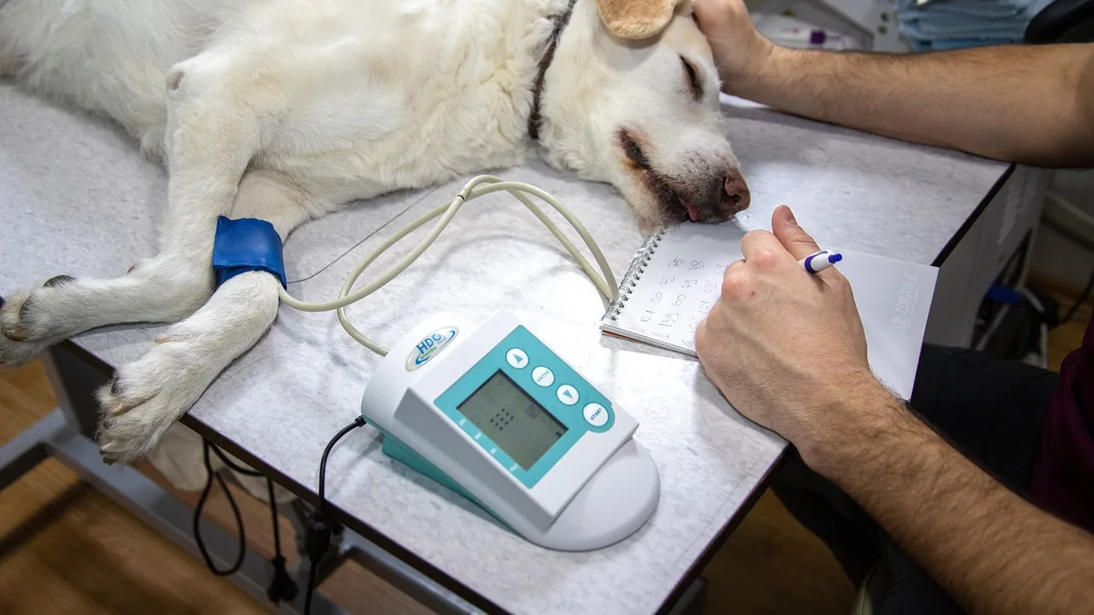 Frequency, timing and content of vomiting narrow the differential faster than any single test. Photo: mirkosajkov via Pixabay.
Error: Extubating too early or too late. Extubation timing in brachycephalic dogs requires individualization. Extubating at the first swallow risks laryngospasm and obstruction from residual sedation. Extubating only when the dog is fully awake risks airway obstruction while the endotracheal tube is removed and the dog cannot coordinate breathing. The corrective action is to extubate when the dog demonstrates a strong palpebral reflex and begins to chew or swallow, then immediately position the head in a neutral to slightly extended position with the tongue gently pulled forward. Have an emergency airway kit at the recovery station before extubation.
Error: Treating the SpO2 reading instead of the patient. A brachycephalic dog with an SpO2 of 88% during recovery may have a displaced tongue, a kinked endotracheal tube, or a collapsed lung lobe. The novice response is to increase oxygen flow. The correct response is a systematic check: airway patency, breathing effort, auscultation, and end-tidal carbon dioxide if available. Increasing oxygen without addressing the underlying problem delays recognition of post-obstructive pulmonary edema or aspiration.
Error: Discharging based on time instead of criteria. The survey of perianesthetic management across US practices found wide variation in recovery and discharge practices, suggesting that objective discharge criteria are not universally applied Crosby et al., 2026. Discharge readiness requires sustained sternal recumbency, normal respiratory rate and effort on room air, ability to maintain body temperature, and absence of regurgitation for at least one hour after extubation.
Limitations of Current Evidence
The evidence base for brachycephalic anesthetic complications has significant gaps. The 2018 cohort study matched brachycephalic and nonbrachycephalic dogs by procedure but was limited to routine surgery and diagnostic imaging, excluding emergency and trauma cases Gruenheid et al., 2018. The 2024 palatoplasty study was retrospective and single-center, with complication rates that may not generalize to practices with different caseloads or surgeon experience Miller et al., 2024.
Expert opinion still differs on several points. The optimal timing of extubation remains contested, with some authorities advocating early extubation while the dog is still deeply anesthetized to allow smooth airway control, and others recommending late extubation to ensure protective reflexes have returned. There is no prospective trial comparing these approaches in brachycephalic dogs. Similarly, the role of prophylactic antiemetics is debated. Some clinicians administer maropitant or metoclopramide to all brachycephalic dogs preoperatively, while others reserve these drugs for dogs with a history of regurgitation or gastroesophageal reflux. The evidence does not currently support a universal recommendation.
The survey data confirm that perianesthetic management varies widely across practices, with differences in premedication, induction, monitoring, and recovery protocols Crosby et al., 2026. This variation reflects both the absence of definitive evidence and the need for protocols tailored to individual patient risk.
Escalation and Referral Criteria
Referral to a specialist anesthesiologist or criticalist is warranted when a brachycephalic dog requires mechanical ventilation, develops post-obstructive pulmonary edema that does not respond to initial oxygen therapy, or has repeated regurgitation episodes despite appropriate positioning and pharmacologic management. Dogs with known aspiration pneumonia, pre-existing pulmonary hypertension, or a history of previous anesthetic complications should be referred for elective procedures when possible.
Laboratory involvement is indicated when a brachycephalic dog develops unexplained hypoxemia, hypercapnia, or metabolic derangements during recovery. Arterial blood gas analysis, thoracic radiography, and serial lactate measurement help distinguish aspiration pneumonitis from pulmonary edema and guide ventilator settings.
Regulatory reporting is rarely required for anesthetic complications in companion animals. However, if a drug reaction or equipment failure is suspected, the manufacturer and the relevant national pharmacovigilance program should be notified. The AVMA provides practice resources on adverse event reporting and professional liability considerations AVMA Practice Resources.
| Observation | Likely Cause | Discriminating Check |
|---|---|---|
| SpO2 falls in recovery despite oxygen | Airway obstruction, atelectasis, pulmonary edema | Auscultation, end-tidal CO2, response to repositioning |
| Frequent swallowing or lip licking | Esophageal reflux or regurgitation | Pharyngeal inspection, esophageal auscultation, thoracic radiographs if aspiration suspected |
| Prolonged recumbency after extubation | Residual anesthetic effect, hypothermia, hypoglycemia | Serial temperature, blood glucose, response to stimulation |
| Tachypnea with normal SpO2 | Pain, hyperthermia, early pulmonary edema | Pain scoring, temperature, serial respiratory rate trending |
| Cyanosis with normal pulse oximetry | Methemoglobinemia, poor perfusion, probe artifact | Co-oximetry, blood pressure, probe site change |
Frequently Asked Questions
How should I adjust my anesthetic plan when advanced monitoring equipment is unavailable?
Prioritize continuous visual assessment and basic monitoring. Pulse palpation, capillary refill time, mucous membrane color, and thoracic auscultation provide meaningful trend data. Capnography is strongly preferred for brachycephalic dogs because tidal volumes are small and airway resistance is high, but when it is unavailable, maintain a higher index of suspicion for hypoventilation and adjust ventilator settings or manual ventilation accordingly. Extend recovery observation time and use a standardized complication checklist. The AAHA anesthesia and monitoring guidelines describe minimum monitoring standards that remain achievable in most practice settings.
What documentation should I include in the medical record for a brachycephalic dog's anesthetic event?
Record the preoperative respiratory examination findings, including stertor, exercise intolerance, and any history of regurgitation or syncope. Document the airway grade at intubation, endotracheal tube size and cuff pressure, and any difficulty encountered. Note every episode of hypoxemia, hypercapnia, regurgitation, or arrhythmia with the time, duration, and intervention. In recovery, record respiratory rate and effort at 15 minute intervals until extubation and then at 30 minute intervals until discharge criteria are met. This documentation supports clinical decision making and provides defensible records if complications arise. The AVMA practice resources offer guidance on medical record content standards.
How do I discuss the added anesthetic risk with an owner who is focused on a routine procedure?
Frame the discussion around the dog's conformation instead of the procedure's complexity. Explain that brachycephalic dogs carry a higher postanesthetic complication rate than nonbrachycephalic dogs, with one retrospective cohort study reporting postanesthetic complications in 13.9% of brachycephalic dogs versus 3.6% of controls. Describe the specific risks of airway swelling, regurgitation, and difficult recovery. Offer concrete mitigation steps such as extended monitoring, preoxygenation, and a longer hospitalization. Provide a written estimate that includes additional recovery time and possible overnight observation. This approach converts an abstract risk discussion into a concrete care plan the owner can approve.
What should I do when a brachycephalic dog regurgitates during recovery despite an empty stomach?
Position the dog in sternal recumbency with the head elevated and slightly extended. Suction the oropharynx immediately and assess the endotracheal tube cuff if the tube is still in place. Obtain thoracic radiographs if aspiration is suspected, recognizing that early changes may be absent. Institute oxygen supplementation and monitor pulse oximetry continuously for at least 12 hours. Postoperative regurgitation occurs in a meaningful proportion of brachycephalic dogs after airway surgery, and aspiration pneumonia is an uncommon but recognized sequel. If respiratory signs develop, escalate to active suctioning, bronchodilator therapy, and further diagnostic imaging as indicated.
How does the approach differ for a brachycephalic cat or a nonbrachycephalic dog with similar airway signs?
Brachycephalic cats share many of the same risks, including post-obstructive pulmonary edema and difficult recovery, but they are more prone to laryngospasm and require additional caution with airway instrumentation. Nonbrachycephalic dogs with acquired airway obstruction, such as laryngeal paralysis, have similar physiologic vulnerabilities but different underlying pathology and a different expected response to anti-inflammatory therapy. The monitoring principles remain the same across species, but the threshold for overnight hospitalization and the choice of rescue medications should be adjusted to the specific patient. The MSD Veterinary Manual provides species-specific guidance on airway anatomy and anesthetic considerations.
How should I respond when a supervisor or referring veterinarian resists extended monitoring for a brachycephalic patient?
Present the evidence directly. A survey of perianesthetic management across United States practices found wide variation in monitoring and recovery protocols for brachycephalic dogs despite well-documented risks. Cite the specific complication rates from the retrospective cohort study and note that brachycephalic status was an independent risk factor for perianesthetic complications. Offer a time-limited compromise, such as 2 hours of extended recovery observation, and document the recommendation in the medical record. Frame the request as a patient safety standard instead of a preference, and reference the AAHA anesthesia and monitoring guidelines as the professional benchmark.
Related Clinical & Scientific Guides
- Anesthetic Machine Leak Testing and Pressure Checks: A Step-by-Step Protocol
- Anesthetic Depth Assessment: Reflexes, Eye Position, and Ventilation
- Anesthesia for Patients with Obesity: Challenges and Solutions
References and Further Reading
- Complications and outcome following staphylectomy and folded flap palatoplasty in dogs with brachycephalic obstructive airway syndrome.. 2024.
- A survey of perianesthetic management of brachycephalic dogs identifies wide variation across United States veterinary practices despite well-documented risks.. 2026.
- Treatment of canine spontaneous chronic corneal epithelial defects (SCCEDs) with corneal thermal cautery: A retrospective study of 89 cases.. 2023.
- Risk of anesthesia-related complications in brachycephalic dogs.. 2018.
- Dental composite for the fixation of mandibular fractures and luxations in 11 cats and 6 dogs.. 1994.
- Syndromic surveillance of the frequency and severity of respiratory compromise of brachycephalic dogs in ICUs.. 2022.
- AAHA Anesthesia and Monitoring Guidelines for Dogs and Cats. AAHA.
- WSAVA Global Pain Council Guidelines. WSAVA.
- MSD Veterinary Manual, Professional Edition. MSD Veterinary Manual.
Related Articles
- Brachycephalic Airway Syndrome and Anesthetic Management
- Anesthetic Complications: Recognition and Initial Management
- Anesthetic Complications in Cats: Recognition and Salvage
- Anesthetic Complications in Rabbits: Emergency Management
- Anesthetic Risk Assessment: Beyond the ASA Score
This article is educational professional reference material for veterinary audiences. It is not a substitute for veterinary diagnosis, individual clinical judgment, current product labeling, or applicable regulatory requirements.