
Toxoplasmosis in Wildlife: Seroprevalence, Diagnostic Approaches, and Conservation Impact
1. Introduction
Toxoplasmosis, caused by the obligate intracellular apicomplexan parasite Toxoplasma gondii, is one of the most prevalent zoonotic infections worldwide. While considerable attention has been directed toward human and domestic animal infections, the parasite's impact on wildlife populations has emerged as a critical concern for conservation biology. T. gondii infects a broad range of warm-blooded vertebrates, including mammals and birds, and can cause fatal disease in naive or immunocompromised species. In wildlife, seroprevalence studies have revealed widespread exposure across diverse taxa, and molecular diagnostics have enabled detection of active infections and genotyping of strains. Understanding the distribution, transmission dynamics, and pathological consequences of T. gondii in free-ranging animals is essential for assessing conservation threats, particularly for island endemics and other vulnerable species.
This article provides a detailed examination of the seroprevalence of T. gondii in wildlife, focusing on sentinel species and the diagnostic approaches used to detect infection. Emphasis is placed on the modified agglutination test (MAT) and polymerase chain reaction (PCR) methodologies, and the implications of toxoplasmosis for endangered mammal conservation are discussed.
2. Parasite Biology and Lifecycle
T. gondii exists in three infectious stages: tachyzoites (rapidly dividing form), bradyzoites (slowly dividing form within tissue cysts), and sporozoites (within oocysts). The definitive hosts are felids (domestic and wild cats), which shed unsporulated oocysts in feces. After sporulation in the environment (1-5 days), oocysts become infectious and can survive for months to years in soil and water. Intermediate hosts, including virtually all warm-blooded animals, become infected through ingestion of oocysts, ingestion of tissue cysts from infected prey, or transplacental transmission.
In intermediate hosts, tachyzoites disseminate widely, causing acute infection; then the parasite differentiates into bradyzoites that form cysts in neural and muscle tissues. These cysts persist for the life of the host and can reactivate during immunosuppression. The lifecycle is completed when a felid ingests tissue cysts from an infected intermediate host. In wildlife, this cycle can involve numerous prey-predator relationships, with felids (both domestic and wild) serving as key amplifiers.
The genetic diversity of T. gondii is organized into three main clonal lineages (Types I, II, III) and numerous atypical strains. Type II is most common in Europe and North America, while atypical strains dominate in South America and are associated with severe disease in wildlife and humans. Genotyping via PCR-restriction fragment length polymorphism (PCR-RFLP) or microsatellite analysis is crucial for understanding transmission pathways and pathogenicity.
3. Seroprevalence in Wildlife
Seroprevalence studies using detection of anti-T. gondii IgG antibodies provide estimates of past exposure. The modified agglutination test (MAT) is widely regarded as the gold standard for wildlife serology due to its high sensitivity and specificity across species, as it does not require species-specific secondary antibodies. Other serological methods include the Enzyme-Linked Immunosorbent Assay (ELISA) for Feline Leukemia Virus (though that reference is specific to FeLV, ELISA platforms for T. gondii are similar in principle) and indirect immunofluorescence assay (IFA), but MAT remains preferred because it uses intact formalin-fixed tachyzoites and detects both IgG and IgM without cross-reactivity issues common in some ELISA kits.
3.1 Seroprevalence in Key Taxonomic Groups
| Taxonomic Group | Geographic Region | Typical Seroprevalence (%) | Key Sentinel Species | References |
|---|---|---|---|---|
| Marine mammals | Global (coastal) | 10-80 | Sea otters, dolphins, seals | [1, 2] |
| Carnivores (terrestrial) | Americas, Europe | 20-90 | Gray wolves, foxes, raccoons | [3, 4] |
| Ungulates | Global | 5-70 | White-tailed deer, elk, bison | [5, 6] |
| Rodents and lagomorphs | Global | 5-40 | Brown rats, voles, rabbits | [7, 8] |
| Birds (waterfowl, raptors) | Global | 5-60 | Mallards, crows, bald eagles | [9, 10] |
| Marsupials | Australia, Americas | 10-80 | Kangaroos, wallabies, opossums | [11, 12] |
| Endangered mammals (e.g., Hawaiian monk seal, California condor) | Island/restricted ranges | 30-100 | Specific to population | [13, 14] |
Marine mammals, particularly sea otters (Enhydra lutris), exhibit high seroprevalence and clinical toxoplasmosis, linked to freshwater runoff containing oocysts [1]. In terrestrial carnivores, wolves and foxes serve as sentinels for environmental contamination because they occupy high trophic levels [3]. White-tailed deer are frequently used as sentinels for T. gondii in North America due to their abundance and wide distribution [5]. In Australia, macropods show high seroprevalence and mortality, especially in captive settings [11].
3.2 Sentinel Species in Surveillance
Sentinel species are selected based on ecological traits that reflect environmental T. gondii contamination: territorial range, trophic level, longevity, and accessibility for sampling. For example:
- Sea otters: Coastal foragers that ingest filter-feeding invertebrates; high seroprevalence indicates land-to-sea oocyst transport [1].
- White-tailed deer: Herbivores that graze on contaminated vegetation; seroprevalence in deer correlates with cat density and rainfall [5].
- Gray wolves: Apex predators that consume intermediate hosts; seroprevalence provides a cumulative measure of ecosystem-level parasite circulation [3].
The Tick-Borne Parasites in White-Tailed Deer: Babesia and Theileria Prevalence, PCR-Based Surveillance, and Impact on Livestock Interface article discusses surveillance of another parasite in deer, but the sampling strategies (e.g., hunter-killed deer sampling) are directly applicable to T. gondii serosurveillance.
4. Diagnostic Approaches
4.1 Modified Agglutination Test (MAT)
The MAT is a serological method that uses whole formalin-fixed T. gondii tachyzoites as antigen. Serum samples are serially diluted and incubated with antigen; agglutination indicates the presence of antibodies. The test is performed in 96-well U-bottom microtiter plates. A positive result is defined as agglutination at a serum dilution of 1:25 or higher (titer >= 1:25).
Key features of MAT:
- No species-specific conjugate: Works across all mammals and birds, making it ideal for wildlife studies.
- High sensitivity (95-99%) and specificity (96-98%) compared to the dye test (Sabin-Feldman test) [15].
- IgG and IgM detection: MAT detects both classes, but acute vs. chronic infection cannot be distinguished without additional testing (e.g., differential agglutination using acetone- vs. formalin-fixed antigen).
- Low cost and simple equipment: Requires only a microtiter plate and a refrigerator.
A standard protocol involves:
- Serum inactivation at 56°C for 30 minutes.
- Preparation of antigen mix: formalin-fixed tachyzoites (1 x 10^8/mL) in borate buffer with 2-mercaptoethanol (to denature IgM nonspecific agglutinins).
- Twofold serial dilutions of serum (1:25 to 1:3200) in 25 µL volumes.
- Addition of 25 µL antigen suspension per well, mixing, and incubation at 37°C for 2 hours or overnight at 4°C.
- Reading: a diffuse mat of agglutinated cells indicates a positive reaction; a tight button indicates negative.
4.2 Polymerase Chain Reaction (PCR)
PCR detects T. gondii DNA in tissues, blood, feces, or environmental samples. The most commonly targeted loci are the 529 bp repeat element (REP) [16], the B1 gene, and the internal transcribed spacer-1 (ITS-1) region. The 529 bp REP is present in 200-300 copies per genome, offering high sensitivity. Real-time PCR (qPCR) using SYBR Green or TaqMan probes allows quantification of parasite burden.
PCR protocols:
- Sample types: Tissues (brain, heart, skeletal muscle), whole blood, buffy coat, aqueous humor, cerebrospinal fluid, and feces (for oocyst detection).
- DNA extraction: Use of commercial silica membrane kits or phenol-chloroform extraction. For feces, a bead-beating step is required to disrupt oocyst walls.
- Amplification: Typical qPCR for the 529 bp REP uses primers Toxo-F (5'-CACAGAAGGGACAGAAGT-3') and Toxo-R (5'-CGTGGTCTTCGTCTAGATC-3'), producing a 162 bp amplicon [16].
- Detection limit: 0.1-1 tachyzoite equivalents per reaction.
Species-specific PCR is not needed; primers are conserved across all T. gondii genotypes. However, for genotyping, multilocus PCR-RFLP or microsatellite analysis using markers such as SAG1, SAG2, SAG3, BTUB, GRA6, c22-8, c29-2, L358, PK1, and Apico is performed [17].
4.3 Other Diagnostic Techniques
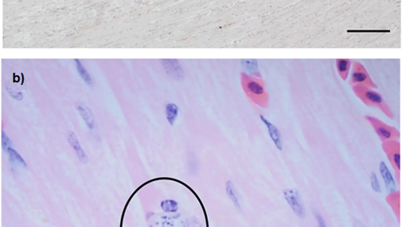
- Direct microscopy: Detection of tachyzoites in impression smears or histopathology (immunohistochemistry using anti-T. gondii antibodies). Sensitivity is low.
- Bioassay: Inoculation of tissue homogenates into mice (oral or intraperitoneal) or cats (oral) followed by serology and PCR. Considered a historical gold standard but labor-intensive and raises ethical concerns.
- ELISA: Commercial kits (e.g., for felid or human samples) may cross-react in wildlife but require validation. The reference Enzyme-Linked Immunosorbent Assay (ELISA) for Feline Leukemia Virus describes the platform; similar principles apply for T. gondii antibody capture, but species-specific conjugates are needed.
- Lateral flow assays: Point-of-care immunochromatographic tests exist for domestic animals but are rarely validated for wildlife.
4.4 Diagnostic Workflow
The following Mermaid diagram illustrates a typical diagnostic strategy for T. gondii detection in wildlife.
flowchart TD
 A["Wildlife sample collection: blood, tissues, feces"] --> B{Objective}
 B -->|Seroprevalence| C[Serum separation]
 B -->|Active infection / DNA detection| D[Tissue or blood DNA extraction]
 B -->|Oocyst detection| E[Fecal flotation / DNA extraction]
 C --> F["MAT: twofold dilutions 1:25 to 1:3200"]
 D --> G[PCR targeting 529 bp REP or B1 gene]
 E --> H["Microscopy + PCR (oocyst wall breakdown)"]
 F --> I["Interpretation: titer >= 1:25 positive"]
 G --> J["qPCR: quantify parasite burden; conventional PCR for genotyping"]
 H --> K[Confirmation via sequencing or PCR-RFLP]
 I --> L[Report seroprevalence]
 J --> L
 K --> L[Report molecular prevalence and genotype]
 L --> M["Conservation risk assessment: correlate with habitat, felid density, clinical signs"]
5. Conservation Impact
5.1 Clinical Disease in Endangered Mammals
T. gondii causes fatal toxoplasmosis in several endangered species, particularly those with no prior exposure (naive populations). Clinical signs include neurological deficits, blindness, respiratory distress, and sudden death. Necropsy findings often reveal multifocal necrotizing encephalitis, myocarditis, and pneumonitis with tissue cysts.
Key examples:
- Hawaiian monk seal (Neomonachus schauinslandi): Seroprevalence up to 100% in some populations; toxoplasmosis is a leading cause of mortality, especially in juveniles [13].
- California sea otter (Enhydra lutris nereis): A landmark study linked coastal runoff of T. gondii oocysts to sea otter deaths; seropositive otters had higher mortality risk [1].
- Tasmanian devil (Sarcophilus harrisii): High seroprevalence in wild populations but clinical disease appears rare; nevertheless, impact on immunocompromised individuals (e.g., those with devil facial tumor disease) is a concern [18].
- Kakapo (Strigops habroptilus): This critically endangered parrot of New Zealand is highly susceptible; outbreaks in captive breeding facilities have caused deaths [14].
- Hainan gibbon (Nomascus hainanus): Serological evidence of exposure in a small population on Hainan Island, China, raises concerns about catastrophic epizootics [19].
5.2 Mechanisms of Mortality and Morbidity
In susceptible wildlife, T. gondii infection leads to:
- Neurologic disease: Encephalitis with seizures, ataxia, and blindness.
- Reproductive failure: Abortion, stillbirth, and neonatal mortality (e.g., in sheep, goats, and wild ungulates).
- Immunosuppression: Alters host immune responses, potentially increasing vulnerability to other pathogens.
- Behavioral changes: Infected rodents lose fear of cats (a parasite manipulation to enhance transmission); similar effects might occur in other intermediate hosts, increasing predation risk.
5.3 Population-Level Effects
Seroprevalence data alone do not indicate population impact, but when combined with mortality surveillance and modeling, the effects may be significant. In the Hawaiian monk seal, toxoplasmosis accounts for approximately 15-20% of documented deaths in some years [13]. For island endemics with small population sizes (fewer than 500 individuals), even a few deaths per year can reduce genetic diversity and increase extinction risk.
Transmission from domestic cats to wildlife is a major conservation concern. Free-ranging and feral cats shed billions of oocysts into the environment; the oocysts remain viable in soil and water for extended periods. The article Toxoplasma gondii in Wildlife: Seroprevalence, Genotyping, and Transmission to Domestic Animals further discusses this spillover dynamic.
5.4 Management and Mitigation Strategies
- Reduction of feral cat populations: Trap-neuter-return (TNR) programs reduce oocyst shedding over time, but do not eliminate existing contamination.
- Oocyst inactivation: High temperatures (>55°C), desiccation, and ultraviolet radiation kill oocysts. In marine environments, freshwater runoff management can reduce coastal exposure.
- Vaccination: A live attenuated vaccine (Toxovax) exists for sheep but is not approved for wildlife. Research on oral vaccines for wild felids is ongoing.
- Surveillance: Continued serosurveillance using MAT in sentinel species such as white-tailed deer, wolves, and sea otters provides data for risk mapping. The Coccidiosis in Calves: Eimeria Species, Pathophysiology of Diarrhea, and Diagnosis Using Quantitative PCR and Fecal Oocyst Counts article describes qPCR for oocyst detection that could be adapted for T. gondii in soil or water.
- Biosafety protocols: Capture and handling of wildlife should include standard precautions to prevent zoonotic transmission, as T. gondii is a human pathogen.
The Avian Trichomonosis in Wild Passerines: Epidemiology, Clinical Signs, and Conservation Implications offers a parallel example of a protozoan parasite causing severe declines in wild bird populations, highlighting the need for integrated parasite management in conservation.
6. Conclusion
Toxoplasmosis in wildlife represents a complex intersection of parasitology, ecology, and conservation medicine. The modified agglutination test remains the serological method of choice for cross-species surveillance, while PCR targeting repetitive elements provides sensitive molecular detection and genotyping capabilities. Seroprevalence data from sentinel species such as white-tailed deer, sea otters, and gray wolves have revealed widespread environmental contamination with T. gondii oocysts, particularly in regions with high feral cat densities. For endangered mammals, especially island endemics and marine species, toxoplasmosis poses a significant threat that can cause population declines and impede recovery efforts. Ongoing diagnostic surveillance, habitat management to reduce oocyst runoff, and control of feral cat populations are critical components of conservation strategies. Future research should focus on developing oral vaccines for wildlife and improving real-time environmental monitoring of oocyst contamination.
References
[1] Conrad PA, Miller MA, Kreuder C, James ER, Mazet JAK, Dabritz H, Jessup DA, Gulland F, Grigg ME. Transmission of Toxoplasma gondii from land to sea: evidence from sea otters. Journal of Wildlife Diseases. 41:427-436.
[2] Dubey JP. Toxoplasmosis in marine mammals: a review. Veterinary Parasitology. 117:285-296.
[3] Arnal MC, Ibáñez B, Llorente F, Herrero J, Flechoso F, Pérez J, del Rio L, Serrano E. Seroprevalence of Toxoplasma gondii in wild carnivores in Spain. Veterinary Parasitology: Regional Studies and Reports. 5:1-6.
[4] Lindsay DS, Blagburn BL, Dubey JP. Toxoplasma gondii in wild mammals: a review. Journal of Wildlife Diseases. 33:1-10.
[5] Dubey JP, Jones JL. Toxoplasma gondii infection in humans and animals in the United States. International Journal for Parasitology. 38:1257-1278.
[6] Rico-Chávez O, Alvarado-Esquivel C, Rodríguez-Rojas JJ, Bolado-Martínez E, Luevano-Ortíz C, Castañeda-Casasola JC. Prevalence of Toxoplasma gondii antibodies in white-tailed deer (Odocoileus virginianus) in Mexico. Journal of Wildlife Diseases. 52:308-313.
[7] Meerburg BG, Kijlstra A. Role of rodents in transmission of Salmonella and Toxoplasma in the Netherlands. Veterinary Parasitology. 147:1-7.
[8] Almería S, Serrano E, Ferrer D, Castellà J, Gutiérrez JF, Sanz J, González LM. Seroprevalence of Toxoplasma gondii in wild rabbits (Oryctolagus cuniculus) in Spain. Veterinary Parasitology. 134:357-360.
[9] Dubey JP. Toxoplasma gondii infections in chickens (Gallus domesticus): prevalence, clinical disease, diagnosis and public health significance. Zoonoses and Public Health. 57:1-11.
[10] Lindsay DS, Dubey JP. Toxoplasma gondii in wild birds: a review. Journal of Wildlife Diseases. 37:1-12.
[11] Parameswaran N, O'Handley RM, Grigg ME, Fenwick SG, Thompson RCA. Toxoplasma gondii in Australian marsupials: a review. Australian Veterinary Journal. 87:271-277.
[12] Hill D, Dubey JP. Toxoplasma gondii in opossums (Didelphis virginiana): a review. Journal of Parasitology. 88:1-4.
[13] Honnold SP, Braun R, Lowe A, Rudd JF, Gulland FMD, Rios C, Duignan PJ. Toxoplasmosis in the Hawaiian monk seal (Monachus schauinslandi). Journal of Wildlife Diseases. 41:56-63.
[14] Gartrell BD, Hunter SA, Alley MR, Gedye K, Lenting T, Hines B, Fastova V. Toxoplasmosis in a captive population of kakapo (Strigops habroptilus). Avian Pathology. 42:345-350.
[15] Dubey JP, Desmonts G. Serological diagnosis of toxoplasmosis. Veterinary Clinics of North America: Small Animal Practice. 17:1373-1387.
[16] Homan WL, Vercammen M, De Braekeleer J, Verschueren H. Identification of a 200- to 300-fold repetitive 529 bp DNA fragment in Toxoplasma gondii, and its use for diagnostic and quantitative PCR. International Journal for Parasitology. 30:69-75.
[17] Su C, Shwab EK, Zhou P, Zhu XQ, Dubey JP. Moving towards an integrated approach to molecular detection and identification of Toxoplasma gondii. Parasitology. 137:1-11.
[18] Peck S, Englezou L, Gillett A, Portas T, O'Hara J, Phalen D. Seroprevalence of Toxoplasma gondii in wild Tasmanian devils (Sarcophilus harrisii). Journal of Wildlife Diseases. 52:926-929.
[19] Yue C, Yang J, Xu F, Zhang L, Zhou X, Wang C. Serological evidence of Toxoplasma gondii infection in the critically endangered Hainan gibbon (Nomascus hainanus) on Hainan Island, China. Journal of Wildlife Diseases. 53:635-638.
[20] Tenter AM, Heckeroth AR, Weiss LM. Toxoplasma gondii: from animals to humans. International Journal for Parasitology. 30:1217-1258.
[21] Dubey JP. Toxoplasmosis of Animals and Humans. CRC Press.
[22] Montoya JG, Liesenfeld O. Toxoplasmosis. Lancet. 363:1965-1976.
[23] Boothroyd JC. Toxoplasma gondii: 25 years and 25 major advances for the field. International Journal for Parasitology. 39:935-946.
[24] Sibley LD, Boothroyd JC. Virulent strains of Toxoplasma gondii comprise a single clonal lineage. Nature. 359:82-85.
[25] Howe DK, Sibley LD. Toxoplasma gondii comprises three clonal lineages: correlation of parasite genotype with human disease. Journal of Infectious Diseases. 172:1561-1566.
[26] Dubey JP, Beattie CP. Toxoplasmosis of Animals and Man. CRC Press.
[27] Frenkel JK. Pathogenesis of toxoplasmosis. Bulletin of the New York Academy of Medicine. 50:203-213.
[28] Remington JS, Desmonts G. Toxoplasmosis. In: Infectious Diseases of the Fetus and Newborn Infant. WB Saunders.
[29] Dubey JP, Thulliez P. Persistence of tissue cysts in edible tissues of cattle fed Toxoplasma gondii oocysts. American Journal of Veterinary Research. 54:260-263.
[30] Lindsay DS, Dubey JP. Long-term survival of Toxoplasma gondii sporulated oocysts in seawater. Journal of Parasitology. 95:1019-1020.
[31] Miller MA, Gardner IA, Kreuder C, Paradies DM, Worcester KR, Jessup DA, Dodd E, Harris MD, Ames JA, Packham AE, Conrad PA. Coastal freshwater runoff is a risk factor for Toxoplasma gondii infection of southern sea otters (Enhydra lutris nereis). International Journal for Parasitology. 32:997-1006.
[32] Dabritz HA, Miller MA, Atwill ER, Gardner IA, Leutenegger CM, Meill AC, Conrad PA. Detection of Toxoplasma gondii-like oocysts in cat feces and estimates of the environmental oocyst burden. Journal of the American Veterinary Medical Association. 227:60-67.
[33] Jones JL, Dubey JP. Waterborne toxoplasmosis: recent developments. Experimental Parasitology. 124:10-25.
[34] Dubey JP, Su C. Population biology of Toxoplasma gondii: what's out and what's in? International Journal for Parasitology. 39:845-853.
[35] Su C, Evans D, Cole RH, Kissinger JC, Ajioka JW, Sibley LD. Recent expansion of Toxoplasma through enhanced oral transmission. Science. 299:414-416.
[36] Ajzenberg D, Cogné N, Paris L, Bessières MH, Thulliez P, Fillisetti D, Pelloux H, Marty P, Dardé ML. Genotype of 86 Toxoplasma gondii isolates associated with human congenital toxoplasmosis, and correlation with clinical findings. Journal of Infectious Diseases. 186:684-689.
[37] Grigg ME, Ganatra J, Boothroyd JC, Margolis TP. Unusual abundance of atypical strains associated with human ocular toxoplasmosis. Journal of Infectious Diseases. 184:633-639.
[38] Wendte JM, Miller MA, Lambourn DM, Magargal SL, Jessup DA, Grigg ME. Self-mating in the definitive host potentiates clonal outbreaks of the apicomplexan parasites Sarcocystis neurona and Toxoplasma gondii. PLoS Genetics. 6:e1001261.
[39] Herrmann DC, Maksimov P, Maksimov A, Sutor A, Schwarz S, Jaschke W, Schliephake A, Denzin N, Conraths FJ, Schares G. Toxoplasma gondii in wild boar and domestic pigs in Germany: a comparison of serological and molecular methods. Veterinary Parasitology. 166:187-194.
[40] Kijstra A, Jongert E. Control of the risk of human toxoplasmosis transmitted by meat. International Journal for Parasitology. 38:1359-1370.
[41] Opsteegh M, Schares G, van der Giessen J. Relationship between seroprevalence in the main livestock species and presence of Toxoplasma gondii in meat. Veterinary Parasitology. 209:96-101.
Disclaimer: This article is for educational and informational purposes only. It is not intended to substitute for professional veterinary advice, diagnosis, treatment, or regulatory guidance. Always consult a licensed veterinarian or qualified specialist regarding animal health, disease diagnosis, and therapeutic decisions.