Tritrichomonas foetus (Feline)
By Dr. Zubair Khalid, DVM, MS, PhD ·
Key Takeaways
- Tritrichomonas foetus is a flagellated protozoan parasite that colonizes the feline gastrointestinal tract, primarily the large intestine, causing chronic, foul-smelling diarrhea, tenesmus, and fecal incontinence.
- Transmission occurs predominantly via the fecal-oral route through ingestion of trophozoites from contaminated environments such as litter boxes and food bowls; venereal transmission is not significant in cats.
- Accurate diagnosis relies on molecular methods, with nested PCR and RT-qPCR demonstrating high sensitivity and specificity, surpassing conventional microscopy and culture which have limited diagnostic yield.
- Ronidazole is the primary therapeutic agent, administered orally at 30 mg/kg once daily for 14 days, though neurotoxicity is a potential adverse effect, and metronidazole is less effective.
- Control measures emphasize stringent hygiene, including daily disinfection of litter boxes with bleach or ethanol solutions, and isolation of infected individuals to prevent environmental contamination and further spread.
- Recent advancements include a chromosome-level genome assembly and proteomic data, facilitating the identification of potential drug targets and vaccine candidates, alongside the development of rapid point-of-care diagnostic assays.
Introduction
Tritrichomonas foetus is a flagellated, anaerobic protozoan parasite that infects the gastrointestinal tract of domestic cats, causing chronic large-bowel diarrhea [<a href="#ref-1">1</a>, <a href="#ref-2">2</a>]. Although originally recognized as a venereal pathogen of cattle, T. foetus has emerged as a significant enteric pathogen in felines worldwide [<a href="#ref-3">3</a>]. The organism colonizes the ileum, cecum, and colon, leading to clinical signs that include chronic foul-smelling diarrhea, tenesmus, and fecal incontinence [<a href="#ref-4">4</a>]. Infection is most prevalent in young cats, particularly those from multi-cat environments such as shelters and breeding catteries [<a href="#ref-2">2</a>, <a href="#ref-4">4</a>]. Accurate diagnosis relies on molecular methods, as conventional microscopy and culture have limited sensitivity [<a href="#ref-5">5</a>]. Treatment options are limited, with ronidazole being the most effective agent, although resistance and adverse effects have been reported [<a href="#ref-3">3</a>]. This article provides a detailed, evidence-based review of T. foetus infection in cats, integrating recent genomic, proteomic, and diagnostic advances.
Taxonomy and Morphology
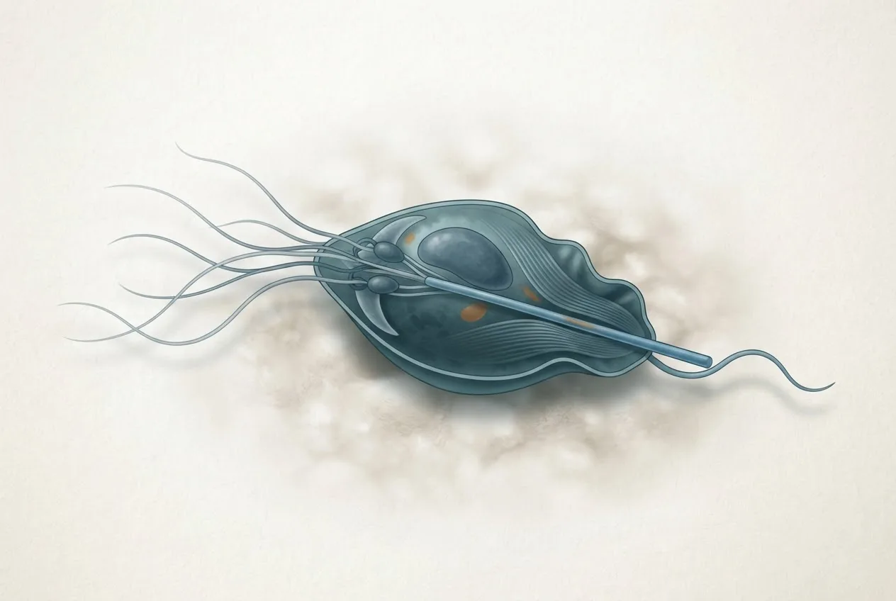
Tritrichomonas foetus belongs to the order Tritrichomonadida within the phylum Parabasalia [<a href="#ref-6">6</a>]. It is closely related to Trichomonas vaginalis, the human urogenital pathogen, but is genetically distinct. The parasite exists only as a trophozoite; no true cyst stage has been confirmed, although pseudocyst and cyst-like structures have been observed under stress conditions. The trophozoite is pyriform, 10-25 µm in length, and possesses three anterior flagella and a single posterior flagellum that forms an undulating membrane [<a href="#ref-7">7</a>]. The costa, a striated fiber of the mastigont system, is a unique cytoskeletal structure that provides structural support [<a href="#ref-7">7</a>]. Three costa-associated proteins (costain-1, costain-2, and costain-3) have been identified and localized using expansion microscopy and immunocytochemistry [<a href="#ref-7">7</a>]. The plasma membrane contains a diverse array of proteins involved in adhesion, nutrient acquisition, and immune evasion. The genome of T. foetus is approximately 148 Mb in size, organized into five chromosomes, and encodes over 41,000 protein-coding genes [<a href="#ref-6">6</a>]. Comparative membrane proteomics has identified common antigenic proteins across isolates, which may serve as targets for novel diagnostics or vaccines.
Life Cycle and Transmission
T. foetus replicates by binary fission under optimal conditions, but also undergoes multiple fission and DNA endoreplication under nutritional stress, leading to the formation of multinucleated or polyploid dormant forms. These resistant forms may facilitate survival in the environment. In cats, transmission occurs primarily via the fecal-oral route, through ingestion of trophozoites shed in diarrheic feces [<a href="#ref-1">1</a>, <a href="#ref-4">4</a>]. Contaminated litter boxes, food bowls, and water sources serve as fomites. The parasite can survive for several days in moist environments, including water and fecal material, likely through pseudocyst formation. Unlike in cattle, venereal transmission is not a significant route in cats [<a href="#ref-3">3</a>]. The incubation period is typically 1-2 weeks, after which trophozoites colonize the crypts of the large intestine, adhering to the mucosal epithelium via surface lectins and adhesins.
Epidemiology
Feline T. foetus infection has a global distribution. Prevalence rates vary widely depending on the population studied and diagnostic method used. In a large-scale study in Poland, 10.6% (170/1606) of cats with recent diarrhea tested positive by nested PCR, with the highest prevalence (14.9%) in cats under one year of age [<a href="#ref-2">2</a>]. In Nanchang, China, the prevalence was 40.5% (45/111) in cats, significantly higher than in dogs (5.8%) [<a href="#ref-1">1</a>]. A study in Henan Province, China, reported a prevalence of 6.01% (54/898) in pet cats, with higher rates in young, unweaned, unimmunized, and diarrheic individuals [<a href="#ref-4">4</a>]. In northeastern Brazil, the first report of T. foetus in cats documented infection in both diarrheic and asymptomatic animals [<a href="#ref-8">8</a>]. Risk factors include multi-cat housing, young age, and concurrent gastrointestinal infections [<a href="#ref-2">2</a>, <a href="#ref-4">4</a>]. The parasite has also been detected in raccoon dogs (Nyctereutes procyonoides) in China, suggesting a broader host range.
Clinical Signs and Pathogenesis
The primary clinical manifestation of feline T. foetus infection is chronic or recurrent large-bowel diarrhea, characterized by increased frequency, mucus, fresh blood, and a foul odor [<a href="#ref-2">2</a>, <a href="#ref-4">4</a>]. Tenesmus and fecal incontinence are common, and the perianal area may become inflamed [<a href="#ref-4">4</a>]. Affected cats often remain systemically well, with normal appetite and activity, unless co-infections are present [<a href="#ref-4">4</a>]. The pathogenesis involves adherence of trophozoites to the colonic epithelium, leading to disruption of tight junctions, increased mucosal permeability, and a neutrophilic inflammatory response. Bovine neutrophils kill T. foetus via trogocytosis, a process of membrane nibbling, and similar mechanisms are likely operative in cats. The parasite expresses ectophosphatases that may modulate host cell signaling and phosphate acquisition [<a href="#ref-9">9</a>]. Iron and calcium concentrations in the intestinal microenvironment influence the expression of immunogenic membrane proteins that activate macrophages through the TLR4/MyD88 pathway, promoting a pro-inflammatory cytokine response. Infection also alters the gut microbiota, with increased abundance of Bacteroidetes and Proteobacteria and decreased Firmicutes/Bacteroidetes ratio, which may contribute to diarrhea [<a href="#ref-4">4</a>].
Host Immune Response
The immune response to T. foetus in cats is incompletely understood. In cattle, infection elicits both humoral and cell-mediated responses, but the parasite employs immune evasion strategies such as antigenic variation and surface protein shedding [<a href="#ref-3">3</a>, <a href="#ref-10">10</a>]. In cats, serum IgG and mucosal IgA antibodies are produced, but their protective role is unclear. The parasite's membrane proteins can activate macrophages and neutrophils, leading to production of nitric oxide and reactive oxygen species. However, T. foetus can survive intracellularly within macrophages for short periods, potentially contributing to chronicity. Vaccine development for feline T. foetus is limited; most research has focused on bovine vaccines, including whole-cell killed, subunit, and cell fraction formulations [<a href="#ref-10">10</a>, <a href="#ref-11">11</a>]. A lyophilized, Quil-A-adjuvanted inactivated vaccine (Trichobovis) improved genital clearance and calving intervals in cattle, but no feline vaccine is commercially available.
Diagnostic Methods
Accurate diagnosis of feline T. foetus infection is essential for appropriate management. Several diagnostic modalities are available, each with advantages and limitations.
| Diagnostic Method | Target | Sensitivity | Specificity | Turnaround Time | Comments |
|---|---|---|---|---|---|
| Direct microscopy | Trophozoites | Low (30-50%) | Moderate | Immediate | Requires fresh, wet-mount preparation; motile trophozoites may be confused with other flagellates [<a href="#ref-2">2</a>]. |
| InPouch culture | Trophozoites | Moderate (60-80%) | High | 2-5 days | Requires viable organisms; false negatives due to delayed transport or prior treatment [<a href="#ref-12">12</a>]. |
| Conventional PCR | ITS1-5.8S rRNA-ITS2 | High (>90%) | High | 24-48 hours | Detects DNA; cannot distinguish live from dead organisms [<a href="#ref-1">1</a>, <a href="#ref-13">13</a>]. |
| Nested PCR | 18S rRNA | Very high (>95%) | Very high | 24-48 hours | Increased sensitivity over conventional PCR; gold standard for research [<a href="#ref-2">2</a>, <a href="#ref-5">5</a>]. |
| RT-qPCR | 5.8S rRNA | Very high (LOD 1 organism) | Very high | 2-4 hours | Targets RNA; can indicate viable organisms; suitable for pooled samples. |
| RPA-CRISPR/Cas12a | 18S rRNA | High (LOD 50 copies/µL) | High | 40 minutes | Point-of-care; visual lateral-flow readout; no thermocycler needed [<a href="#ref-5">5</a>]. |
The diagnostic algorithm for suspected feline T. foetus infection is presented below.
flowchart TD
 A["Cat with chronic large-bowel diarrhea"] --> B["Collect fresh fecal sample"]
 B --> C{"Direct wet-mount microscopy"}
 C -->|"Motile trophozoites seen"| D["Presumptive positive"]
 C -->|"No trophozoites seen"| E["Perform molecular test"]
 D --> F["Confirm with PCR or RT-qPCR"]
 E --> G{"Choose PCR method"}
 G --> H["Conventional PCR (ITS1-5.8S)"]
 G --> I["Nested PCR (18S rRNA)"]
 G --> J["RT-qPCR (5.8S rRNA)"]
 H --> K["Positive?"]
 I --> K
 J --> K
 K -->|"Yes"| L["Diagnose T. foetus infection"]
 K -->|"No"| M["Consider other causes: Giardia, Cryptosporidium, Toxoplasma, bacterial enteritis"]
 L --> N["Initiate treatment and hygiene measures"]
 M --> O["Further diagnostic workup"]
Sample collection and transport are critical. Fecal samples should be fresh (collected within 2 hours) and kept moist. For RT-qPCR, phosphate-buffered saline (PBS) or 0.9% sterile saline can be used as transport media without significant loss of sensitivity. Samples are stable for up to 5 days at 4°C and for 7-14 days at -20°C, although RNA degradation occurs at low organism concentrations. Pooling of up to five samples is feasible for RT-qPCR without loss of sensitivity, reducing costs in surveillance programs.
Treatment and Control
Ronidazole, a nitroimidazole, is the treatment of choice for feline T. foetus infection [<a href="#ref-3">3</a>]. The recommended dosage is 30 mg/kg orally once daily for 14 days. However, neurotoxicity (ataxia, seizures) has been reported, particularly at higher doses or in cats with renal impairment. Metronidazole is less effective, and resistance has been documented [<a href="#ref-3">3</a>]. Alternative therapies under investigation include plant-derived compounds such as citronella grass oil (Cymbopogon nardus), which demonstrated an EC50 of 0.4 µg/mL against T. foetus trophozoites in vitro. Proteasome inhibitors (e.g., bortezomib) and gold(I) compounds have shown activity in murine models, but their safety and efficacy in cats are unknown. Supportive care includes dietary modification (high-fiber or easily digestible diets) and probiotics to restore gut microbiota balance [<a href="#ref-4">4</a>].
Control measures focus on hygiene: daily cleaning of litter boxes with bleach or ethanol solutions, which are trophocidal at appropriate concentrations. Chlorhexidine gluconate is also effective in vitro (EC50 in micromolar range) and may be used for surface disinfection. Acetic acid and hydrogen peroxide have limited utility due to lower efficacy or tissue toxicity. Infected cats should be isolated from other cats, and all in-contact animals should be tested [<a href="#ref-2">2</a>, <a href="#ref-4">4</a>].
Disinfection and Prevention
T. foetus trophozoites are susceptible to common disinfectants. In vitro studies have shown that 70% ethanol and 0.5% sodium hypochlorite (bleach) kill trophozoites within 1 minute of contact. Chlorhexidine gluconate (0.05%) is also effective, with the added advantage of being safe for topical use on mucous membranes. Acetic acid (5%) and hydrogen peroxide (3%) require longer contact times and are less reliable. For environmental decontamination, thorough cleaning of organic material followed by application of bleach or ethanol is recommended. Litter boxes should be cleaned with hot water and detergent, then soaked in bleach solution for at least 10 minutes. Food and water bowls should be washed separately and disinfected. There is no commercially available vaccine for feline T. foetus, although bovine vaccines exist [<a href="#ref-10">10</a>]. Prevention relies on reducing fecal-oral transmission through good hygiene and early detection.
Future Directions
Recent advances in omics technologies have provided a chromosome-level genome assembly and comprehensive membrane proteome of T. foetus, enabling identification of novel drug targets and vaccine candidates [<a href="#ref-3">3</a>, <a href="#ref-6">6</a>]. The development of point-of-care diagnostics such as RPA-CRISPR/Cas12a assays offers rapid, field-deployable testing [<a href="#ref-5">5</a>]. Understanding the role of the gut microbiome in disease progression may lead to probiotic interventions [<a href="#ref-4">4</a>]. Further research is needed to elucidate the mechanisms of drug resistance and to develop safe, effective therapies for feline trichomonosis. The use of murine vaginal infection models for drug screening against T. foetus may accelerate discovery of new compounds.
Frequently Asked Questions
What is Tritrichomonas foetus in cats?
Tritrichomonas foetus is a protozoan parasite that infects the large intestine of cats, causing chronic diarrhea, tenesmus, and fecal incontinence [<a href="#ref-1">1</a>, <a href="#ref-2">2</a>].
How do cats become infected with Tritrichomonas foetus?
Cats acquire infection through the fecal-oral route by ingesting trophozoites from contaminated litter boxes, food bowls, or water sources [<a href="#ref-4">4</a>].
What are the clinical signs of feline trichomonosis?
The most common sign is chronic, foul-smelling, large-bowel diarrhea with mucus and fresh blood, often accompanied by tenesmus and perianal inflammation [<a href="#ref-2">2</a>, <a href="#ref-4">4</a>].
How is Tritrichomonas foetus diagnosed in cats?
Diagnosis is confirmed by molecular methods such as PCR or RT-qPCR on fresh fecal samples; direct microscopy and culture have lower sensitivity [<a href="#ref-2">2</a>, <a href="#ref-5">5</a>].
What is the treatment for Tritrichomonas foetus in cats?
Ronidazole (30 mg/kg orally once daily for 14 days) is the recommended treatment, but it carries a risk of neurotoxicity [<a href="#ref-3">3</a>].
Can Tritrichomonas foetus be prevented?
Prevention relies on strict hygiene, including daily disinfection of litter boxes with bleach or ethanol, and isolation of infected cats.
Is Tritrichomonas foetus zoonotic?
There is no evidence that feline T. foetus infects humans; the parasite is host-specific, with distinct genotypes in cats and cattle [<a href="#ref-3">3</a>].
What is the prognosis for cats with Tritrichomonas foetus?
With appropriate treatment, most cats resolve clinical signs, although reinfection can occur in multi-cat environments [<a href="#ref-2">2</a>, <a href="#ref-4">4</a>].
Related Clinical & Scientific Guides
- Tick-Borne Diseases in Dogs: Pathogens, Clinical Signs, Diagnosis, and Prevention
- Toxoplasmosis in Cats and the Risk of Brain Infection in Humans
- Dog Heartworm and Tick-Borne Disease Prevention