
Trichostrongylus axei: Abomasal Hairworm of Cattle and Sheep (Gastric Parasitism)
Taxonomic Classification and Morphology
Trichostrongylus axei (Cobbold, 1879) is a slender, hair-like nematode belonging to the family Trichostrongylidae, order Strongylida. It is a common abomasal parasite of cattle, sheep, and goats, and occasionally infects horses, pigs, and deer [1, 2]. The adult worms are small, measuring 3-5 mm in length (males) and 4-6 mm (females), with a filariform appearance that gives rise to the common name "hairworm" [3]. Male worms possess a well-developed copulatory bursa with two large lateral lobes and a small dorsal lobe, supported by characteristic ray patterns used for species identification [4]. The spicules are short, stout, and curved, often with a distinctive barb at the tip [5].
The cuticle bears fine longitudinal ridges (synlophe) that vary among trichostrongylid species but are less pronounced than in Ostertagia or Haemonchus [6]. Eggs of T. axei are thin-shelled, morulated, and measure approximately 75-95 µm by 35-46 µm, indistinguishable from those of other trichostrongylids by standard flotation [7]. Larvae must be cultured to the third stage (L3) for species-level identification using morphological keys [8, 9].
Host Range and Geographic Distribution
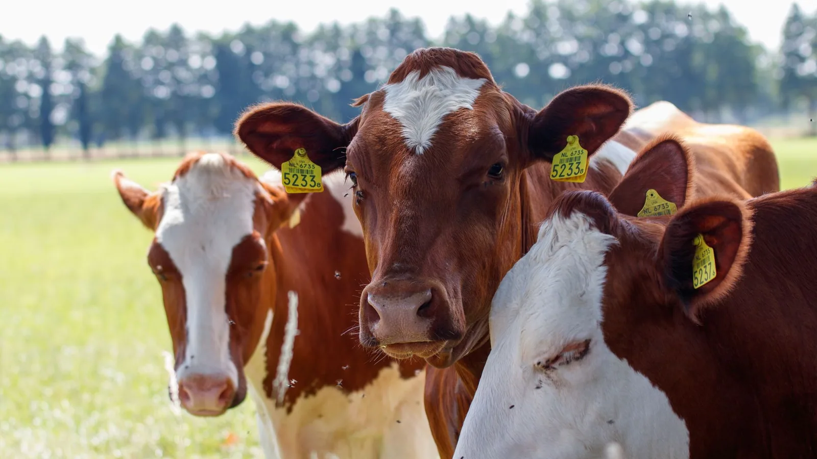
Trichostrongylus axei has a broad host range, primarily infecting ruminants. In cattle, it is frequently found co-infecting the abomasum along with Ostertagia ostertagi and Haemonchus placei [10, 11]. In sheep, it commonly coexists with Teladorsagia circumcincta and Trichostrongylus colubriformis [12, 13]. The parasite has a worldwide distribution, with prevalence reported in temperate and subtropical regions. In Europe, surveys have recorded T. axei in grazing dairy cattle in Germany and Austria [2], in dairy heifers in England and Wales [14], and in Austrian beef cattle [15]. In North America, it is prevalent in cattle from the United States and Canada [16, 17]. In Japan, it is part of the abomasal nematode complex in dairy cattle in Hokkaido [18]. In tropical regions such as The Gambia, T. axei has been identified in N'Dama cattle, often in a state of hypobiosis during dry seasons [19].
Life Cycle and Epidemiology
The life cycle of T. axei is direct, similar to other trichostrongylids. Adult worms reside in the abomasum, attached to the mucosal surface or lying free in the mucus [3]. Eggs pass out in feces, and under favorable field conditions (15-30°C, adequate moisture), they hatch to release first-stage larvae (L1) that feed on bacteria and develop through two molts to become infective third-stage larvae (L3) in 7-14 days [1, 20]. The L3 migrate onto herbage and are ingested by grazing animals. The exsheathment of L3 occurs in the rumen or abomasum, triggered by carbon dioxide and pH changes [1, 21]. After exsheathment, the larvae penetrate the abomasal mucosa and undergo two more molts to reach the adult stage. The prepatent period is approximately 18-21 days in cattle and sheep [3, 17].
Environmental factors profoundly influence transmission. Cool, moist conditions favor survival of L3 on pasture, while hot, dry conditions reduce larval longevity [2, 22]. In temperate regions, transmission peaks in spring and autumn. Hypobiosis (arrested larval development) is an important epidemiological feature observed in T. axei, though it is less pronounced than in O. ostertagi [11, 19]. In the Gambia, hypobiotic larvae have been recovered during the dry season, enabling the parasite to persist through unfavorable periods [19]. In northern Japan, seasonal fluctuations in worm burdens have been documented, with peaks in late autumn [18]. Co-infection with O. ostertagi can modulate the hypobiotic response; experimental studies have shown that concurrent infection with both species increases the proportion of inhibited O. ostertagi larvae, likely due to host immune interactions [11].
The following Mermaid diagram summarizes the life cycle of Trichostrongylus axei in cattle and sheep:
graph TD
 A[Adult worms in abomasum] --> B[Eggs in feces]
 B --> C[L1 in dung pat]
 C --> D[L2 in dung pat]
 D --> E[L3 infective on herbage]
 E --> F[Ingested by host]
 F --> G[Exsheathment in rumen/abomasum]
 G --> H[L4 penetrate mucosa]
 H --> I[L5 develop in lumen]
 I --> A
 H --> J[Hypobiosis arrested L4]
 J --> I
Pathogenesis and Clinical Signs
Trichostrongylus axei is a blood-feeding nematode, though its pathogenic impact is generally less severe than that of Haemonchus contortus or Ostertagia ostertagi [3]. The adult worms irritate the abomasal mucosa, causing a catarrhal inflammation with increased mucus production [11, 23]. Histologically, there is hyperplasia of gastric glands, infiltration of eosinophils and lymphocytes, and occasional sloughing of surface epithelium [11, 24]. The damage leads to a protein-losing gastroenteropathy, reduced digestive efficiency, and impaired growth.
In cattle, high burdens (e.g., >50,000 worms) can cause diarrhea, weight loss, submandibular edema ("bottle jaw"), and reduced milk yield in dairy cows [2, 25]. In young calves, experimental infections have shown reduced appetite and growth rates [17]. In sheep, the clinical syndrome is similar to that caused by Teladorsagia circumcincta, with diarrhea, poor fleece quality, and loss of condition [12]. The parasite can contribute to the "parasitic gastroenteritis" complex, especially when co-occurring with other species.
In contrast to Ostertagia ostertagi, T. axei does not typically cause the raised "Morocco leather" lesions characteristic of ostertagiosis type I, but rather diffuse inflammatory changes [11, 24]. Experimental single-species infections in calves have demonstrated that T. axei induces an eosinophilic infiltration and mucosal thickening, but without the severe nodular hyperplasia seen with O. ostertagi [11].
Diagnosis
Diagnosis of T. axei infection relies on detection of eggs in feces using standard flotation techniques (e.g., saturated sodium chloride, specific gravity 1.20) [26]. Because eggs are morphologically indistinguishable from those of other trichostrongylids, identification to species requires larval culture and differentiation of third-stage larvae (L3) using morphological keys [8, 27]. The Baermann technique is not required for abomasal nematodes, as larvae are not pulmonary stages. However, for differential diagnosis, fecal egg count reduction tests (FECRT) can be used to monitor anthelmintic efficacy, and identification of cultured larvae to species can improve interpretation of resistance patterns [27, 28].
Necropsy and abomasal worm counts remain the gold standard for quantifying burdens [3, 5]. The abomasum is opened, washed, and the contents and digest of the mucosa are examined for adult worms. Speciation of adult males relies on spicule morphology and bursal ray patterns [5]. For field surveys, composite sedimentation techniques are used [14, 15].
Molecular diagnostics are increasingly employed for species-specific detection and quantification. PCR-based nemabiome sequencing targeting the ITS-2 rDNA region can identify T. axei from fecal samples with high specificity [29, 30]. This nemabiome approach allows simultaneous profiling of the entire gastrointestinal nematode community and is particularly useful for mixed infections.
Treatment and Control
Anthelmintic treatment for T. axei follows the same guidelines as for other abomasal nematodes. Macrocyclic lactones (e.g., ivermectin, eprinomectin), benzimidazoles (e.g., fenbendazole, albendazole), and imidazothiazoles (e.g., levamisole) are effective [5, 7, 31, 32, 33]. Ivermectin and eprinomectin have demonstrated high efficacy against adult and larval stages [5, 31]. A sustained-release bolus of ivermectin has shown efficacy against T. axei in cattle [33]. However, resistance to benzimidazoles and macrocyclic lactones is emerging in trichostrongylid populations globally, and FECRT should be performed to confirm product efficacy in a herd or flock [27, 34].
Control strategies integrate pasture management, stocking density reduction, and strategic anthelmintic treatments to minimize pasture contamination. Rotational grazing can reduce exposure to infective larvae, though the effect may be less pronounced for T. axei than for other species due to its shorter survival on pasture [35]. In sheep, combining grazing with cattle or horses can take advantage of host specificity; T. axei is a generalist, but cross-grazing with resistant species may still reduce overall larval burden [36]. Biological control using nematophagous fungi (e.g., Duddingtonia flagrans) has been evaluated for T. axei and other trichostrongylids, with some success in reducing larval numbers on pasture [37, 38]. Immunological control is not yet available, but vaccine development against Trichostrongylus colubriformis has shown some cross-protection in experimental models.
The following table summarizes recommended diagnostic and control measures:
| Aspect | Recommendation | Key References |
|---|---|---|
| Fecal examination | Flotation with saturated NaCl; egg counts using McMaster technique | [26] |
| Larval differentiation | Culture at 25°C for 7-10 days; morphological keys | [8, 27] |
| Molecular detection | ITS-2 nemabiome PCR for species composition | [29, 30] |
| Anthelmintic class | Macrocyclic lactone, benzimidazole, levamisole; use FECRT to confirm efficacy | [5, 31, 32, 33, 34] |
| Pasture management | Rotational grazing; rest periods >6 weeks; mixed-species grazing | [35, 36] |
| Biocontrol | Nematophagous fungi (D. flagrans) on pasture; preliminary results | [37, 38] |
| Resistance monitoring | Annual FECRT; culture and species-level differentiation | [27, 34] |
Drug Resistance
Anthelmintic resistance in T. axei has been reported, although less extensively than in Haemonchus contortus or Teladorsagia circumcincta. Resistance to benzimidazoles has been documented in sheep flocks in several countries, and macrocyclic lactone resistance is suspected but not as well characterized [27, 34]. The FECRT, with a reduction threshold of <95% (mean) and lower 95% confidence interval <90%, is the field standard for detecting resistance [27]. When resistance is suspected, larval culture and differentiation are essential to attribute resistance to a particular species, as mixed infections are common [27].
Public Health Considerations
Trichostrongylus axei is primarily a veterinary parasite and rarely infects humans. Accidental human infections have been reported following ingestion of contaminated raw vegetables or water, but such cases are sporadic and of minor clinical significance. No specific public health control measures are required beyond standard food hygiene. This article focuses strictly on veterinary aspects.
Conclusion
Trichostrongylus axei is a widely distributed abomasal nematode of cattle and sheep that contributes to production losses through impaired growth, reduced milk yield, and predisposition to parasitic gastroenteritis. Its small size and morphological similarity to other trichostrongylids necessitate careful larval differentiation and, increasingly, molecular nemabiome approaches for species-level diagnosis. Anthelmintic resistance is emerging, making integrated control strategies and regular efficacy testing imperative.
References
[1] Hertzberg H, Huwyler U, Kohler L et al. Kinetics of exsheathment of infective ovine and bovine strongylid larvae in vivo and in vitro. Parasitology. 2002. https://pubmed.ncbi.nlm.nih.gov/12166522/
[2] Rehbein S, Hamel D, Lackerschmid J et al. Multispecies helminth parasitism of grazing dairy cows in Germany and Austria, examined in the housing period. Vet Parasitol Reg Stud Reports. 2023. https://pubmed.ncbi.nlm.nih.gov/37068863/
[3] Cardoso CP, Silva BF, Trinca LA et al. Resistance against gastrointestinal nematodes in Crioulo Lageano and crossbred Angus cattle in southern Brazil. Vet Parasitol. 2013. https://pubmed.ncbi.nlm.nih.gov/23177359/
[4] Rehbein S, Visser M, Winter R. [Helminth infection in cattle from Schleswig-Holstein (Germany) after one grazing season]. Berl Munch Tierarztl Wochenschr. 2003. https://pubmed.ncbi.nlm.nih.gov/12592928/
[5] Yazwinski TA, Johnson EG, Thompson DR et al. Nematocidal efficacy of eprinomectin, delivered topically, in naturally infected cattle. Am J Vet Res. 1997. https://pubmed.ncbi.nlm.nih.gov/9185967/
[6] Ndao M, Pandey VS, Zinsstag J et al. Helminth parasites and hypobiosis of nematodes in N'Dama cattle during the dry season in The Gambia. Vet Parasitol. 1995. https://pubmed.ncbi.nlm.nih.gov/8644452/
[7] Williams JC, Plue RE. Efficacy of ivermectin delivered from a sustained-release bolus against inhibited early fourth-stage larvae of Ostertagia ostertagi and other nematodes in cattle. Am J Vet Res. 1992. https://pubmed.ncbi.nlm.nih.gov/1524310/
[8] Fukumoto S, Etani K, Toi K et al. Epidemiology of abomasal nematodes of dairy cattle in Hokkaido, northern Japan. Nihon Juigaku Zasshi. 1990. https://pubmed.ncbi.nlm.nih.gov/2348599/
[9] Marnu W, Wintersteller E, Prosl H. Monthly and seasonal fluctuations in abomasal nematode worm burden of naturally infected cattle in Austria. Vet Parasitol. 1987. https://pubmed.ncbi.nlm.nih.gov/3564352/
[10] Vercruysse J, Dorny P, Berghen P et al. Abomasal parasitism in dairy cows in Belgium. Vet Parasitol. 1986. https://pubmed.ncbi.nlm.nih.gov/3564332/
[11] Snider TG 3rd, Williams JC, Karns PA et al. Synergistic influence of Ostertagia ostertagi and Trichostrongylus axei on Ostertagia ostertagi larval inhibition and abomasal lesions in cattle. Am J Vet Res. 1985. https://pubmed.ncbi.nlm.nih.gov/4037502/
[12] Snider TG 3rd, Williams JC, Knox JW et al. Persistence of dead Ostertagia ostertagi in the abomasal mucosa following anthelmintic treatment. Vet Rec. 1985. https://pubmed.ncbi.nlm.nih.gov/3838393/
[13] Hong C, Lancaster MB, Michel JF. Worm burdens of dairy heifers in England and Wales. Vet Rec. 1981. https://pubmed.ncbi.nlm.nih.gov/7197843/
[14] Lyons ET, Tolliver SC, Drudge JH et al. Efficacy of levamisole against abomasal nematodes and lungworms in dairy calves: preliminary tests indicating reduced activity for Ostertagia ostertagi. Am J Vet Res. 1981. https://pubmed.ncbi.nlm.nih.gov/7271028/
[15] Lyons ET, Tolliver SC, Drudge JH et al. Ivermectin: controlled test of anthelmintic activity in dairy calves with emphasis on Dictyocaulus viviparus. Am J Vet Res. 1981. https://pubmed.ncbi.nlm.nih.gov/6455949/
[16] Williams JC, Knox JW, Baumann BA et al. Further studies on the efficacy of fenbendazole against inhibited larvae of Ostertagia ostertagi. Vet Rec. 1981. https://pubmed.ncbi.nlm.nih.gov/7257120/
[17] Herlich H. Infection and immune kinetics of Trichostrongylus axei in calves. Am J Vet Res. 1979. https://pubmed.ncbi.nlm.nih.gov/475128/
[18] Slocombe JO. Abomasal nematodes in cattle in Ontario. Can J Comp Med. 1974. https://pubmed.ncbi.nlm.nih.gov/4272952/
[19] Ross JG, Purcell DA, Todd JR. Experimental infections of calves with Trichostrongylus axei investigations using abomasal cannulae. Res Vet Sci. 1969. https://pubmed.ncbi.nlm.nih.gov/4896772/
[20] Borkowski EA, Redman EM, Chant R et al. Comparison of ITS-2 rDNA nemabiome sequencing with morphological identification to quantify gastrointestinal nematode community species composition in small ruminant feces. Vet Parasitol. 2020. https://pubmed.ncbi.nlm.nih.gov/32446107/
[21] Knoll S, Dessì G, Tamponi C et al. Practical guide for microscopic identification of infectious gastrointestinal nematode larvae in sheep from Sardinia, Italy, backed by molecular analysis. Parasit Vectors. 2021. https://pubmed.ncbi.nlm.nih.gov/34583765/
[22] van Wyk JA, Mayhew E. Morphological identification of parasitic nematode infective larvae of small ruminants and cattle: a practical lab guide. Onderstepoort J Vet Res. 2013. https://pubmed.ncbi.nlm.nih.gov/23718204/
[23] Andronicos NM, McNally J, Kotze AC et al. Trichostrongylus colubriformis larvae induce necrosis and release of IL33 from intestinal epithelial cells in vitro: implications for gastrointestinal nematode vaccine design. Int J Parasitol. 2012. https://pubmed.ncbi.nlm.nih.gov/22366550/
[24] Colvin AF, Walkden-Brown SW, Knox MR. Role of host and environment in mediating reduced gastrointestinal nematode infections in sheep due to intensive rotational grazing. Vet Parasitol. 2012. https://pubmed.ncbi.nlm.nih.gov/21924833/
[25] Fan B, Bryant RH, Greer AW. Behavioural patterns of grazing ram lambs exposed to gastrointestinal nematode parasitism. Vet Parasitol. 2026. https://pubmed.ncbi.nlm.nih.gov/42000447/
[26] Leathwick D, Green P, Bouchet C et al. The faecal egg count reduction test: Will identification of larvae to species improve its utility? Int J Parasitol Drugs Drug Resist. 2025. https://pubmed.ncbi.nlm.nih.gov/40158261/
[27] Dai HW, Xu Q, Wang BB et al. In Vitro, in Vivo, and Interaction Studies of Nematophagous Fungus Arthrobotrys thaumasia (Monacrosporium thaumasium) with the Larvae of Trichostrongylides of Sheep. J Parasitol. 2017. https://pubmed.ncbi.nlm.nih.gov/28953417/
[28] Wang BB, Liu W, Chen MY et al. Isolation and Characterization of China Isolates of Duddingtonia flagrans, a Candidate of the Nematophagous Fungi for Biocontrol of Animal Parasitic Nematodes. J Parasitol. 2015. https://pubmed.ncbi.nlm.nih.gov/25978186/
[29] Rocha LOD, Lemos GCDS, Vieira IJC et al. Chemical characterization and in vitro biological activity of Cymbopogon citratus extracts against Haemonchus spp. and Trichostrongylus spp. nematodes from sheep. Parasitology. 2020. https://pubmed.ncbi.nlm.nih.gov/32741411/
[30] de Godoi SN, Gressler LT, de Matos AFIM et al. Eucalyptus oil nanoemulsions against eggs and larvae of Haemonchus contortus. Exp Parasitol. 2022. https://pubmed.ncbi.nlm.nih.gov/35985513/
[31] George MM, Lopez-Soberal L, Storey BE et al. Motility in the L3 stage is a poor phenotype for detecting and measuring resistance to avermectin/milbemycin drugs in gastrointestinal nematodes of livestock. Int J Parasitol Drugs Drug Resist. 2018. https://pubmed.ncbi.nlm.nih.gov/29274827/
[32] Costa-Junior LM, Silva CR, Soares AMS et al. Assessment of biophysical properties of Haemonchus contortus from different life cycle stages with atomic force microscopy. Ultramicroscopy. 2020. https://pubmed.ncbi.nlm.nih.gov/31707231/
[33] Acevedo-Ramírez PMDC, Hallal-Calleros C, Flores-Pérez I et al. Anthelmintic effect and tissue alterations induced in vitro by hydrolysable tannins on the adult stage of the gastrointestinal nematode Haemonchus contortus. Vet Parasitol. 2019. https://pubmed.ncbi.nlm.nih.gov/30736941/
[34] Andre WP, Ribeiro WL, Cavalcante GS et al. Comparative efficacy and toxic effects of carvacryl acetate and carvacrol on sheep gastrointestinal nematodes and mice. Vet Parasitol. 2016. https://pubmed.ncbi.nlm.nih.gov/26872928/
[35] Dos Santos MC, Amarante MRV, Amarante AFT. Is there competition between Haemonchus contortus and Haemonchus placei in a pasture grazed by only sheep? Vet Parasitol. 2020. https://pubmed.ncbi.nlm.nih.gov/32065932/
[36] Santos MC, Amarante MRV, Amarante AFT. Establishment of co-infection and hybridization of Haemonchus contortus and Haemonchus placei in sheep. J Helminthol. 2019. https://pubmed.ncbi.nlm.nih.gov/30176949/ *** Disclaimer: This article is for educational and informational purposes only. It is not intended to substitute for professional veterinary advice, diagnosis, treatment, or regulatory guidance. Always consult a licensed veterinarian or qualified specialist regarding animal health, disease diagnosis, and therapeutic decisions.