
Strongylus vulgaris: Equine Strongyle Arteritis and Colic, Pathogenesis, Diagnosis, and Control
Introduction
Strongylus vulgaris, commonly referred to as the equine bloodworm, is historically the most pathogenic intestinal helminth parasite of horses [1, 20]. This large strongyle nematode causes significant vascular damage through its larval migration within the mesenteric arterial system, leading to verminous arteritis, thrombosis, and non-strangulating intestinal infarction (NSII) [2, 3, 4]. Colic episodes attributable to S. vulgaris infection range from mild transient discomfort to fatal intestinal ischemia. Despite decades of intensive anthelmintic use that dramatically reduced its prevalence in managed populations, recent epidemiological evidence confirms that S. vulgaris remains enzootic across many regions and continues to cause serious abdominal disease [5, 1, 6, 19]. This article provides a comprehensive clinical reference on the etiology, epidemiology, pathogenesis, clinical presentation, diagnostic modalities, treatment, and control of Strongylus vulgaris equine strongyle arteritis colic horse.
Etiology and Life Cycle
Strongylus vulgaris is a member of the family Strongylidae (subfamily Strongylinae) and is among the largest of the equine strongyles. Adult worms reside in the large intestine, primarily the cecum and ventral colon, where they feed on blood. The life cycle is monoxenous and involves both free-living and parasitic stages.
Eggs are passed in feces. Under favorable environmental conditions (temperatures above 15 deg C, adequate moisture), first-stage larvae (L1) hatch, molt to second-stage larvae (L2), and then to third-stage infective larvae (L3) within approximately one to two weeks [7]. Horses acquire infection by ingesting L3 larvae from contaminated pasture. The prepatent period is approximately six to seven months, a prolonged interval that historically permitted the parasite to survive on pastures despite intermittent anthelmintic treatments.
After ingestion, L3 larvae exsheath in the small intestine, penetrate the intestinal mucosa, and migrate within the submucosa to reach small arterioles. From there, they travel via the arterial system to the cranial mesenteric artery and its major branches. Larvae undergo two molts (L3 to L4, L4 to L5) within the arterial intima over several months. Young adult worms (L5) eventually return to the intestinal lumen via small arterioles, completing the cycle [1, 20].
Epidemiology
The global distribution of S. vulgaris has shifted markedly in response to anthelmintic practices. In the mid-20th century, infection was nearly universal in domestic horses. Frequent use of macrocyclic lactones (ivermectin, moxidectin) and other broad-spectrum drugs in subsequent decades drove prevalence down to very low levels in many managed herds [1, 33, 38]. However, the parasite has never been eradicated, and recent surveys document a clear resurgence in areas where selective therapy replaced routine interval deworming.
A large-scale molecular survey using ITS-1 PCR on horses in Al-Najaf Province, Iraq, reported a molecular prevalence of 30.0% (30/100), with significantly higher infection in adults (42.0%) than foals (27.0%) and peak detection in winter months [2]. In Alberta, Canada, nemabiome sequencing of feral horses (naive to deworming) revealed an exceptional S. vulgaris prevalence of 85.91% (128/149), with species-specific fecal egg counts strongly influenced by host age and season [5]. A retrospective postmortem study in Alberta further confirmed cranial mesenteric arteritis attributable to S. vulgaris in a notable proportion of equine necropsies from 2010 to 2022 [3].
In Europe, a Swedish national survey conducted ten years after prescription-only anthelmintic regulations found an individual-level prevalence of 28% and farm-level prevalence of 61%, with a threefold increase compared with 1999 estimates. In Germany, serological testing for anti-S. vulgaris antibodies using an ELISA detected 21.2% of horses as seropositive, far exceeding the 1.3% molecular detection rate by larval culture PCR [6]. This discrepancy strongly suggests that many horses are exposed to migrating larvae that do not reach patency due to regular treatments. In Prince Edward Island, Canada, 60% of horses tested positive for S. vulgaris antibodies. Similarly, a German case-control study of clinic patients found a seroprevalence of 32.3%.
Key risk factors consistently identified across studies include low age (young horses have higher egg counts), increasing pasture access, longer intervals since last deworming, and use of selective therapy based on fecal egg count (FEC) alone rather than combined FEC and larval culture [6, 19]. Feral and untreated populations serve as reservoirs that can transmit infection to neighboring domestic herds [5].
Pathogenesis
Larval Migration and Arterial Damage
The pathognomonic lesion of S. vulgaris infection is verminous arteritis of the cranial mesenteric artery and its branches, caused by the intimal migration of L4 and L5 larvae. Larvae induce mechanical injury to the endothelium, triggering platelet aggregation, fibrin deposition, and thrombus formation [4, 11, 26]. Ultrastructural studies have demonstrated that larvae burrow through the intima and subjacent media, creating tunnels lined by fibrin, neutrophils, and eosinophils [4, 11]. This process results in intimal thickening, smooth muscle hyperplasia, fibrosis, and aneurysm formation [4].
The organized thrombi associated with larval tunnels can occlude the arterial lumen partially or completely. Thromboemboli, dislodged from these vegetative lesions, travel distally to occlude smaller intestinal arteries, causing focal ischemia. The resulting lesion is termed non-strangulating intestinal infarction (NSII), a condition distinct from strangulating obstructions and strongly associated with S. vulgaris infection [1]. NSII most commonly affects the cecum, colon, or ileum and may present as single or multiple infarcts.
Impact on Intestinal Perfusion and Colic
Ischemic injury to the intestinal wall compromises mucosal barrier function, leading to peritonitis (often septic), endotoxemia, and systemic inflammation. The clinical manifestation of colic in S. vulgaris infection is primarily attributable to NSII [1]. A prospective study of colic and non-colic equine clinic patients, while not identifying a direct statistical association between serostatus and colic, did find that recent anthelmintic treatment (within seven days) was associated with a 2.4-fold increased risk of colic signs. This observation may reflect the acute death and disintegration of adult worms in the arterial system following treatment, with consequent thromboembolism.
Hemostatic and Immunologic Alterations
Natural infection with S. vulgaris in foals is associated with measurable changes in hemostatic indices, including increased plasma concentrations of D-dimer and thrombin-antithrombin complexes, indicative of enhanced coagulation and fibrinolysis. Cytokine profiling has shown a Th1/TNF-alpha bias in infected animals, with modulation of the Th2 (IL-4) response depending on larval stage [23, 37]. Antibody responses against larval excretory-secretory antigens develop early and form the basis of serological diagnostics [23, 43].
Clinical Signs
Clinical signs of Strongylus vulgaris equine strongyle arteritis colic horse depend on the stage of infection and the severity of arterial damage.
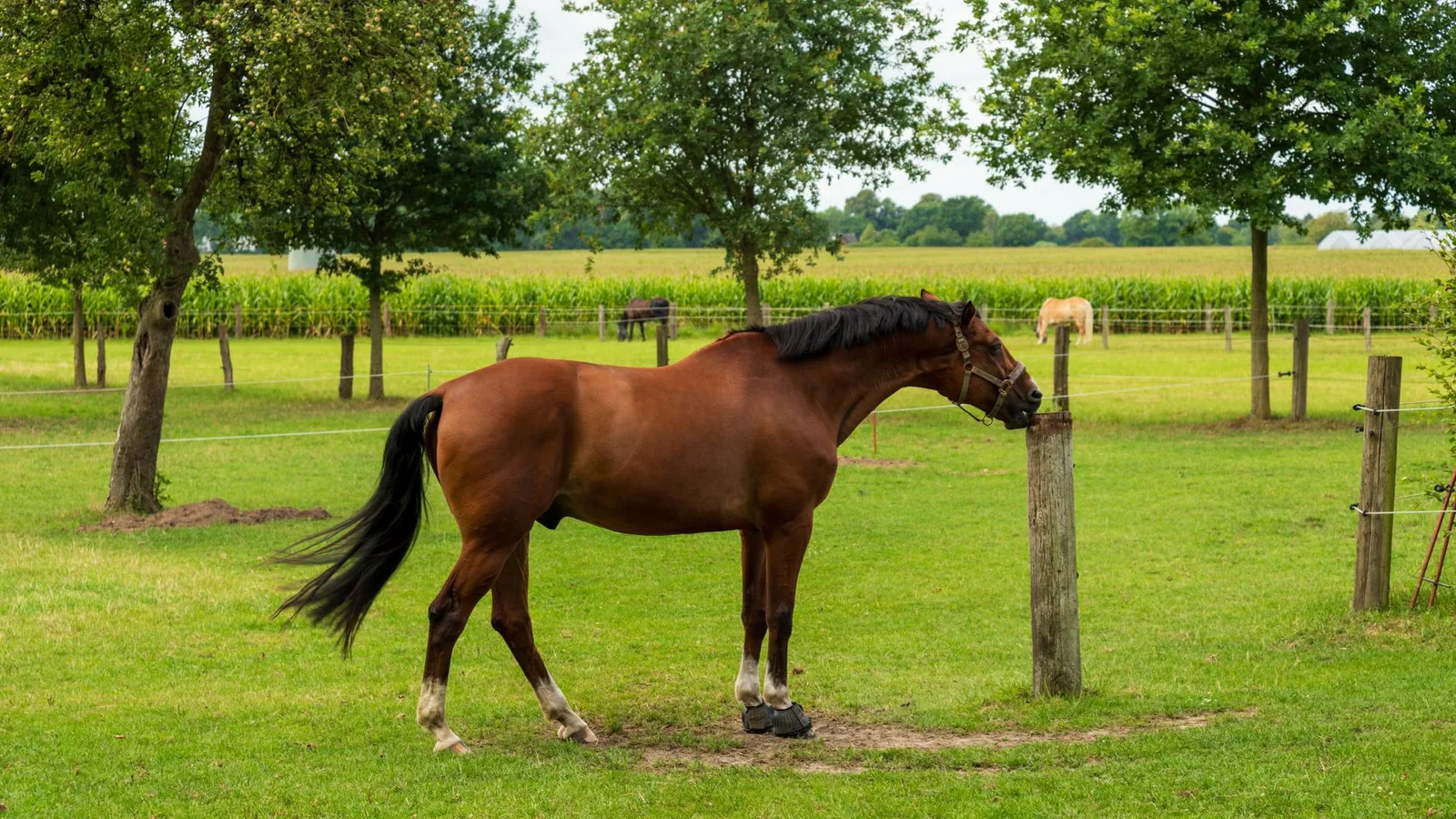
Colic manifestations: Recurrent, mild to moderate colic episodes (lethargy, inappetence, flank watching, pawing, rolling) are common in horses with patent infections and ongoing NSII. Acute severe colic with signs of shock occurs in cases of massive intestinal infarction.
Non-colonic signs: Fever, depression, weight loss, poor coat quality, and peripheral edema may accompany systemic inflammation. Chronically infected horses may exhibit unthriftiness and reduced performance.
Prepatent infection: The most clinically dangerous period is the prepatent phase (approximately months 2 to 5 post infection), when migrating larvae are most numerous in the mesenteric arteries and thromboembolic risk is highest. Diagnostic tools for this stage remain limited [1, 43].
Pathology
Macroscopic Findings
At necropsy, the cranial mesenteric artery and its major branches (ileocecocolic artery, cecal artery, colic arteries) show profound enlargement, thickening, and firmness on palpation. The arterial lumen contains adherent, organizing thrombi that are often vermiform and may contain remnants of larvae. Aneurysmal dilation of the cranial mesenteric artery (historically termed "aneurysm of the anterior mesenteric artery") is a classic but less common finding.
In horses with NSII, affected segments of cecum or colon are dark, edematous, and friable, with a clear demarcation between viable and necrotic tissue. The overlying serosa may be dull with fibrinous adhesions. Peritoneal fluid is typically serosanguinous to purulent.
Histologic and Ultrastructural Findings
Histologic examination of affected arteries shows intimal hyperplasia, fibrosis, and infiltration by eosinophils, macrophages, and lymphocytes. Fibrinoid necrosis of the media may be present. Larvae, when identified, are surrounded by a zone of necrosis and inflammatory cells [4, 11]. Ultrastructurally, the cuticular surface of the larvae interacts closely with the damaged intima, and thrombi incorporate platelets, fibrin, and abundant cellular debris [11, 26].
Diagnosis
Diagnosis of S. vulgaris infection relies on a combination of parasitological, serological, and molecular methods. No single test is optimal for all stages of infection. The diagnostic approach should account for the high sensitivity of serology for exposure and the low sensitivity of copromicroscopy for low-level patent infections.
Parasitological Methods
Fecal egg count (FEC): Quantitative FEC (e.g., McMaster or Mini-FLOTAC) detects eggs of the strongyle group but cannot differentiate S. vulgaris from cyathostomins. It is useful for estimating total strongyle egg shedding and for identifying high-shedding horses (>500 eggs per gram).
Larval culture and morphological identification: Third-stage larvae (L3) from coprocultures can be identified by light microscopy based on specific features (e.g., long, slightly curved tail with a pointed tip for S. vulgaris). However, morphological identification has poor inter-rater reliability when compared with molecular methods. A comparative study in Germany found that morphological examination detected S. vulgaris in 0% of samples, whereas PCR identified 0.8%. Dice-PCR and sodium nitrate flotation sedimentation time also affect recovery.
Molecular Methods
Conventional and real-time PCR: Species-specific PCR targeting the ITS-1 region (290 bp) or ITS-2 region provides reliable detection of S. vulgaris DNA from egg pellets, fecal samples, or L3 cultures [2, 10, 14, 18, 41]. Real-time quantitative PCR (qPCR) enables semi-quantification and has high analytical sensitivity [14, 18]. ITS-2 gene sequencing from individual worms in Egypt showed >95% identity with German, Chinese, and Turkish isolates.
High-resolution melting (HRM) PCR: This method can differentiate among S. vulgaris, S. edentatus, S. equinus, and S. asini without sequencing [6, 10].
Nemabiome deep amplicon sequencing: Metabarcoding of the internal transcribed spacer 2 (ITS-2) or cytochrome c oxidase subunit I (COI) gene regions from pooled fecal DNA allows comprehensive characterization of strongyle communities [5, 8, 22]. This approach has revealed that Strongylus spp. are more abundant in untreated equine populations, while cyathostomins dominate in regularly treated herds [8]. The COI marker provides superior species-level resolution for some species [8].
Serological Methods
ELISA for anti-S. vulgaris antibodies: Several commercial and in-house ELISAs detect IgG(T) antibodies against larval antigens. These assays have moderate sensitivity (approximately 43%) but high specificity (approximately 96%) at conservative cut-offs and reliably indicate exposure [6, 43]. Seropositivity is consistently higher than copromicroscopic or PCR detection, reflecting exposure to prepatent or aborted larval stages [6, 12, 34]. A study on Danish horses demonstrated a significant association between seropositivity and selective therapy practices.
Diagnostic Imaging
Transabdominal ultrasonography: In skilled hands, cranial mesenteric arteritis can be visualized as a thickened, dilated, hypoechoic arterial wall in the dorsal abdomen [1]. This is not a routine clinical tool and requires advanced training.
Exploratory laparotomy or laparoscopy: Direct visualization of infarcted intestine and thickened mesenteric arteries remains the definitive intra vitam assessment for NSII [1].
Comparison of Diagnostic Methods
| Method | Target | Stage Detected | Sensitivity (relative) | Specificity | Interpretation | |, - |, - |, - |, - |, - |, - | | Fecal egg count (McMaster) | Strongyle eggs | Adult (patent) | Low for low shedders | Low (species not identified) | Quantifies total strongyle burden | | Larval culture + morphology | L3 larvae | Adult (patent) | Very low | Moderate | Species identification; poor reliability | | Conventional/real-time PCR | DNA (ITS-1, ITS-2) | All parasitic stages | High | High | Confirms S. vulgaris; species specific | | HRM PCR | DNA (28S) | All parasitic stages | High | High | Multiplex species differentiation | | Nemabiome (ITS-2 or COI) amplicon sequencing | DNA | All parasitic stages | Very high | High | Community-level species profiling | | Anti-S. vulgaris ELISA | Host antibodies | Prepatent and patent | Moderate | High | Indicates exposure; cannot distinguish active from past infection |
Diagnostic Decision Tree
The following Mermaid diagram presents a clinical diagnostic workflow for suspected Strongylus vulgaris equine strongyle arteritis colic horse.
flowchart TD
 A[Equine patient with colic, fever, or unexplained peritonitis] --> B{Strongyle FEC and larval culture};
 B --> C["FEC positive: Strongyle eggs detected"];
 B --> D["FEC negative: No eggs detected"];
 C --> E[Larval culture with molecular PCR / HRM];
 D --> F{"Serology: anti-S. vulgaris ELISA"};
 E --> G[S. vulgaris DNA positive];
 E --> H[S. vulgaris DNA negative];
 G --> I[Confirm diagnosis of patent S. vulgaris infection];
 H --> J[Infection likely cyathostomin or low-level large strongyle];
 F --> K[Seropositive];
 F --> L[Seronegative];
 K --> M[Strongly suggests recent larval exposure, possible prepatent infection];
 L --> N[Low probability of S. vulgaris infection];
 M --> O[Consider nemabiome or RT-PCR on fecal DNA for confirmation];
 I --> P["Treatment: moxidectin or ivermectin based on age and risk"];
 P --> Q[Post-treatment FEC + larval culture at 14 days to confirm clearance];
Antenortem Diagnosis of Non-Strangulating Intestinal Infarction
Presently, no non-invasive test definitively diagnoses NSII. A combination of clinical suspicion, seropositivity, elevated peritoneal fluid lactate and protein, and typical ultrasonographic findings supports the diagnosis. Exploratory laparotomy remains the gold standard [1].
Treatment
The goals of treatment for S. vulgaris infection are two-fold: elimination of adult worms from the intestinal lumen (patent infection) and killing of migrating larvae in the arterial system (prepatent infection) to halt progressive vascular damage.
Anthelmintic Options
Macrocyclic lactones (MLs): Ivermectin (200 mcg/kg, oral paste or injection) and moxidectin (400 mcg/kg, oral gel) are highly efficacious against both adults and arterial L4 larvae of S. vulgaris [44, 45, 46]. A controlled trial in Brazil found ivermectin and moxidectin to have high effectiveness (>98% FEC reduction), whereas fenbendazole showed resistance. The strongyle egg reappearance period after moxidectin treatment is longer than after ivermectin (approximately eight weeks vs. four weeks) in some populations, though this may vary regionally.
Pyrantel: Pyrantel pamoate (6.6 mg/kg, oral) is effective against adult S. vulgaris but has poor penetration into arterial larvae. It is not recommended for treatment of suspected larval arteritis.
Benzimidazoles: Fenbendazole at 10 mg/kg daily for five consecutive days is sometimes used for larvicidal therapy, but widespread resistance in cyathostomins and variable efficacy against S. vulgaris limit its utility.
Ivermectin + praziquantel: Combination products are not necessary for S. vulgaris but may be used if concurrent tapeworm infection is suspected.
Considerations for NSII Cases
In horses presenting with acute colic and confirmed or highly suspected NSII, anthelmintic therapy should be administered with caution. Rapid death of large numbers of larvae in the arteries may exacerbate thromboembolic events. Supportive care includes fluid therapy, anti-endotoxemic treatment (e.g., polymyxin B, flunixin meglumine), and surgical intervention (resection of infarcted intestine) if indicated.
Anthelmintic Resistance
Critically, S. vulgaris has not been reported to develop resistance to any anthelmintic class [1, 44]. This is in stark contrast to cyathostomins, which exhibit widespread benzimidazole and emerging macrocyclic lactone resistance. However, validated methods for species-specific efficacy evaluation in S. vulgaris are currently lacking, and continued vigilance is warranted [1, 35].
Control and Prevention
The re-emergence of S. vulgaris in many managed populations necessitates a re-evaluation of control strategies. The core principles of modern parasite control in horses apply, with specific adaptations for this pathogenic species.
Targeted Selective Therapy (TST)
Also known as selective therapy, TST involves treating only horses with FEC above a threshold (typically >200 eggs per gram) rather than deworming all horses at fixed intervals. While TST reduces drug selection pressure on cyathostomins, it has been associated with increased S. vulgaris risk in several studies [19, 17]. In Sweden, farms using FEC-only TST had a 2.9-fold higher odds of S. vulgaris infection than farms combining FEC with larval culture or practicing regular interval deworming. Accordingly, TST programs should incorporate periodic larval culture or molecular screening for S. vulgaris at the farm level.
Quarantine and Biosecurity
Quarantining new arrivals and treating them with an effective larvicidal drug (moxidectin or ivermectin) before introduction to the resident herd significantly reduces the risk of introducing S. vulgaris [1]. A negative fecal egg count alone should not rule out the parasite because prepatent infections produce no eggs.
Pasture Management
- Remove feces from pastures at least twice weekly during the grazing season.
- Rotate pastures or rest pastures for at least six months (longer in cool climates) to reduce L3 survival.
- Co-graze with cattle or sheep (which are not hosts for equine strongyles) to dilute contamination.
Strategic Deworming of Foals and Weanlings
Young horses (under three years) are at highest risk for S. vulgaris infection and severe pathology [2, 6]. A program of three to four annual treatments with an ML (ivermectin or moxidectin) is recommended for this age group in endemic areas, with the first treatment at two to three months of age [1, 20].
Monitoring and Surveillance
- Annual or semi-annual FEC for all horses (to identify high shedders).
- Periodic S. vulgaris-specific testing (ELISA or PCR) of at least 10-15% of the herd or all new arrivals.
- Nemabiome profiling using ITS-2 or COI deep amplicon sequencing provides a high-resolution view of strongyle community composition and is increasingly available through academic and commercial reference laboratories [5, 8, 22].
- In feral populations, serological surveillance provides a snapshot of exposure [5].
Role of the Veterinary Practitioner
The veterinarian must integrate farm-level risk assessment (pasture hygiene, biosecurity, treatment history) with individual horse diagnostics to design a sustainable control plan. Regular testing for S. vulgaris using molecular or serological methods should be built into the annual health program.
Conclusion
Strongylus vulgaris remains a critically important equine pathogen despite decades of suppression. The ability of this parasite to cause severe vascular damage and life-threatening colic through the pathogenesis of verminous arteritis and non-strangulating intestinal infarction makes it a primary target of equine parasite control programs globally. The recent publication of multiple large-scale molecular and serological surveys confirms that S. vulgaris is undergoing a resurgence in areas where selective therapy is practiced without adequate species-specific surveillance. The adoption of sensitive diagnostic tools, including real-time PCR, nemabiome metabarcoding, and validated ELISA, is essential for accurate detection. Integrated control strategies that combine targeted selective therapy with pasture management, quarantine protocols, and strategic deworming of high-risk age groups offer the best prospect for maintaining low prevalence and preventing life-threatening disease.
References
[1] Nielsen, M.K. (2025). Strongylus vulgaris, An ancient threat revisited. Equine Veterinary Education. URL: https://www.semanticscholar.org/paper/214c01f0b59e2193b47c541805c36d804442a998
[2] Hamid, M.A., Albayati, H. (2026). First Molecular Identification and Phylogenetic Analysis of Strongylus vulgaris in Horses from Al-Najaf Province, Iraq. South Asian Research Journal of Agriculture and Fisheries. URL: https://www.semanticscholar.org/paper/c970e6a856abd75a86aae6718f218e58528def6e
[3] Domshy, K.A., Whitehead, A.E., Poissant, J., et al. (2024). A retrospective study of the prevalence in equine postmortems of cranial mesenteric arteritis caused by Strongylus vulgaris in Alberta (2010 to 2022). The Canadian Veterinary Journal. URL: https://www.semanticscholar.org/paper/fa69d76a89313893d50d1da30d02c450fcceb3e3
[4] Morgan, S., Stromberg, P., Storts, R., et al. (1991). Histology and morphometry of Strongylus vulgaris-mediated equine mesenteric arteritis. Journal of Comparative Pathology. URL: https://www.semanticscholar.org/paper/6ccb37d97c0020275d6c12d8da350d036bd4b324
[5] Ochigbo, G.O., Ahn, S., Belhumeur, K.A., et al. (2025). Nemabiome sequencing reveals seasonal and age associated patterns of strongyle infection and high prevalence of Strongylus vulgaris in Alberta feral horses. International Journal for Parasitology: Parasites and Wildlife. URL: https://www.semanticscholar.org/paper/e2e3a8c9bdcfecbb3c67909c57ae27c234636a17
[6] Jürgenschellert, L., Krücken, J., Bousquet, E., et al. (2022). Occurrence of Strongylid Nematode Parasites on Horse Farms in Berlin and Brandenburg, Germany, With High Seroprevalence of Strongylus vulgaris Infection. Frontiers in Veterinary Science. URL: https://www.semanticscholar.org/paper/20eecb16c5c8e1b86981726a0d1462d1b61de24b
[7] Shite, A., Admassu, B., Abere, A. (2015). Large Strongyle Parasites in Equine: A Review. Journal. URL: https://www.semanticscholar.org/paper/987cff02b8f1f2239879868fc51e548b8ce35cb2
[8] Krücken, J., Diekmann, I., Andreotti, S., et al. (2025). Cytochrome c oxidase I deep amplicon sequencing for metabarcoding of equine strongyle communities: unexpectedly high Strongylus spp. burden in treated horses. bioRxiv. URL: https://www.semanticscholar.org/paper/a76ee007f22034f154bdc5e12b9f894f0de7fb74
Disclaimer: This article is for educational and informational purposes only. It is not intended to substitute for professional veterinary advice, diagnosis, treatment, or regulatory guidance. Always consult a licensed veterinarian or qualified specialist regarding animal health, disease diagnosis, and therapeutic decisions.