
Setaria digitata in Cattle and Aberrant Migration in Horses: Cerebrospinal Nematodiasis
Introduction
Setaria digitata is a filarial nematode parasite primarily found in cattle and other bovids, where adult worms reside in the peritoneal cavity with minimal pathological consequences. The parasite is of significant veterinary importance not only due to its direct effects in cattle but also because of its capacity for aberrant migration in aberrant hosts, particularly horses, goats, and sheep. In these hosts, migrating larvae can enter the central nervous system (CNS) and cause cerebrospinal nematodiasis, a condition characterized by progressive neurological deficits, ocular lesions, and sometimes fatal outcomes. This article provides a comprehensive reference on Setaria digitata, covering its etiology, epidemiology, clinical manifestations, pathology, diagnostic approaches, treatment, and control strategies, with an emphasis on cattle as the definitive host and horses as the primary aberrant host for cerebrospinal nematodiasis.
Etiology and Taxonomy
Setaria digitata belongs to the family Onchocercidae, order Spirurida, and class Chromadorea. The genus Setaria includes several species that parasitize the peritoneal cavity of ungulates. Setaria digitata is morphologically distinguished from congeners such as Setaria marshalli and Setaria labiatopapillosa by features including the shape and arrangement of peribuccal teeth, the length of the spicules in males, and the structure of the tail tip in females [1, 2, 27]. Sodium dodecyl sulfate polyacrylamide gel electrophoresis (SDS-PAGE) has been used to differentiate Setaria species based on protein profiles, providing a biochemical adjunct to morphological identification.
The adult female worms measure approximately 50 to 120 mm in length, while males are smaller, ranging from 30 to 60 mm [1, 3]. The anterior end bears a characteristic peribuccal ring with cuticular teeth, and the tail of the female terminates in a bulbous structure with small caudal projections. Microfilariae are sheathed and measure approximately 240 to 280 micrometers in length. They circulate in the peripheral blood of the definitive host and are taken up by intermediate hosts during blood feeding.
The genome of Setaria digitata has been sequenced and analyzed, revealing a compact filarial genome of approximately 75 Mb in size [4]. Comparative genomic analyses have demonstrated that S. digitata is closely related to human filarial parasites such as Brugia malayi and Wuchereria bancrofti, making it a useful model organism for studying filarial biology and drug target identification [4]. Several molecular characterization studies have identified novel parasite-specific proteins, including a hypodermally expressed SXP/RAL2 protein [5] and a parasitic nematode-specific protein localized in longitudinal muscles, reproductive systems, and developing embryos [1, 6, 7]. Heat shock protein 70 (hsp70) has also been characterized at the molecular level.
Epidemiology and Transmission
Setaria digitata is widely distributed across Asia, including India, Sri Lanka, Thailand, the Republic of Korea, Indonesia, and Northeast India [1, 8, 9, 10, 2, 11, 27]. The parasite has been documented in domestic cattle (Bos taurus and Bos indicus) and in semi-wild bovids such as Bos frontalis (gayal) in Northeast India [9]. Prevalence rates in cattle vary considerably depending on geographic location, herd management practices, and vector density. Microfilaraemia levels can be high in endemic regions, with a significant proportion of adult cattle showing patent infections.
Transmission is mediated by mosquito vectors belonging to several genera, including Culex, Aedes, Mansonia, and Anopheles. Vector competence has been experimentally demonstrated for Culex quinquefasciatus, which supports the full development of S. digitata from microfilariae to infective third-stage larvae (L3) [10]. Mosquitoes feeding on microfilaraemic cattle ingest microfilariae, which penetrate the mosquito midgut, migrate to the thoracic musculature, and undergo two molts to become L3 larvae. The L3 larvae then migrate to the head and proboscis of the mosquito, from which they are transmitted to a new vertebrate host during a subsequent blood meal [12, 10].
Surveys of potential vectors in Thailand have identified multiple mosquito species harboring filarial nematodes, including S. digitata, S. labiatopapillosa, and Brugia pahangi [12]. The role of different mosquito species in the transmission of S. digitata may vary regionally, and the ecological overlap between mosquito populations and cattle herds influences the intensity of transmission.
Unlike some filarial nematodes of medical importance, Setaria digitata does not harbor Wolbachia endosymbionts in at least some geographic isolates. A study of Sri Lankan isolates demonstrated the absence of Wolbachia endobacteria, suggesting that this parasite does not rely on Wolbachia for essential biological processes [13]. This finding has implications for treatment strategies, as anti-Wolbachia therapies (e.g., doxycycline) would not be effective against S. digitata.
Clinical Signs in Cattle
In the definitive bovine host, adult Setaria digitata worms reside in the peritoneal cavity and typically cause no clinical signs. The host-parasite relationship is well adapted, with adult worms eliciting minimal inflammatory response under normal circumstances. However, heavy parasite burdens can occasionally be associated with mild peritonitis or abdominal discomfort.
Ocular infections have been reported in cattle, where adult worms or L3 larvae migrate into the anterior chamber of the eye. This condition, termed ocular setariosis, presents with corneal edema, conjunctival hyperemia, photophobia, and visual impairment [3]. Worms may be visualized within the anterior chamber during ophthalmic examination. Ocular setariosis is more frequently reported in horses and goats but can occur in cattle as well.
Cerebrospinal nematodiasis has been documented in cattle infected with Setaria digitata and Setaria marshalli [2]. Though less common than in horses, aberrant migration into the CNS of cattle can occur and produces neurological signs including ataxia, circling, head pressing, and paralysis. The clinical presentation depends on the location of the migrating larvae within the brainstem, cerebellum, or spinal cord [2].
Microfilaraemia is the most common laboratory finding in infected cattle. Microfilarial density in peripheral blood can fluctuate with diurnal periodicity, a pattern that may be influenced by the feeding behavior of local mosquito vectors. The immune response to infection includes both humoral and cellular components, with parasite-specific IgG antibodies detectable in serum. Parasite burdens are positively correlated with microfilaraemia levels, and immune responses are modulated by parasite-derived factors that promote a Th1-type response [14, 11].
Aberrant Migration in Horses: Cerebrospinal Nematodiasis
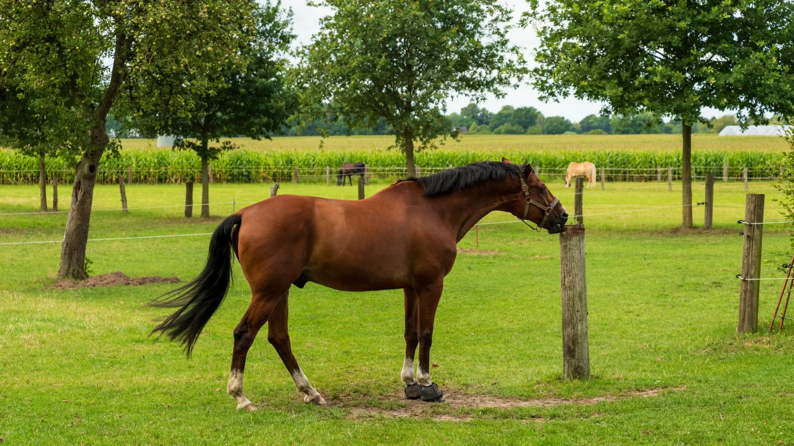
Horses are aberrant hosts for Setaria digitata. When L3 larvae are transmitted by mosquitoes to horses, the larvae do not complete normal development to adult worms in the peritoneal cavity. Instead, the larvae undergo aberrant migration, often entering the CNS. This condition is known as cerebrospinal nematodiasis or cerebrospinal setariosis.
The first documented cases of equine cerebral setariosis caused by S. digitata were reported in the Republic of Korea, where horses presented with acute onset of neurological signs including ataxia, circling, head tilt, nystagmus, and blindness [8]. In these cases, nematode larvae were identified within brain tissue upon histopathological examination. The clinical presentation is often acute and progressive, with some horses developing complete recumbency and requiring euthanasia.
Ocular involvement is a hallmark of aberrant S. digitata migration in horses. Worms migrating through the optic nerve or entering the anterior chamber cause acute blindness, corneal edema, uveitis, and hyphema. In the Korean case series, blindness was a prominent clinical feature, and nematodes were observed within the anterior chamber of affected eyes [8]. Ocular setariosis in horses may be unilateral or bilateral and carries a guarded prognosis for vision recovery even after worm removal.
The pathogenesis of cerebrospinal nematodiasis involves mechanical damage from migrating larvae, associated inflammation, and tissue necrosis. As larvae burrow through neural parenchyma, they create characteristic linear tracts of malacia and hemorrhage. The host inflammatory response includes eosinophilic infiltration, gliosis, and edema. The location of the lesions determines the clinical signs: cerebellar involvement produces ataxia and dysmetria; brainstem lesions cause cranial nerve deficits and altered mental status; and spinal cord involvement leads to paresis and paralysis.
Cerebrospinal nematodiasis has also been documented in goats and sheep, where S. digitata larvae cause similar neurological and ocular signs. In these small ruminant hosts, the condition is frequently fatal if untreated, and early diagnosis is critical for successful intervention.
Pathology and Pathogenesis
The pathological changes associated with Setaria digitata infection differ between the definitive bovine host and aberrant hosts.
In cattle, adult worms in the peritoneal cavity induce a mild, chronic peritonitis characterized by serosal thickening, fibrous adhesions, and eosinophilic infiltration. Heavy infections may produce granulomatous nodules on the peritoneal surface. Microfilariae in the bloodstream are generally well tolerated, though transient eosinophilia may be observed.
In aberrant hosts, the pathological response to migrating larvae is severe. In the CNS, larvae create hemorrhagic tracts surrounded by malacia, spongiosis, and reactive gliosis. The inflammatory infiltrate is predominantly eosinophilic, with lymphocytes, plasma cells, and macrophages present in chronic lesions. Degenerating larvae elicit a granulomatous response with foreign body giant cells. In the eye, larvae induce fibrinopurulent exudate, corneal endothelial damage, iris edema, and secondary glaucoma [8, 3].
At the molecular level, Setaria digitata produces a range of bioactive molecules that modulate host immune responses. Filarial lipids secreted by adult worms have been shown to modulate interleukin-12 (IL-12) signaling through the JAK-STAT pathway, promoting the development of a Th1-type immune response in the bovine host [14]. The parasite also secretes excretory-secretory (ES) products that include antigens detectable in host circulation and that stimulate antibody production [15]. Glutathione S-transferase (GST) from S. digitata has been biochemically characterized and may play a role in detoxification of host-derived reactive oxygen species [16]. The ubiquinone system, particularly ubiquinone Q8, functions as an antioxidant in adult worms, protecting the parasite from oxidative stress [17, 18].
RNA interference studies using siRNA have been developed to investigate gene function in S. digitata, providing a valuable tool for functional genomics [19]. Novel parasite-specific proteins have been characterized in silico and in vitro, including RNA binding proteins and growth factor-like proteins, which may represent potential drug or vaccine targets [20, 21].
Diagnostics
Accurate diagnosis of Setaria digitata infection requires a combination of parasitological, molecular, and serological methods, with the choice of approach depending on the host species and the clinical presentation.
Morphological Identification
Detection of microfilariae in peripheral blood is the standard method for diagnosing patent infections in cattle. Blood samples (preferably collected during periods of peak microfilarial activity) are examined using wet mount preparations, Knott's technique, or membrane filtration. The modified Knott's technique concentrates microfilariae and preserves their morphology for species identification [22]. Microfilariae of S. digitata are sheathed and measure 240 to 280 micrometers in length, with a characteristic cephalic hook and pointed tail.
Adult worms recovered from the peritoneal cavity at necropsy or during laparoscopy can be identified to species level based on morphological features. Scanning electron microscopy of the peribuccal region and tail structures provides definitive species identification [1].
Molecular Diagnostics
Polymerase chain reaction (PCR) based assays offer high sensitivity and specificity for detecting Setaria digitata DNA and are particularly valuable for diagnosing cerebrospinal nematodiasis in aberrant hosts. A sensitive PCR assay targeting the internal transcribed spacer (ITS) region of ribosomal DNA has been developed and validated for detecting S. digitata in cerebrospinal fluid (CSF) and tissue samples from horses, goats, and sheep. This assay is capable of detecting DNA from a single larva and can differentiate S. digitata from other filarial nematodes.
For molecular diagnostics in cattle, PCR targeting the mitochondrial cytochrome c oxidase subunit I (COI) gene has been used for species confirmation and phylogenetic analysis [1]. Real-time PCR (qPCR) assays provide quantitative data on parasite burden and can be used to monitor treatment efficacy.
Sequence analysis of PCR amplicons allows for molecular typing and phylogenetic comparison. The COI gene and the ITS region are commonly used targets for molecular identification and population genetic studies [1, 28].
Serological Assays
Serological detection of anti-Setaria antibodies can indicate exposure but does not necessarily differentiate between current and past infection. Enzyme-linked immunosorbent assays (ELISAs) using whole worm extract or recombinant antigens have been developed for research purposes [23, 5]. Cross-reactivity between S. digitata antigens and those of other filarial nematodes, including Brugia malayi, has been described, which can complicate serological interpretation in regions where multiple filarial species are endemic [23].
The SXP/RAL2 family of proteins, which are highly expressed by S. digitata, represent promising candidates for serodiagnostic antigen development [5]. Species-specific epitopes have been identified that may improve diagnostic specificity.
The following table summarizes the main diagnostic approaches for Setaria digitata infection:
| Diagnostic Method | Sample Type | Target | Sensitivity | Specificity | Application |
|---|---|---|---|---|---|
| Blood smear / Knott's technique | Whole blood (EDTA) | Microfilariae | Moderate | Moderate | Routine screening in cattle |
| PCR (ITS region) | CSF, blood, tissue | Ribosomal DNA | High | High | Cerebrospinal nematodiasis diagnosis |
| PCR (COI gene) | Blood, adult worms | Mitochondrial DNA | High | High | Species confirmation, phylogenetics |
| Real-time qPCR | Blood, CSF | Ribosomal or mitochondrial DNA | Very high | High | Quantitative parasite load |
| ELISA | Serum, plasma | Parasite antigens or anti-parasite antibodies | Moderate | Variable | Serosurveillance, exposure history |
| SDS-PAGE | Adult worm protein extract | Protein profiles | Moderate | Moderate | Species differentiation |
| Morphology (adults) | Peritoneal cavity specimens | Cuticular teeth, spicules, tail | High | High | Definitive species identification [1] |
Treatment and Control
Treatment of Setaria digitata infection depends on the host species and the clinical presentation. In cattle with patent infections but no clinical signs, treatment is not routinely indicated. However, in cases of ocular setariosis or cerebrospinal nematodiasis, intervention is necessary.
In horses with cerebrospinal nematodiasis, the therapeutic approach involves a combination of anthelmintic therapy with ivermectin or other macrocyclic lactones, supportive care, and anti-inflammatory medications. Ivermectin is effective against migrating larvae and has been used successfully to treat cerebrospinal setariosis. Corticosteroids such as dexamethasone are often administered concurrently to reduce inflammation and edema associated with larval death in neural tissue. Early diagnosis and treatment improve the prognosis for neurological recovery, though residual deficits may persist [8, 28].
For ocular setariosis in horses, surgical removal of the worm from the anterior chamber may be performed under general anesthesia or standing sedation with topical anesthesia. This is followed by topical and systemic anti-inflammatory therapy.
In cattle with ocular infections, surgical removal of the worm is the treatment of choice, with ivermectin used to eliminate any remaining larvae [3].
Control of Setaria digitata relies on reducing exposure to infected mosquito vectors. Integrated vector management strategies, including environmental modification to eliminate mosquito breeding sites, use of insecticide-treated cattle, and application of mosquito repellents, can reduce transmission pressure. In endemic regions, strategic anthelmintic treatment of cattle with macrocyclic lactones during peak mosquito activity seasons can reduce microfilaraemia levels and lower the force of infection.
The absence of Wolbachia endosymbionts in S. digitata precludes the use of anti-Wolbachia treatments, which are effective against some other filarial nematodes [13]. This necessitates reliance on direct anthelmintic therapy for parasite control.
In vitro studies have explored the potential of plant-derived compounds against S. digitata. Rhizome extracts of Curcuma zedoaria have been shown to induce caspase-dependent apoptosis in adult worms via generation of reactive oxygen species [24]. While not yet developed into commercial products, such natural compounds may represent future therapeutic options.
In horses, prevention of exposure to mosquito vectors is the most effective control strategy. Stabling during peak mosquito feeding times (dusk and dawn), use of fly masks and insect repellents, and management of mosquito breeding habitats around equine facilities can reduce the risk of aberrant migration and cerebrospinal nematodiasis.
For cattle in endemic regions, prophylactic treatment with ivermectin at regular intervals during the transmission season can prevent the establishment of patent infections and reduce the incidence of aberrant migration in horses and other aberrant hosts sharing the environment. Vaccination strategies are not currently available, though the identification of immunogenic parasite-specific proteins has laid the groundwork for future vaccine development [23, 6, 21, 5].
Diagnostic and Clinical Decision Workflow
The following diagram illustrates a clinical decision workflow for the diagnosis and management of suspected Setaria digitata induced cerebrospinal nematodiasis in horses:
flowchart TD
 A["Horse with acute neurological signs (ataxia, circling, blindness)"] --> B{"History of travel or residence in endemic region?"}
 B -->|Yes| C["Perform ophthalmic examination"]
 B -->|No| D["Consider alternative diagnoses (West Nile virus, EPM, trauma)"]
 C --> E{"Worm visualized in anterior chamber?"}
 E -->|Yes| F["Surgical worm removal + ivermectin + dexamethasone"]
 E -->|No| G["Collect CSF and blood for PCR"]
 G --> H{"PCR positive for S. digitata?"}
 H -->|Yes| I["Ivermectin treatment + anti-inflammatory therapy"]
 H -->|No| J["Perform blood microfilaria examination (Knott's technique)"]
 J --> K{"Microfilariae detected?"}
 K -->|Yes| L["Diagnosis: Patent S. digitata infection (unlikely cause of neurological signs) , investigate other causes"]
 K -->|No| M["Reassess for alternative neurological etiologies:<br>Equine herpesvirus myeloencephalopathy<br>Equine protozoal myeloencephalitis<br>Cervical stenotic myelopathy<br>Trauma"]
 I --> N["Monitor clinical response over 48-72 hours"]
 N --> O{"Neurological improvement?"}
 O -->|Yes| P["Continue supportive care; taper steroids"]
 O -->|No| Q["Consider repeat PCR or MRI; investigate concurrent pathology"]
 F --> N
This workflow emphasizes the importance of considering cerebrospinal nematodiasis as a differential diagnosis in horses from endemic regions presenting with acute neurological signs, particularly when ocular involvement is present. Molecular diagnostic confirmation via PCR is strongly recommended before initiating specific therapy.
Conclusions
Setaria digitata is a filarial nematode of cattle that causes minimal pathology in its definitive host but is responsible for severe, often life-threatening cerebrospinal nematodiasis in aberrant hosts such as horses. The parasite is transmitted by mosquito vectors and is endemic across much of Asia. Aberrant migration of larvae into the CNS and eyes of horses produces acute neurological deficits and blindness that require prompt diagnosis and treatment. Molecular diagnostics, particularly PCR targeting ribosomal and mitochondrial DNA sequences, have revolutionized the detection of S. digitata infections and are the methods of choice for confirming cerebrospinal nematodiasis in aberrant hosts. Treatment with macrocyclic lactones combined with anti-inflammatory therapy can be effective when initiated early. Control of the parasite depends on integrated vector management and strategic anthelmintic use in cattle populations. The genomic characterization of S. digitata has opened new avenues for understanding filarial biology and identifying targets for therapeutic intervention, making this parasite an important model for both veterinary and comparative filarial research.
References
[1] Helmi TZ, Farida F, Kasturi A, et al. Unraveling the morphological and molecular profile of Setaria digitata in Aceh cattle. Open Vet J. 2025. URL: https://pubmed.ncbi.nlm.nih.gov/40092195/
[2] Tung KC, Lai CH, Ooi HK, et al. Cerebrospinal setariosis with Setaria marshalli and Setaria digitata infection in cattle. J Vet Med Sci. 2003. URL: https://pubmed.ncbi.nlm.nih.gov/14532689/
[3] Shin SS, Cho KO, Wee SH. Ocular infection of cattle with Setaria digitata. J Vet Med Sci. 2002. URL: https://pubmed.ncbi.nlm.nih.gov/11853150/
[4] Senanayake KS, Söderberg J, Põlajev A, et al. The Genome of Setaria digitata: A Cattle Nematode Closely Related to Human Filarial Parasites. Genome Biol Evol. 2020. URL: https://pubmed.ncbi.nlm.nih.gov/32022853/
[5] Sasisekhar B, Suba N, Sindhuja S, et al. Setaria digitata: identification and characterization of a hypodermally expressed SXP/RAL2 protein. Exp Parasitol. 2005. URL: https://pubmed.ncbi.nlm.nih.gov/15979614/
[6] Rodrigo WW, Dassanayake RS, Weerasena SJ, et al. Novel parasitic nematode-specific protein of bovine filarial parasite Setaria digitata displays conserved gene structure and ubiquitous expression. Trop Biomed. 2014. URL: https://pubmed.ncbi.nlm.nih.gov/25382479/
[7] Rodrigo WW, Dassanayake RS, Voronin D. Novel parasitic nematode-specific protein of Setaria digitata largely localized in longitudinal muscles, reproductive systems and developing embryos. Exp Parasitol. 2014. URL: https://pubmed.ncbi.nlm.nih.gov/24632187/
[8] Shin J, Ahn KS, Suh GH, et al. First Blindness Cases of Horses Infected with Setaria Digitata (Nematoda: Filarioidea) in the Republic of Korea. Korean J Parasitol. 2017. URL: https://pubmed.ncbi.nlm.nih.gov/29320823/
[9] Ronghang B, Roy B. Filarial infections in semi-wild cattle of Northeast India: first record of Setaria digitata (von Linstow 1906) in Bos frontalis (Lambert 1804). J Parasit Dis. 2015. URL: https://pubmed.ncbi.nlm.nih.gov/26688636/
[10] Tung KC, Cheng FP, Lai CH, et al. Demonstration of vector competence of Culex quinquefasciatus (Diptera: Culicidae) for Setaria digitata. Vet Parasitol. 2004. URL: https://pubmed.ncbi.nlm.nih.gov/15325055/
[12] Siriyasatien P, Intayot P, Sawaswong V, et al. Description of potential vectors of zoonotic filarial nematodes, Brugia pahangi, Setaria digitata, and Setaria labiatopapillosa in Thai mosquitoes. Heliyon. 2023. URL: https://pubmed.ncbi.nlm.nih.gov/36846682/
[13] Voronin D, Abeykoon AM, Gunawardene YI, et al. Absence of Wolbachia endobacteria in Sri Lankan isolates of the nematode parasite of animals Setaria digitata. Vet Parasitol. 2015. URL: https://pubmed.ncbi.nlm.nih.gov/25579393/
[14] Muthian G, Pradeep CG, Sargapradeep K, et al. Setaria digitata secreted filarial lipids modulate IL-12 signaling through JAK-STAT pathway leading to the development of Th1 response. Exp Parasitol. 2006. URL: https://pubmed.ncbi.nlm.nih.gov/16647056/
[15] Sundar ST, D'Souza PE. Survival, activity and release of antigenic excretory secretory products and microfilariae of Setaria digitata maintained in artificial media. J Parasit Dis. 2015. URL: https://pubmed.ncbi.nlm.nih.gov/25698871/
[16] Srinivasan L, Mathew N, Karunan T, et al. Biochemical studies on glutathione S-transferase from the bovine filarial worm Setaria digitata. Parasitol Res. 2011. URL: https://pubmed.ncbi.nlm.nih.gov/21207063/
[17] Mohan S, Pradeep CG, Abhilash Kumar R, et al. The adult-specific ubiquinone Q(8) functions as an antioxidant in the filarial parasite, Setaria digitata. Biochem Biophys Res Commun. 2001. URL: https://pubmed.ncbi.nlm.nih.gov/11689001/
[18] Abhilashkumar R, Mohan S, Jayakumar K, et al. Functional importance of the different ubiquinones in the filarial parasite Setaria digitata. Biochem Biophys Res Commun. 2001. URL: https://pubmed.ncbi.nlm.nih.gov/11350076/
[19] Somarathne MBCL, Gunawardene YINS, Chandrasekharan NV, et al. Development of siRNA mediated RNA interference and functional analysis of novel parasitic nematode-specific protein of Setaria digitata. Exp Parasitol. 2018. URL: https://pubmed.ncbi.nlm.nih.gov/29448039/
[20] Nagaratnam N, Karunanayake EH, Tennekoon KH, et al. In silico characterization of a RNA binding protein of cattle filarial parasite Setaria digitata. Bioinformation. 2014. URL: https://pubmed.ncbi.nlm.nih.gov/25258487/
[21] Rodrigo WW, Dassanayake RS, Weerasena SJ, et al. Heterologous expression of uncharacterized parasitic nematode-specific growth factor-like protein of Setaria digitata in Pichia pastoris expression systems. Trop Biomed. 2013. URL: https://pubmed.ncbi.nlm.nih.gov/23959483/
[22] Watanabe Y, Yang CH, Tung KC, et al. Comparison of microfilaria concentration method for Setaria digitata infection in cattle and for Dirofilaria immitis infection in dogs. J Vet Med Sci. 2004. URL: https://pubmed.ncbi.nlm.nih.gov/15187366/
[23] Satapathy AK, Sahoo PK. Species-specific and cross-reactive antigen of filarial worm: Brugia malayi and Setaria digitata. J Vector Borne Dis. 2019. URL: https://pubmed.ncbi.nlm.nih.gov/33269732/
[24] Senathilake KS, Karunanayake EH, Samarakoon SR, et al. Rhizome extracts of Curcuma zedoaria Rosc induce caspase dependant apoptosis via generation of reactive oxygen species in filarial parasite Setaria digitata in vitro. Exp Parasitol. 2016. URL: https://pubmed.ncbi.nlm.nih.gov/27174667/
Disclaimer: This article is for educational and informational purposes only. It is not intended to substitute for professional veterinary advice, diagnosis, treatment, or regulatory guidance. Always consult a licensed veterinarian or qualified specialist regarding animal health, disease diagnosis, and therapeutic decisions.