Sarcoptes scabiei var. suis: Porcine Sarcoptic Mange, Etiology, Diagnosis, and Control
By Dr. Zubair Khalid, DVM, MS, PhD ·
Key Takeaways
- Sarcoptes scabiei var. suis is a host-adapted mite causing significant economic losses in swine production through reduced growth and feed efficiency, with a zoonotic risk to human handlers.
- Diagnosis relies on clinical signs of intense pruritus and characteristic skin lesions (alopecia, lichenification, crusts, especially in ears), confirmed by microscopic examination of skin scrapings or serological detection of IgG antibodies via ELISA.
- The mite's life cycle, from egg to adult, is completed within the porcine epidermis in 10-15 days, with transmission primarily through direct contact, though fomites can play a minor role.
- Effective treatment involves macrocyclic lactones, specifically ivermectin (300 µg/kg SC) and doramectin (300 µg/kg IM), which are highly effective against all parasitic stages and can facilitate herd eradication.
- Pathogenesis involves a type I hypersensitivity reaction to mite antigens, leading to eosinophilic infiltration, altered oxidative stress markers, and systemic hematological and biochemical disturbances.
- Genomic resources for S. scabiei are emerging, enabling the development of more sensitive molecular diagnostics and facilitating research into host adaptation and acaricide resistance mechanisms.
Introduction
Sarcoptic mange in swine is a highly contagious ectoparasitic disease caused by the burrowing mite Sarcoptes scabiei variety suis. This obligate arthropod represents a host-adapted variant of the S. scabiei species complex, which also includes varieties infecting humans (var. hominis), dogs, cattle, and other mammals. The porcine variant is morphologically and genetically distinct, though genomic resources indicate substantial homology between var. suis and var. hominis. Infestations lead to significant economic losses through reduced growth rates, decreased feed efficiency, and increased culling in breeding herds [<a href="#ref-1">1</a>, <a href="#ref-2">2</a>]. In addition, S. scabiei var. suis poses a zoonotic risk to pig handlers and abattoir workers, as the mite can temporarily infest humans, causing transient papular dermatitis [<a href="#ref-1">1</a>, <a href="#ref-3">3</a>].
This reference article integrates findings from serological surveys, treatment trials, diagnostic development, and genomic studies to provide a comprehensive overview of porcine sarcoptic mange. The discussion follows the parasite from its biology to clinical management, emphasizing evidence-based diagnostic and control strategies used in intensive swine production systems.
Taxonomy and Life Cycle
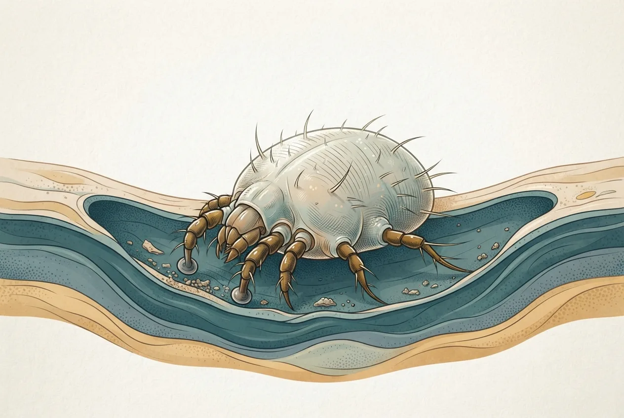
Sarcoptes scabiei (Acari: Sarcoptidae) is a microscopic, astigmatid mite with a rounded body, short legs, and dorsal spines. The female measures approximately 300-500 µm in length, while males are smaller at 200-300 µm [<a href="#ref-1">1</a>]. The entire life cycle, egg, larva, protonymph, tritonymph, and adult, is completed within the epidermis of the porcine host [<a href="#ref-1">1</a>]. Female mites burrow into the stratum corneum, creating tunnels where they deposit eggs. Larvae hatch within 3-5 days, molt through nymphal stages, and become adults in approximately 10-15 days [<a href="#ref-1">1</a>]. Scanning electron microscopy studies have detailed the burrowing behavior, noting that mites preferentially locate in the outer ear canal, perineum, and axillary regions, with hyperkeratotic forms showing massive mite proliferation in thick crusts. Transmission occurs primarily through direct contact with infested pigs, though contaminated fomites and housing environments can sustain mites for short periods. Experimental quantification of transmission among finishing pigs has demonstrated that the basic reproduction ratio (R0) can exceed 2 in susceptible populations, explaining rapid spread in naive herds.
Clinical Signs and Pathogenesis
The clinical presentation of sarcoptic mange in pigs ranges from subclinical carrier states to severe generalized dermatitis. Early clinical signs include intense pruritus, rubbing against pen fixtures, and head shaking [<a href="#ref-2">2</a>, <a href="#ref-4">4</a>, <a href="#ref-5">5</a>]. The rubbing index, defined as the proportion of pigs observed scratching per unit time, is a validated behavioral metric used both in field diagnosis and in monitoring treatment efficacy [<a href="#ref-5">5</a>, <a href="#ref-6">6</a>]. Skin lesions initially appear as small erythematous papules, progressing to alopecia, lichenification, and crust formation, particularly on the ears, flanks, and thighs [<a href="#ref-1">1</a>, <a href="#ref-4">4</a>]. Chronic infestation leads to hyperkeratotic, yellowish-gray crusts, especially in the ear pinnae, which are a classic sign of “ear mange” [<a href="#ref-1">1</a>].
Pathophysiologically, the mite burrowing triggers both innate and adaptive immune responses. Infested pigs develop a type I hypersensitivity reaction to mite antigens, evidenced by elevated IgE and IgG levels [<a href="#ref-4">4</a>, <a href="#ref-7">7</a>]. Histopathology of skin biopsies reveals tunnel-like ulcerated lesions with severe infiltration of eosinophils, lymphocytes, and neutrophils in the dermis [<a href="#ref-1">1</a>]. Oxidative stress markers are significantly altered in infested pigs: malondialdehyde (LPO) and nitric oxide increase, while glutathione (GSH), superoxide dismutase (SOD), and total antioxidant activity (TAA) decrease [<a href="#ref-8">8</a>, <a href="#ref-9">9</a>]. Hematological changes include leukocytosis with eosinophilia and neutrophilia, along with reductions in red blood cell counts, hemoglobin, and packed cell volume [<a href="#ref-8">8</a>]. Biochemical alterations involve elevated serum enzymes such as AST, ALT, ALP, and increased BUN and creatinine, suggesting systemic metabolic disturbance secondary to chronic dermatitis [<a href="#ref-8">8</a>].
Diagnostic Approaches
Clinical diagnosis based on pruritus and skin lesions is suggestive but requires laboratory confirmation due to subclinical carriers and differential diagnoses including dermatophytosis, bacterial pyoderma, and Haemophilus parasuis (Glaesserella parasuis)-associated dermatitis (see Haemophilus parasuis and Glässer's Disease in Swine). Several laboratory methods are employed:
Skin Scraping and Microscopy
Deep skin scrapings from the ear pinna or crusted lesions are digested in 10% potassium hydroxide and examined microscopically to identify mites, eggs, or fecal pellets [<a href="#ref-1">1</a>]. This method has high specificity but limited sensitivity, particularly in chronic carriers with low mite burdens. In a study from Meghalaya, microscopy confirmed infestation in 10.71% of suspected pigs. Sensitivity can be increased by scraping multiple sites and examining the sediment after centrifugation.
Serological Methods
Enzyme-linked immunosorbent assays (ELISAs) detect circulating IgG antibodies against S. scabiei antigens and are widely used for herd-level screening [<a href="#ref-1">1</a>, <a href="#ref-3">3</a>, <a href="#ref-8">8</a>, <a href="#ref-10">10</a>, <a href="#ref-11">11</a>]. Several commercial indirect ELISA kits are available, including the SARCOPTES-ELISA 2001·Pig (AFOSA GmbH, Germany) used in both Italian and Indian studies [<a href="#ref-3">3</a>, <a href="#ref-7">7</a>, <a href="#ref-8">8</a>]. In a seroprevalence study of 370 pigs in Lombardy, Italy, 65.2% of farms were seropositive, with higher prevalence in sows (23.2%) than in fatteners (3.6%) [<a href="#ref-3">3</a>]. Similarly, a wild boar study in Italy found 7.0% seroprevalence for S. scabiei var. suis [<a href="#ref-10">10</a>]. However, substantial variation exists between different commercial ELISA kits, with some showing poor agreement due to differences in antigen preparation and cut-off thresholds [<a href="#ref-7">7</a>]. Bayesian evaluation of ELISA performance using meat juice samples has been conducted, offering an alternative sampling approach for slaughterhouse surveillance [<a href="#ref-12">12</a>]. Additional serological techniques such as passive hemagglutination assays have been developed but are less commonly used [<a href="#ref-13">13</a>]. ELISAs are also useful for evaluating immune response after treatment; antibody titers decline slowly over months, making them suitable for monitoring eradication programs [<a href="#ref-11">11</a>].
Molecular Diagnostics
Nucleic acid amplification tests for S. scabiei DNA have been developed but are not yet standardized for routine swine diagnostics. Genomic resources for var. suis are now available, including draft assemblies and preliminary annotations of 13,226 putative coding sequences, enabling development of species-specific PCR assays. Molecular methods offer the potential to distinguish viable versus non-viable mites and to detect subclinical infestations with greater sensitivity than microscopy.
Diagnostic Workflow
graph TD
 A["Clinical suspicion: pruritus, ear crusts, rubbing"] --> B["Deep skin scraping"]
 B --> C{"Microscopy positive?"}
 C -->|"Yes"| D["Confirm Sarcoptes scabiei var. suis"]
 C -->|"No"| E["Collect serum for ELISA"]
 E --> F{"ELISA positive?"}
 F -->|"Yes"| D
 F -->|"No"| G["Consider alternative diagnoses or low infestation"]
 G --> H["Rule out dermatophytosis, bacterial pyoderma, Streptococcus suis"]
 D --> I["Implement treatment protocol: macrocyclic lactone"]
 I --> J["Post-treatment monitoring: skin lesion score, rubbing index, repeat ELISA at 6 months"]
Treatment and Control
Effective control of sarcoptic mange relies on macrocyclic lactones (avermectins and milbemycins), primarily ivermectin and doramectin [<a href="#ref-6">6</a>, <a href="#ref-14">14</a>, <a href="#ref-15">15</a>, <a href="#ref-16">16</a>]. Ivermectin administered subcutaneously at 300 µg/kg body weight is highly effective against all life stages [<a href="#ref-16">16</a>]. A single dose eliminates mites from most infested pigs, though repeated treatments at 10-14 day intervals may be needed in heavily infected herds to break the cycle [<a href="#ref-16">16</a>]. Doramectin, administered intramuscularly at 300 µg/kg, also shows high efficacy and persistent activity; in a Danish study, a single injection eliminated S. scabiei var. suis from two naturally infested sow herds for 20 months [<a href="#ref-6">6</a>]. Pre-farrowing treatment of sows with doramectin prevents transmission to nursing piglets. In vitro bioassays using S. scabiei var. suis as test organisms have been developed to evaluate acaricidal activity of various compounds, including ivermectin, and plant extracts such as neem (Azadirachta indica) and Brazilian Cerrado bioproducts [<a href="#ref-15">15</a>, <a href="#ref-17">17</a>, <a href="#ref-18">18</a>]. Ethanolic extracts of Stryphnodendron adstringens and Lafoensia pacari show modest acaricidal activity, while oil resins from Copaifera sp. and Pterodon emarginatus achieve 100% mite mortality in vitro [<a href="#ref-18">18</a>]. Neem fruit extracts have been evaluated as a cost-effective alternative in grower pigs [<a href="#ref-17">17</a>]. Polyherbal ointments have shown equivalent efficacy to ivermectin in improving skin lesions and oxidative stress markers by day 15 of therapy [<a href="#ref-8">8</a>].
Environmental treatment is not strictly necessary when all pigs are treated, as mites survive only a few days off the host [<a href="#ref-6">6</a>]. However, cleaning and acaricide application can accelerate break-of-cycle in severe outbreaks [<a href="#ref-6">6</a>]. Biosecurity measures, including quarantine of incoming stock, all-in/all-out production, and maintaining a high sanitary score, are strongly associated with lower seroprevalence [<a href="#ref-3">3</a>, <a href="#ref-19">19</a>]. Successful eradication programs have been reported from farrow-to-finish farms, particularly those using whole-herd treatment with doramectin and rigorous monitoring [<a href="#ref-19">19</a>]. Serological monitoring using ELISA on serum or meat juice permits certification of mange-free status.
Epidemiology and Prevalence
Porcine sarcoptic mange has a worldwide distribution, with seroprevalence varying by management system, geography, and production stage. In Italy, seroprevalence in intensive farms was 11.6% at the individual level and 65.2% at the farm level, with sows showing 23.2% seropositivity compared to 3.6% in fatteners [<a href="#ref-3">3</a>]. In Tripura, India, 11.81% of 1600 screened pigs were seropositive, with higher prevalence in young males and in the winter season [<a href="#ref-1">1</a>]. In Meghalaya, India, microscopy revealed 10.71% prevalence among 196 suspected pigs. In the state of Paraná, Brazil, among certified swine breeder herds, prevalence was very low (0.06-0.12% over 2002-2004), attributed to stringent biosecurity and routine treatment. In wild boars (Sus scrofa) from anthropized areas of Italy, seroprevalence was 7.0% [<a href="#ref-10">10</a>]. Risk factors consistently reported include production category (sows at higher risk), poor biosecurity score, and season (winter peaks due to close confinement) [<a href="#ref-1">1</a>, <a href="#ref-3">3</a>].
Genomic Resources and Future Directions
The availability of draft genomes for Sarcoptes scabiei var. suis and var. hominis has opened avenues for understanding host adaptation, acaricide resistance mechanisms, and antigen discovery. The preliminary annotation identified 13,226 putative coding sequences, many of which are involved in digesting host skin proteins and evading immune responses. These resources enable the development of molecular diagnostics, including real-time PCR assays for quantification of mite DNA in skin scrapings, and for detecting mutations associated with macrocyclic lactone resistance. Future research should focus on standardizing diagnostic protocols across ELISAs [<a href="#ref-7">7</a>], improving sensitivity of microscopy through PCR, and evaluating novel acaricides including plant-derived compounds [<a href="#ref-17">17</a>, <a href="#ref-18">18</a>].
Frequently Asked Questions
What is the primary host range of Sarcoptes scabiei var. suis?
Sarcoptes scabiei var. suis is host-adapted to domestic pigs and wild boars (Sus scrofa), though it can transiently infest humans and other mammals, causing pruritic dermatitis [<a href="#ref-1">1</a>, <a href="#ref-3">3</a>].
How is porcine sarcoptic mange diagnosed in the field?
Diagnosis combines clinical signs (intense pruritus, ear crusts) with microscopic examination of deep skin scrapings and serological testing using commercial indirect ELISA kits [<a href="#ref-1">1</a>, <a href="#ref-3">3</a>, <a href="#ref-8">8</a>, <a href="#ref-10">10</a>, <a href="#ref-11">11</a>].
Which antiparasitic agents are recommended for treatment?
Macrocyclic lactones such as ivermectin (300 µg/kg subcutaneously) and doramectin (300 µg/kg intramuscularly) are highly effective against all parasitic stages [<a href="#ref-6">6</a>, <a href="#ref-14">14</a>, <a href="#ref-16">16</a>].
Can sarcoptic mange be eradicated from a pig herd?
Yes, whole-herd treatment with macrocyclic lactones combined with biosecurity measures can eliminate S. scabiei var. suis, as demonstrated in Danish and Spanish studies [<a href="#ref-6">6</a>, <a href="#ref-19">19</a>].
What is the zoonotic potential of this mite?
S. scabiei var. suis can cause temporary, self-limiting dermatitis in humans, particularly in pig handlers and abattoir workers, but the mites do not complete their life cycle on human skin [<a href="#ref-1">1</a>, <a href="#ref-3">3</a>].
Are there plant-based alternatives for treatment?
Yes, neem fruit extracts and polyherbal ointments have shown acaricidal efficacy comparable to ivermectin in grower pigs, and oil resins from Brazilian trees achieve 100% mite mortality in vitro [<a href="#ref-8">8</a>, <a href="#ref-17">17</a>, <a href="#ref-18">18</a>].
Related Clinical & Scientific Guides
- Tick-Borne Diseases in Dogs: Pathogens, Clinical Signs, Diagnosis, and Prevention
- Toxoplasmosis in Cats and the Risk of Brain Infection in Humans
- Dog Heartworm and Tick-Borne Disease Prevention