Psoroptes ovis (Sheep Scab): Etiology, Pathogenesis, Diagnostics, and Control
By Dr. Zubair Khalid, DVM, MS, PhD ·
Key Takeaways
- Psoroptes ovis is an obligate ectoparasite causing highly contagious sheep scab, characterized by intense pruritus, wool loss, and exudative dermatitis, leading to significant economic losses and welfare concerns. Transmission occurs via direct contact or contaminated fomites, and the mite's life cycle is completed on the host within 10-14 days.
- Traditional diagnosis via microscopy of skin scrapings has low sensitivity, particularly for early or subclinical infestations; molecular diagnostics (ITS2-PCR) and serological assays (ELISA using recombinant antigens like rPsoSP3, rPsoCys, or rPoc-AK) offer superior detection rates and are crucial for identifying subclinical carriers.
- Macrocyclic lactones (MLs) like ivermectin, doramectin, and moxidectin are primary treatment options, but widespread resistance has emerged in P. ovis populations, linked to the overexpression and amplification of CYP and UGT genes.
- Alternative acaricides, including permethrin-based products, isoxazolines (lotilaner), and plant-derived compounds like carvacrol, are being investigated and utilized to combat ML resistance.
- Integrated control strategies are essential, encompassing strict biosecurity, quarantine of new animals, strategic treatment with effective acaricides, and regular monitoring using sensitive diagnostic tools to manage resistance and prevent disease spread.
Introduction
Psoroptes ovis is an astigmatid mite of the family Psoroptidae that causes a highly contagious, exudative dermatitis known as psoroptic mange or sheep scab [<a href="#ref-1">1</a>]. The parasite infests sheep, cattle, rabbits, horses, and other ungulates, leading to substantial economic losses through reduced weight gain, wool damage, and compromised animal welfare [<a href="#ref-1">1</a>, <a href="#ref-2">2</a>, <a href="#ref-3">3</a>]. Sheep scab is endemic in many regions, including the United Kingdom, where it remains a major welfare and production concern [<a href="#ref-4">4</a>, <a href="#ref-5">5</a>]. The disease is characterized by intense pruritus, wool loss, and the formation of thick, yellowish crusts on the skin [<a href="#ref-6">6</a>]. Early and subclinical infestations are difficult to detect by traditional microscopy, prompting the development of molecular and serological diagnostic tools [<a href="#ref-4">4</a>, <a href="#ref-7">7</a>, <a href="#ref-8">8</a>, <a href="#ref-9">9</a>, <a href="#ref-10">10</a>, <a href="#ref-11">11</a>]. The emergence of resistance to macrocyclic lactone (ML) acaricides has further complicated control efforts [<a href="#ref-12">12</a>, <a href="#ref-13">13</a>, <a href="#ref-14">14</a>, <a href="#ref-15">15</a>, <a href="#ref-16">16</a>].
Etiology and Life Cycle
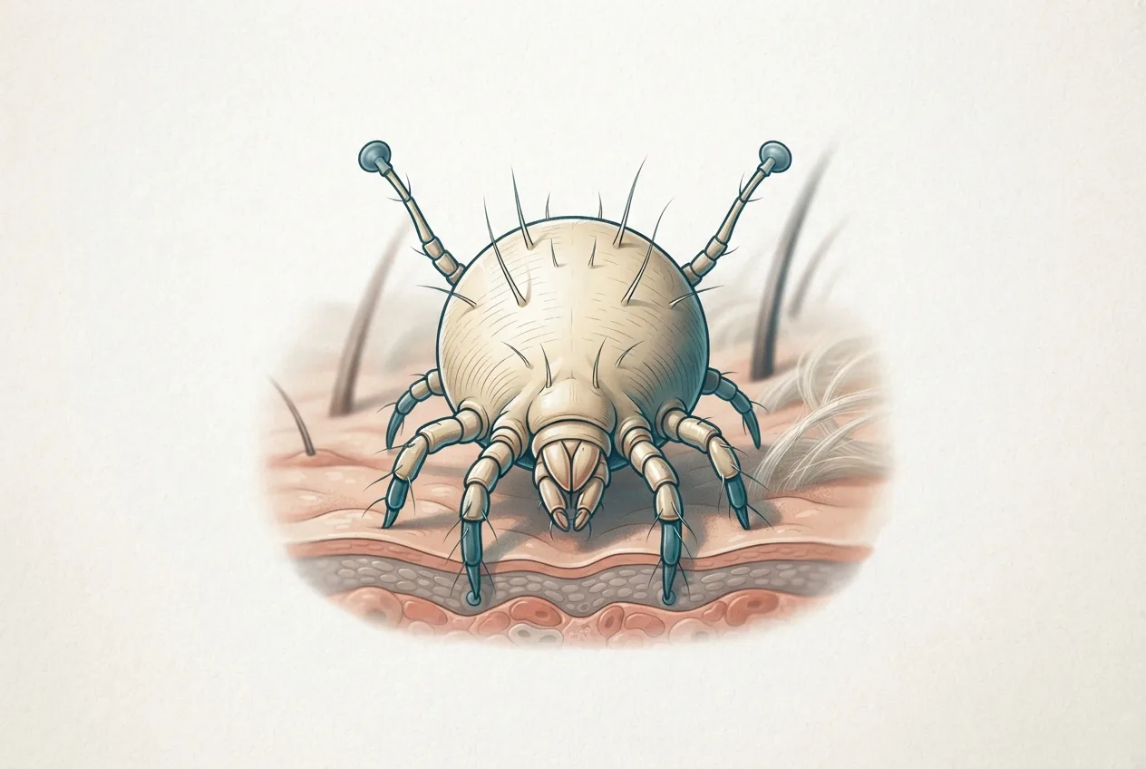
Psoroptes ovis is an obligate, non-burrowing ectoparasite that feeds on skin exudates and lymph [<a href="#ref-1">1</a>]. The mite completes its entire life cycle on the host, typically within 10 to 14 days [<a href="#ref-1">1</a>]. All motile stages (larva, nymph, adult) are found on the skin surface, where they pierce the epidermis to feed, causing an inflammatory serous exudate that dries to form crusts [<a href="#ref-1">1</a>]. Transmission occurs through direct contact between infested and naive animals, or via contaminated fomites such as shearing equipment and bedding [<a href="#ref-1">1</a>, <a href="#ref-5">5</a>]. The mite can survive off the host for several days under favorable conditions, facilitating indirect spread [<a href="#ref-1">1</a>].
Clinical Signs and Pathogenesis
The hallmark of sheep scab is intense pruritus, leading to rubbing, biting, and wool loss [<a href="#ref-6">6</a>]. Affected sheep exhibit restlessness, poor nutritional status, and yellowish crusted skin lesions with edge exudate and erythema [<a href="#ref-6">6</a>]. In a natural outbreak, clinical signs may be absent in a substantial proportion of infested animals; serological testing has revealed that many clinically normal sheep are seropositive, indicating subclinical infestation [<a href="#ref-4">4</a>]. The disease has a significant impact on ewe body condition and lamb birthweight when infestation occurs during mid-pregnancy [<a href="#ref-2">2</a>]. Organ weight analysis in experimentally infested sheep demonstrates reduced omental and perirenal fat, lower thymus weight, and increased liver, adrenal, and peripheral lymph node weights, reflecting heightened metabolic demand, chronic stress, and immune activation [<a href="#ref-3">3</a>]. Histopathological examination reveals eosinophilic pustular dermatitis with marked eosinophilia in peripheral blood [<a href="#ref-6">6</a>]. The immune response is skewed toward a Th2-type profile, with elevated eosinophils and IL-4 levels, particularly in susceptible breeds such as Belgian Blue cattle [<a href="#ref-17">17</a>]. Anti-inflammatory treatment with dexamethasone suppresses lesion development and eosinophil infiltration, confirming the role of host inflammatory responses in pathology [<a href="#ref-17">17</a>].
Diagnosis
Microscopic Examination
The traditional gold standard for diagnosis is microscopic detection of mites or eggs in skin scrapings [<a href="#ref-7">7</a>, <a href="#ref-9">9</a>, <a href="#ref-10">10</a>]. However, this method has poor sensitivity for low-level or early infestations, and false negatives are common [<a href="#ref-4">4</a>, <a href="#ref-7">7</a>, <a href="#ref-9">9</a>, <a href="#ref-10">10</a>]. In a comparative study, microscopy detected only 33.3% of infections at 7 days post-infection (dpi), whereas PCR and ELISA achieved 88.9% and 77.7%, respectively [<a href="#ref-7">7</a>].
Molecular Diagnostics
A PCR assay targeting the internal transcribed spacer 2 (ITS2) region of ribosomal DNA has been developed for P. ovis detection [<a href="#ref-7">7</a>]. The ITS2-PCR has a detection limit of 40.3 pg/μL DNA and shows high specificity [<a href="#ref-7">7</a>]. In experimentally infested rabbits, ITS2-PCR detected infection earlier than microscopy or serology, and its positivity rate dropped to 0% by 7 days post-treatment, making it useful for monitoring treatment success [<a href="#ref-7">7</a>]. Field studies confirmed a higher detection rate with PCR (19.4%) compared to microscopy (11.1%) [<a href="#ref-7">7</a>].
Serological Assays
Several enzyme-linked immunosorbent assays (ELISAs) have been developed using recombinant antigens. An ELISA based on recombinant PsoSP3 (rPsoSP3-iELISA) demonstrated 77.7% sensitivity at 7 dpi and remained 100% positive at 7 days post-treatment, indicating its utility for detecting past exposure [<a href="#ref-7">7</a>]. A more recent indirect ELISA using recombinant cystatin (rPsoCys-iELISA) achieved 95.4% sensitivity and 95.7% specificity, with an area under the ROC curve of 0.991 [<a href="#ref-10">10</a>]. This assay outperformed microscopy in detecting low-level and early infestations (90% vs. 40% at 3 weeks post-infestation) [<a href="#ref-10">10</a>]. Another ELISA based on recombinant arginine kinase (rPoc-AK) showed 94.4% sensitivity and 88.2% specificity, detecting infection as early as 1 week post-infection [<a href="#ref-11">11</a>]. Immunoproteomic approaches using two-dimensional gel electrophoresis coupled with LC-MS/MS have identified additional early diagnostic antigens, including PsoSP3, Pso14-3-3(1), and Pso14-3-3(2) [<a href="#ref-9">9</a>]. An improved ELISA technique for cattle has also been described [<a href="#ref-8">8</a>, <a href="#ref-18">18</a>]. Serological testing is particularly valuable for identifying subclinically infested animals that may act as reservoirs for disease spread [<a href="#ref-4">4</a>].
Differential Diagnosis
Sheep scab must be differentiated from other causes of pruritus and dermatitis, including lice infestations (e.g., Damalinia ovis, Linognathus ovillus), sheep ked (Melophagus ovinus), and fungal infections [<a href="#ref-1">1</a>, <a href="#ref-19">19</a>]. The presence of mites in skin scrapings confirms the diagnosis [<a href="#ref-19">19</a>].
Treatment and Resistance
Macrocyclic Lactones
Macrocyclic lactones (MLs), including ivermectin, doramectin, and moxidectin, are widely used for treatment and control of sheep scab [<a href="#ref-5">5</a>, <a href="#ref-13">13</a>, <a href="#ref-14">14</a>, <a href="#ref-15">15</a>, <a href="#ref-20">20</a>, <a href="#ref-21">21</a>, <a href="#ref-22">22</a>, <a href="#ref-23">23</a>]. However, resistance has emerged in both sheep and cattle populations [<a href="#ref-12">12</a>, <a href="#ref-13">13</a>, <a href="#ref-14">14</a>, <a href="#ref-15">15</a>, <a href="#ref-16">16</a>]. In the UK, resistance to moxidectin, ivermectin, and doramectin has been documented in P. ovis from outbreak populations [<a href="#ref-14">14</a>, <a href="#ref-16">16</a>]. In Belgian Blue cattle, unambiguous treatment failure was detected on 12 of 16 farms, with resistance confirmed by in vivo efficacy parameters [<a href="#ref-15">15</a>]. Pharmacokinetic-pharmacodynamic analysis indicates that both peak plasma concentration (Cmax) and area under the concentration-time curve (AUC) relative to minimal inhibitory concentration (MIC) are critical for efficacy against resistant mites [<a href="#ref-13">13</a>]. Doramectin 1% and moxidectin 1% achieved 100% efficacy in one study, while long-acting formulations showed delayed responses [<a href="#ref-13">13</a>]. Oral doramectin (200 μg/kg) has been effective in rabbits [<a href="#ref-24">24</a>]. Ivermectin administered as two subcutaneous injections (1 mL/33 kg) at a 10-day interval resolved an outbreak in Brazil [<a href="#ref-6">6</a>]. Controlled-release ivermectin boluses provide prolonged prophylaxis [<a href="#ref-23">23</a>, <a href="#ref-25">25</a>].
Alternative Acaricides
Permethrin-based products, including combinations with dinotefuran and pyriproxyfen, have shown efficacy against P. ovis in rabbits [<a href="#ref-26">26</a>, <a href="#ref-27">27</a>]. Lotilaner, an isoxazoline, achieved 100% parasitological efficacy by day 7 in naturally infested rabbits [<a href="#ref-28">28</a>]. Oral ivermectin (400 μg/kg) was 100% effective against P. ovis in rabbits by day 7 [<a href="#ref-29">29</a>]. Plant-derived essential oil components, particularly carvacrol, demonstrate strong acaricidal activity in vitro and in vivo, reducing mite counts by 98.5% in cattle after three weekly topical applications [<a href="#ref-30">30</a>]. The entomopathogenic fungus Metarhizium pingshaense combined with peppermint essential oil or its constituents (menthol, menthone) also shows acaricidal potential [<a href="#ref-31">31</a>]. A deltamethrin-based product (Delcid 7.5) provided persistent activity for 21 days after a single treatment in sheep [<a href="#ref-32">32</a>].
Resistance Mechanisms
Genomic analysis of ML-resistant P. ovis from cattle has identified constitutive overexpression of a cytochrome P450 monooxygenase (CYP) gene and two tandemly located UDP-glucuronosyltransferase (UGT) genes [<a href="#ref-12">12</a>]. Copy number variation at these loci is associated with resistance: the CYP gene is amplified in resistant populations, while the UGT genes are massively amplified in all populations, with a weaker selection signal [<a href="#ref-12">12</a>]. These findings suggest distinct mechanisms of amplification and gene regulation underlying ML resistance [<a href="#ref-12">12</a>].
Integrated Control
Effective control of sheep scab requires a combination of biosecurity, quarantine, strategic treatment, and monitoring [<a href="#ref-5">5</a>]. All incoming sheep should be inspected and, ideally, treated with an effective acaricide before introduction to the flock [<a href="#ref-5">5</a>]. Dipping with organophosphate or synthetic pyrethroid formulations remains an option in some regions [<a href="#ref-5">5</a>]. The use of serological testing can identify subclinically infested animals that might otherwise perpetuate the disease [<a href="#ref-4">4</a>]. Resistance management should include rotation of acaricide classes, targeted treatment based on diagnosis, and avoidance of underdosing [<a href="#ref-5">5</a>, <a href="#ref-14">14</a>]. The development of molecular markers for resistance, such as CYP and UGT copy number variants, may facilitate surveillance and guide treatment choices [<a href="#ref-12">12</a>].
Frequently Asked Questions
What is the causative agent of sheep scab?
Sheep scab is caused by the astigmatid mite Psoroptes ovis, which feeds on skin exudates and causes a severe exudative dermatitis [<a href="#ref-1">1</a>].
How is sheep scab transmitted?
Transmission occurs primarily through direct contact between infested and naive animals, but indirect spread via contaminated fomites (e.g., shearing equipment, bedding) is also possible [<a href="#ref-1">1</a>, <a href="#ref-5">5</a>].
What are the clinical signs of sheep scab?
Intense pruritus, wool loss, yellowish crusted skin lesions, erythema, and restlessness are typical [<a href="#ref-6">6</a>]. Subclinical infestations are common and can only be detected by serological or molecular methods [<a href="#ref-4">4</a>].
How is sheep scab diagnosed?
Diagnosis is confirmed by microscopic identification of mites in skin scrapings, but PCR (ITS2-PCR) and ELISA (e.g., rPsoSP3, rPsoCys, rPoc-AK) offer higher sensitivity, especially for early or low-level infestations [<a href="#ref-7">7</a>, <a href="#ref-9">9</a>, <a href="#ref-10">10</a>, <a href="#ref-11">11</a>].
What treatments are available for sheep scab?
Macrocyclic lactones (ivermectin, doramectin, moxidectin) are commonly used, but resistance is widespread [<a href="#ref-14">14</a>, <a href="#ref-15">15</a>, <a href="#ref-16">16</a>]. Alternative acaricides include permethrin, lotilaner, and plant-derived compounds such as carvacrol [<a href="#ref-27">27</a>, <a href="#ref-28">28</a>, <a href="#ref-30">30</a>].
Is resistance to macrocyclic lactones a problem?
Yes, resistance to ivermectin, doramectin, and moxidectin has been documented in sheep and cattle populations in the UK and Europe [<a href="#ref-12">12</a>, <a href="#ref-13">13</a>, <a href="#ref-14">14</a>, <a href="#ref-15">15</a>, <a href="#ref-16">16</a>]. Resistance is associated with overexpression and copy number amplification of CYP and UGT genes [<a href="#ref-12">12</a>].
Can sheep scab be prevented?
Prevention relies on biosecurity, quarantine of new animals, strategic treatment, and monitoring using sensitive diagnostic tests [<a href="#ref-5">5</a>]. Serological screening can identify subclinical carriers that might introduce the disease into a naive flock [<a href="#ref-4">4</a>].
Mermaid Diagram: Diagnostic Workflow for Sheep Scab
flowchart TD
 A["Sheep with pruritus, wool loss, crusts"] --> B{"Clinical suspicion of sheep scab"}
 B --> C["Perform skin scraping"]
 C --> D{"Microscopy"}
 D -->|"Mites/eggs detected"| E["Confirmed diagnosis"]
 D -->|"Negative"| F["Consider PCR or serology"]
 F --> G["ITS2-PCR on skin scraping or exudate"]
 G -->|"Positive"| E
 G -->|"Negative"| H["ELISA (rPsoSP3, rPsoCys, rPoc-AK)"]
 H -->|"Positive"| I["Subclinical or recent exposure"]
 H -->|"Negative"| J["Consider alternative diagnoses"]
 E --> K["Treatment based on susceptibility"]
 K --> L["Monitor post-treatment with PCR or microscopy"]
 L -->|"Negative"| M["Clearance confirmed"]
 L -->|"Positive"| N["Re-treat or switch acaricide class"]
Related Clinical & Scientific Guides
- Tick-Borne Diseases in Dogs: Pathogens, Clinical Signs, Diagnosis, and Prevention
- Toxoplasmosis in Cats and the Risk of Brain Infection in Humans
- Dog Heartworm and Tick-Borne Disease Prevention