Ostertagia ostertagi: Comprehensive Reference on Bovine Ostertagiosis
By Dr. Zubair Khalid, DVM, MS, PhD ·
Key Takeaways
- Ostertagia ostertagi, the brown stomach worm, is a globally prevalent abomasal nematode causing significant production losses in cattle, ranging from subclinical growth impairment to severe gastroenteritis (ostertagiosis).
- The parasite's life cycle involves free-living larval stages on pasture and a parasitic phase in the abomasum, with infective third-stage larvae (L3) ingested by grazing cattle; hypobiosis (arrested development) at the L4 stage is a critical factor contributing to Type II ostertagiosis.
- Diagnosis integrates fecal egg counts (FEC), coproculture for larval identification, serological tests (ELISA on serum or milk) for herd-level exposure, and plasma pepsinogen levels as a biomarker for abomasal damage.
- Anthelmintic resistance is a growing concern, with confirmed resistance to ivermectin and fenbendazole, often linked to specific genetic mutations (e.g., F200Y in the β-tubulin gene), necessitating integrated resistance monitoring using FECRT and molecular tools.
- Control strategies emphasize integrated pasture management and strategic anthelmintic use, including targeted selective treatment (TST) and potentially vaccination, as host immunity develops slowly and is often incomplete.
- Economic impacts stem from reduced weight gain, decreased milk yield, and treatment costs, with subclinical infections significantly affecting productivity even in the absence of overt clinical signs.
Introduction
Ostertagia ostertagi, commonly known as the brown stomach worm, is a highly prevalent abomasal nematode of cattle worldwide and a major cause of production losses in grazing herds [<a href="#ref-1">1</a>, <a href="#ref-2">2</a>]. The parasite induces a spectrum of disease from subclinical growth impairment to severe gastroenteritis, termed ostertagiosis [<a href="#ref-1">1</a>, <a href="#ref-3">3</a>]. Understanding its biology, epidemiology, and host interactions is critical for designing effective control programs, particularly in the face of emerging anthelmintic resistance [<a href="#ref-4">4</a>, <a href="#ref-5">5</a>, <a href="#ref-6">6</a>]. This article provides a detailed reference on O. ostertagi for veterinary professionals, diagnosticians, and computational biologists.
Taxonomy and Morphology
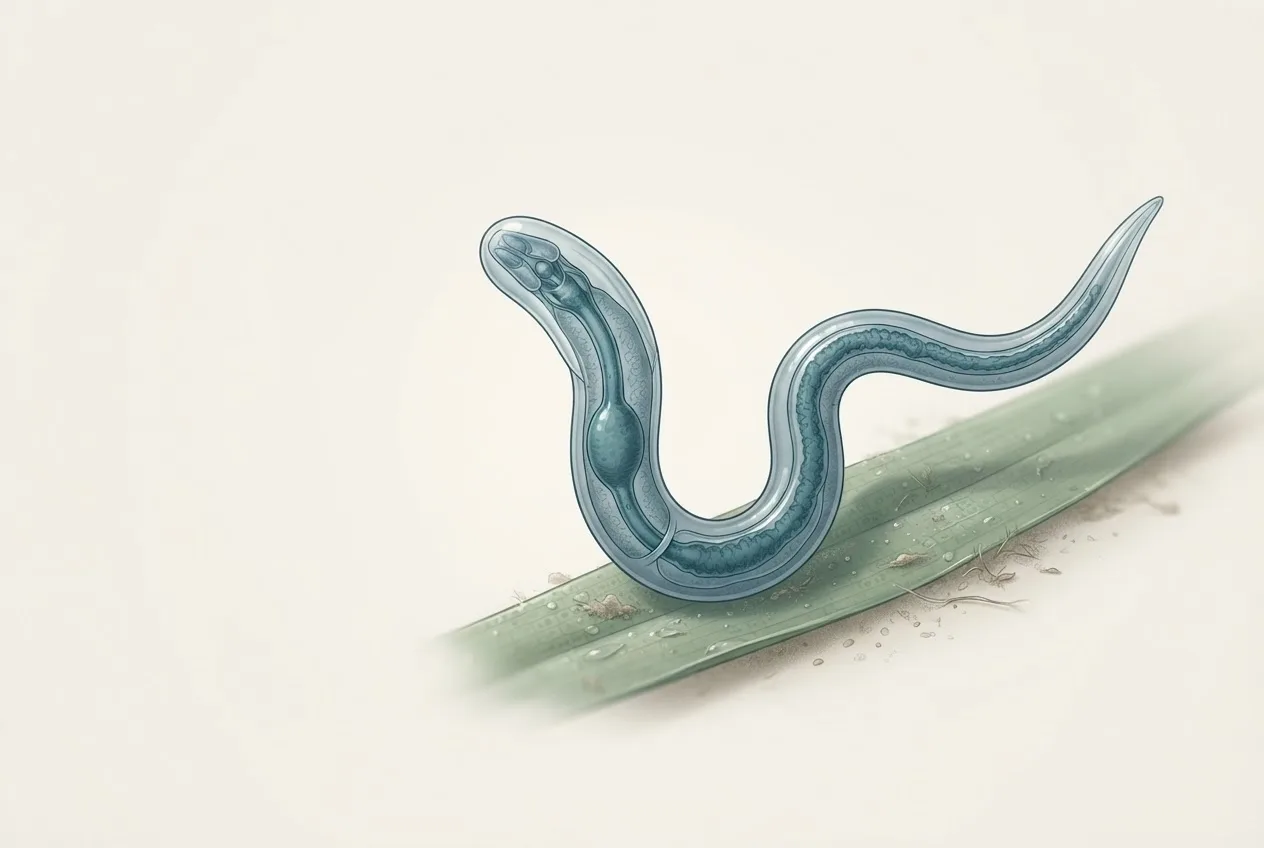
O. ostertagi belongs to the family Trichostrongylidae, order Strongylida [<a href="#ref-1">1</a>]. Adult worms are slender, reddish-brown, and measure 6-9 mm in length. Males possess a prominent copulatory bursa with two spicules, while females have a vulval flap and produce thin-shelled, morulated eggs (approximately 70-90 µm by 35-45 µm) [<a href="#ref-1">1</a>]. The third-stage larvae (L3) are the infective stage, ensheathed and resistant to environmental extremes [<a href="#ref-1">1</a>, <a href="#ref-7">7</a>].
Life Cycle
The life cycle is direct, with a free-living phase on pasture and a parasitic phase within the bovine abomasum [<a href="#ref-1">1</a>, <a href="#ref-8">8</a>]. Eggs are passed in feces and develop through first (L1) and second (L2) larval stages to the infective L3 within 1-3 weeks under favorable conditions [<a href="#ref-1">1</a>]. L3 migrate onto herbage and are ingested by grazing cattle. After ingestion, exsheathment occurs in the rumen, and L3 penetrate the abomasal mucosa, molting to L4 and then to adults [<a href="#ref-1">1</a>, <a href="#ref-7">7</a>]. Adults emerge onto the mucosal surface, where they feed on tissue fluids and produce eggs [<a href="#ref-1">1</a>]. The prepatent period is approximately 18-21 days [<a href="#ref-1">1</a>].
A key feature is hypobiosis (arrested development) at the early L4 stage, triggered by environmental cues such as decreasing temperature or seasonal changes [<a href="#ref-1">1</a>, <a href="#ref-4">4</a>, <a href="#ref-9">9</a>]. Hypobiotic larvae can resume development months later, contributing to type II ostertagiosis [<a href="#ref-1">1</a>, <a href="#ref-10">10</a>].
graph TD
 A["Adult worms in abomasum"] --> B["Eggs in feces"]
 B --> C["L1 larvae in feces"]
 C --> D["L2 larvae"]
 D --> E["L3 infective larvae on pasture"]
 E --> F["Ingestion by cattle"]
 F --> G["Exsheathment in rumen"]
 G --> H["L3 penetrate abomasal mucosa"]
 H --> I["L4 larvae in mucosa"]
 I --> J["Adult worms emerge"]
 J --> A
 I --> K["Hypobiotic L4 arrested development"]
 K --> I
Epidemiology
O. ostertagi is distributed globally in temperate and subtropical regions [<a href="#ref-1">1</a>, <a href="#ref-2">2</a>, <a href="#ref-11">11</a>]. Infection prevalence in dairy herds is high; serological surveys using bulk tank milk ELISA have demonstrated widespread exposure across Europe [<a href="#ref-12">12</a>]. In Norway, a cross-sectional study found high variability in individual antibody ODR values within herds, with bulk tank milk ODR useful only for herds with very high or low exposure [<a href="#ref-13">13</a>]. Seasonal dynamics show peak larval availability on pasture in late summer and autumn, with hypobiosis during winter [<a href="#ref-2">2</a>, <a href="#ref-14">14</a>]. In Hokkaido, Japan, fecal egg counts decreased in winter and increased in warmer months, with O. ostertagi dominating coprocultures in autumn [<a href="#ref-2">2</a>]. Risk factors include high stocking rate, continuous grazing, and lack of anthelmintic treatment history [<a href="#ref-14">14</a>, <a href="#ref-15">15</a>].
Pathogenesis and Clinical Signs
Pathogenesis results from abomasal mucosal damage caused by emerging L4 and adult worms [<a href="#ref-1">1</a>, <a href="#ref-16">16</a>]. The parasite induces hyperplasia of mucus-secreting cells, loss of parietal cells, and increased abomasal pH, leading to hypergastrinemia and elevated plasma pepsinogen levels [<a href="#ref-16">16</a>, <a href="#ref-17">17</a>, <a href="#ref-18">18</a>]. These changes impair protein digestion and reduce feed intake [<a href="#ref-3">3</a>, <a href="#ref-16">16</a>]. Clinical ostertagiosis is classified into type I (acute) and type II (chronic). Type I occurs in first-season grazers with high L3 intake, presenting with diarrhea, weight loss, anorexia, and hypoproteinemia [<a href="#ref-1">1</a>, <a href="#ref-19">19</a>]. Type II results from synchronous emergence of hypobiotic larvae, causing severe abomasitis and high mortality [<a href="#ref-1">1</a>, <a href="#ref-10">10</a>]. Subclinical infections are more common and cause reduced weight gain and milk production [<a href="#ref-2">2</a>, <a href="#ref-20">20</a>, <a href="#ref-21">21</a>].
Transcriptomic analyses of abomasal mucosa reveal that infection elicits host immune responses but also induces immunosuppressive pathways, including T cell exhaustion, which may explain slow development of protective immunity [<a href="#ref-22">22</a>]. Peripheral blood mononuclear cell transcriptomics show stage-specific immune reactions [<a href="#ref-23">23</a>]. The rumen microbiome is also altered; subclinical infection enriches opportunistic pathogens like Listeria and depletes Filifactor, while affecting methane metabolism pathways [<a href="#ref-24">24</a>].
Diagnosis
Diagnosis relies on a combination of methods. Fecal egg count (FEC) using modified McMaster or FLOTAC techniques detects eggs, but sensitivity is low in low-shedding animals [<a href="#ref-13">13</a>, <a href="#ref-25">25</a>]. Coproculture and larval differentiation are required to speciate O. ostertagi from other strongyles [<a href="#ref-4">4</a>]. Serological diagnosis using indirect ELISA to detect anti-O. ostertagi antibodies in serum, milk, or bulk tank milk is widely used for herd-level exposure assessment [<a href="#ref-12">12</a>, <a href="#ref-13">13</a>, <a href="#ref-20">20</a>, <a href="#ref-26">26</a>]. The optical density ratio (ODR) correlates with infection intensity and can predict milk production response to anthelmintic treatment [<a href="#ref-20">20</a>, <a href="#ref-26">26</a>]. Plasma pepsinogen levels are elevated in abomasal damage and serve as a biomarker [<a href="#ref-17">17</a>]. Coproantigen detection by ELISA offers an alternative for patent infections [<a href="#ref-25">25</a>].
Molecular diagnostics include quantitative PCR (qPCR) targeting L1 larvae for species identification and detection of anthelmintic resistance alleles [<a href="#ref-5">5</a>, <a href="#ref-27">27</a>]. ITS-2 rDNA nemabiome metabarcoding integrated with FECRT provides species- and stage-specific resistance profiles [<a href="#ref-4">4</a>]. Transcriptome analysis of different life stages has identified stage-specific gene expression, enabling development of molecular markers [<a href="#ref-7">7</a>].
Anthelmintic Resistance
Resistance in O. ostertagi is a growing concern. Ivermectin resistance has been confirmed in New Zealand, western Canada, and the western United States [<a href="#ref-4">4</a>, <a href="#ref-6">6</a>, <a href="#ref-28">28</a>]. In New Zealand, FECRT and controlled efficacy trials showed ivermectin efficacy as low as 13% against O. ostertagi, while moxidectin remained >99% effective [<a href="#ref-6">6</a>]. In western Canada, ivermectin treatment resulted in 82.4% FECR at 14 days, but recrudescence of O. ostertagi L3 in coprocultures 3-6 months post-treatment indicated resistance in hypobiotic larvae [<a href="#ref-4">4</a>]. Fenbendazole resistance has been documented in a field isolate from the UK, with 0% efficacy in a controlled test and high frequency of F200Y and F167Y SNPs in the β-tubulin isotype 1 gene [<a href="#ref-5">5</a>]. Rapid selection for F200Y alleles on pasture has been demonstrated [<a href="#ref-29">29</a>]. Dual resistance to macrocyclic lactones and albendazole has been reported [<a href="#ref-6">6</a>]. Levamisole has historically shown variable efficacy against O. ostertagi [<a href="#ref-30">30</a>]. Monitoring resistance requires integration of FECRT with molecular tools [<a href="#ref-4">4</a>, <a href="#ref-5">5</a>, <a href="#ref-27">27</a>].
Control Strategies
Control relies on integrated pasture management and strategic anthelmintic use. Targeted selective treatment (TST) based on individual animal performance (e.g., average daily gain) can reduce selection for resistance while maintaining productivity [<a href="#ref-15">15</a>]. Stochastic models indicate that stocking rate has a larger impact on parasitological outcomes than initial pasture contamination [<a href="#ref-8">8</a>]. The "dose and move" strategy, where calves are treated and moved to clean pasture, is effective but may select for resistance [<a href="#ref-8">8</a>]. Repeated drug-truncated infections (rDTI) have been used experimentally to induce partial protective immunity, suggesting potential for vaccination [<a href="#ref-31">31</a>]. Vaccination with whole worm or excretory-secretory antigens elicits humoral and cell-mediated responses and reduces fecal egg counts [<a href="#ref-24">24</a>, <a href="#ref-31">31</a>]. However, no commercial vaccine is currently available.
Economic Impact
Economic losses from O. ostertagi arise from reduced weight gain, decreased milk yield, and treatment costs. In German dairy herds, modeling using Paracalc estimated significant annual losses per cow [<a href="#ref-21">21</a>]. In Europe, bulk tank milk antibody levels are associated with milk production losses [<a href="#ref-12">12</a>, <a href="#ref-20">20</a>]. Subclinical infections in lactating cows reduce milk yield even when fecal egg counts are negative [<a href="#ref-2">2</a>].
Frequently Asked Questions
What is the prepatent period of Ostertagia ostertagi?
The prepatent period is approximately 18 to 21 days from ingestion of L3 to appearance of eggs in feces [<a href="#ref-1">1</a>].
How does hypobiosis affect disease presentation?
Hypobiosis is an arrested development at the early L4 stage, triggered by seasonal cues [<a href="#ref-1">1</a>, <a href="#ref-9">9</a>]. Synchronous resumption of development leads to type II ostertagiosis, characterized by severe abomasitis and high mortality, often occurring in winter or early spring [<a href="#ref-1">1</a>, <a href="#ref-10">10</a>].
Which diagnostic method is most sensitive for herd-level exposure?
Bulk tank milk ELISA measuring anti-O. ostertagi antibodies is widely used for herd-level surveillance, but it has poor predictive ability for individual animal ODR values [<a href="#ref-12">12</a>, <a href="#ref-13">13</a>]. For individual diagnosis, serum or milk ELISA combined with fecal egg count is recommended [<a href="#ref-13">13</a>, <a href="#ref-26">26</a>].
What anthelmintic resistance mechanisms are known in O. ostertagi?
Resistance to macrocyclic lactones (ivermectin, moxidectin) and benzimidazoles (fenbendazole, albendazole) has been confirmed [<a href="#ref-4">4</a>, <a href="#ref-5">5</a>, <a href="#ref-6">6</a>, <a href="#ref-28">28</a>]. Benzimidazole resistance is associated with SNPs F167Y, E198A, and F200Y in the β-tubulin isotype 1 gene [<a href="#ref-5">5</a>, <a href="#ref-29">29</a>]. Ivermectin resistance in hypobiotic larvae has been demonstrated via recrudescence after treatment [<a href="#ref-4">4</a>].
Can cattle develop protective immunity against O. ostertagi?
Protective immunity develops slowly and is often incomplete [<a href="#ref-1">1</a>, <a href="#ref-22">22</a>]. Repeated drug-truncated infections can induce partial protection with strong humoral and cell-mediated responses [<a href="#ref-31">31</a>]. Transcriptomic data suggest parasite-induced immunosuppression, including T cell exhaustion, hinders immunity [<a href="#ref-22">22</a>].
What is the role of the rumen microbiome in O. ostertagi infection?
Subclinical infection alters the rumen microbiome, enriching opportunistic pathogens like Listeria and depleting Filifactor [<a href="#ref-24">24</a>]. Methane metabolism pathways are affected, and vaccination partially reverses some microbial changes [<a href="#ref-24">24</a>].
How is anthelmintic resistance monitored in O. ostertagi?
Resistance is monitored using the fecal egg count reduction test (FECRT) combined with coproculture and larval differentiation [<a href="#ref-4">4</a>, <a href="#ref-6">6</a>]. Molecular methods such as ITS-2 rDNA nemabiome metabarcoding and qPCR for resistance-associated SNPs enhance detection [<a href="#ref-4">4</a>, <a href="#ref-5">5</a>, <a href="#ref-27">27</a>].