Fasciola gigantica: The Tropical Liver Fluke of Livestock in Africa and Asia
By Dr. Zubair Khalid, DVM, MS, PhD ·
Key Takeaways
- Fasciola gigantica is the primary cause of tropical fasciolosis in livestock across Africa and Asia, leading to significant economic losses through reduced productivity, liver condemnation, and mortality.
- The parasite's life cycle is indirect, requiring specific lymnaeid snail species (e.g., Radix natalensis, Radix auricularia rubiginosa) as intermediate hosts, with transmission occurring via ingestion of metacercariae on aquatic vegetation.
- Diagnosis relies on detecting eggs in feces (chronic cases), serological ELISAs for antibodies, and molecular PCR methods (e.g., ITS1 RFLP) for definitive species identification, especially to differentiate from F. hepatica.
- Triclabendazole is the current drug of choice for treatment, though resistance is emerging; research into plant-based anthelmintics with ovicidal and larvicidal properties is ongoing.
- Pathogenesis is driven by proteases, particularly cathepsin L and B families, which facilitate tissue invasion and immune evasion, and are key targets for vaccine development.
- Control strategies necessitate an integrated approach, including anthelmintic treatment, molluscicidal interventions, and improved grazing and water management practices to minimize contact with infected snails and vegetation.
Introduction
Fasciola gigantica is a trematode parasite of the family Fasciolidae and the primary causative agent of tropical fasciolosis in livestock across Africa and Asia [<a href="#ref-1">1</a>, <a href="#ref-2">2</a>]. This parasite imposes a substantial economic burden on the cattle, buffalo, sheep, and goat industries through reduced productivity, liver condemnation at slaughter, and mortality in acute cases [<a href="#ref-3">3</a>]. Unlike its temperate counterpart Fasciola hepatica, F. gigantica is adapted to warmer climatic zones and uses different species of lymnaeid snails as intermediate hosts [<a href="#ref-2">2</a>, <a href="#ref-4">4</a>, <a href="#ref-5">5</a>]. The parasite is also a significant zoonotic pathogen, though this article focuses exclusively on its veterinary and biological aspects [<a href="#ref-6">6</a>, <a href="#ref-7">7</a>].
Taxonomy and Morphology
Fasciola gigantica belongs to the phylum Platyhelminthes, class Trematoda, order Fasciolida, and family Fasciolidae [<a href="#ref-8">8</a>]. The adult fluke is characterized by a large, leaf-shaped, dorsoventrally flattened body that is distinctly longer and narrower than that of F. hepatica [<a href="#ref-1">1</a>, <a href="#ref-8">8</a>]. Adult specimens measure 30 to 75 mm in length and 5 to 12 mm in width, with a cephalic cone that is less pronounced than in F. hepatica [<a href="#ref-1">1</a>, <a href="#ref-8">8</a>]. The tegument is covered in spines, and the parasite possesses a blind-ending bifurcate gut, two suckers (oral and ventral), and a complex reproductive system [<a href="#ref-1">1</a>, <a href="#ref-9">9</a>].
The eggs of F. gigantica are operculated, ovoid, and measure approximately 150 to 190 µm in length by 75 to 90 µm in width [<a href="#ref-1">1</a>]. They are morphologically indistinguishable from those of F. hepatica using light microscopy alone, necessitating molecular methods for definitive species identification [<a href="#ref-7">7</a>].
Life Cycle
The life cycle of F. gigantica is indirect and involves a definitive mammalian host and an intermediate snail host [<a href="#ref-1">1</a>, <a href="#ref-10">10</a>, <a href="#ref-11">11</a>]. The complete developmental chronology has been experimentally characterized [<a href="#ref-1">1</a>].
Definitive Host Stages
Adult flukes reside in the bile ducts of the definitive host, where they produce eggs that are carried with bile into the intestine and excreted in feces [<a href="#ref-1">1</a>]. Eggs must reach freshwater to embryonate. At an optimal temperature of approximately 29 degrees Celsius, embryonation and hatching of the miracidium occur within 11 to 12 days [<a href="#ref-1">1</a>]. The free-swimming miracidium is ciliated and must locate and penetrate a compatible lymnaeid snail within a few hours [<a href="#ref-1">1</a>, <a href="#ref-12">12</a>].
Intermediate Host Stages
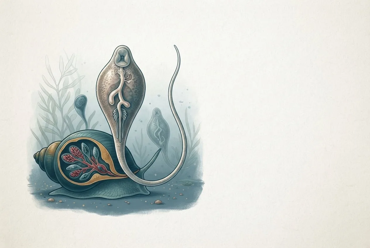
Upon penetration of the snail, the miracidium transforms into a sporocyst, which then gives rise to rediae [<a href="#ref-1">1</a>, <a href="#ref-10">10</a>, <a href="#ref-11">11</a>]. The redial generation undergoes several cycles of asexual reproduction, producing daughter rediae and ultimately cercariae [<a href="#ref-1">1</a>, <a href="#ref-10">10</a>]. The prepatent period within the snail is approximately 39 days post-infection under experimental conditions [<a href="#ref-1">1</a>]. Cercariae are released from the snail and swim to aquatic vegetation, where they shed their tails and encyst as metacercariae on plant surfaces [<a href="#ref-1">1</a>, <a href="#ref-2">2</a>].
Transmission to the Definitive Host
The definitive host acquires infection by ingesting metacercariae-contaminated vegetation or water [<a href="#ref-1">1</a>, <a href="#ref-2">2</a>]. In the small intestine, the metacercaria excysts, and the juvenile fluke penetrates the intestinal wall, migrates through the peritoneal cavity, and penetrates the liver capsule [<a href="#ref-1">1</a>]. The juvenile migrates through the liver parenchyma for several weeks before entering the bile ducts, where it matures into an adult [<a href="#ref-1">1</a>, <a href="#ref-13">13</a>]. The prepatent period in the definitive host is approximately 42 days [<a href="#ref-1">1</a>].
graph TD
 A["Adult flukes in bile ducts"] --> B["Eggs in feces"]
 B --> C["Embryonated egg in water"]
 C --> D["Miracidium"]
 D --> E["Penetrates snail host"]
 E --> F["Sporocyst"]
 F --> G["Rediae"]
 G --> H["Cercariae"]
 H --> I["Metacercariae on vegetation"]
 I --> J["Ingested by definitive host"]
 J --> K["Excystment in small intestine"]
 K --> L["Migration through liver parenchyma"]
 L --> A
Intermediate Host Snails
The intermediate hosts for F. gigantica are freshwater snails of the family Lymnaeidae [<a href="#ref-2">2</a>, <a href="#ref-4">4</a>, <a href="#ref-5">5</a>]. Key species include Radix natalensis in Africa, Radix auricularia rubiginosa in Southeast Asia, and Radix acuminata (syn. Lymnaea acuminata) in the Indian subcontinent [<a href="#ref-1">1</a>, <a href="#ref-4">4</a>, <a href="#ref-5">5</a>, <a href="#ref-14">14</a>]. The life history traits of these snails, including fecundity and survival, are strongly influenced by water temperature, which in turn affects the transmission dynamics of the parasite [<a href="#ref-4">4</a>]. Snail population management is considered a critical control point, as the molluscan host represents the weakest link in the life cycle [<a href="#ref-15">15</a>].
Epidemiology and Geographic Distribution
Fasciola gigantica is endemic throughout tropical and subtropical regions of Africa and Asia [<a href="#ref-2">2</a>, <a href="#ref-16">16</a>]. Geographic information system (GIS) models based on climate data have been used to predict the potential distribution and abundance of F. gigantica in East Africa, highlighting the role of temperature and moisture in defining suitable habitats for the snail intermediate host [<a href="#ref-16">16</a>]. In Southeast Asia, F. gigantica and its hybrids with F. hepatica are prevalent, complicating both diagnosis and control [<a href="#ref-2">2</a>, <a href="#ref-7">7</a>].
Prevalence in livestock can be extremely high. In northern Vietnam, a cross-sectional study reported a Fasciola spp. prevalence of 52% by copro-microscopy and 54% by serology in buffaloes and cattle [<a href="#ref-6">6</a>]. In Western Uttar Pradesh, India, epizootiological surveys have documented significant infection rates in cattle and buffaloes [<a href="#ref-3">3</a>]. The parasite has also been documented in donkeys in Karakalpakstan, with a prevalence of 6.4% and an intensity of 3 to 19 specimens per animal [<a href="#ref-8">8</a>]. Risk factors for infection include age of the animal, access to snail-infested water bodies, and management practices such as grazing on flooded pastures [<a href="#ref-3">3</a>, <a href="#ref-6">6</a>].
Molecular Biology and Genomics
Comparative omics approaches have revealed the complex protease and anti-protease profiles that underpin the virulence and pathogenicity of F. gigantica [<a href="#ref-17">17</a>]. The parasite expresses a diverse array of cathepsin peptidases, which are critical for tissue invasion, feeding, and immune evasion [<a href="#ref-17">17</a>, <a href="#ref-18">18</a>]. The expression of cathepsin L1 is developmentally regulated, with high levels observed in the migratory juvenile stages and in adult flukes [<a href="#ref-18">18</a>]. Comparative genomics has shown that F. gigantica possesses a reduced number of cathepsin peptidase genes relative to F. hepatica, providing insights into the evolution of these gene families [<a href="#ref-17">17</a>].
The parasite also expresses antioxidant enzymes, such as 2-Cys peroxiredoxin, which protect against host-derived oxidative stress. A type-1 nuclear receptor (FgNR1) has been identified and shown to be activated by bile salts, suggesting a role in the adaptation to the bile duct environment [<a href="#ref-19">19</a>]. Nitric oxide synthase activity has been purified and characterized from F. gigantica, indicating a potential role for nitric oxide in parasite biology [<a href="#ref-20">20</a>].
Proteases and Virulence Factors
Proteases are central to the pathogenesis of fasciolosis [<a href="#ref-17">17</a>]. The major proteases belong to the cathepsin L and cathepsin B families [<a href="#ref-17">17</a>, <a href="#ref-18">18</a>]. These enzymes degrade host extracellular matrix components, facilitating migration through the liver parenchyma [<a href="#ref-17">17</a>]. They also cleave host immunoglobulins and interfere with immune cell function, contributing to immune evasion [<a href="#ref-17">17</a>]. The parasite also expresses a range of protease inhibitors (anti-proteases) that regulate endogenous protease activity and may modulate host inflammatory responses [<a href="#ref-17">17</a>].
Excretory/secretory (E/S) products, which include these proteases and other molecules, are highly immunogenic and have been localized to the tegument, gut, and spines of various life cycle stages [<a href="#ref-9">9</a>, <a href="#ref-21">21</a>]. In infected cattle, E/S antigens have been detected in the liver, gall bladder, spleen, kidney, heart, and lungs, as well as in peripheral blood mononuclear cells [<a href="#ref-9">9</a>].
Immunology and Vaccine Development
The host immune response to F. gigantica is complex and involves both humoral and cellular components [<a href="#ref-9">9</a>, <a href="#ref-21">21</a>]. The parasite has evolved sophisticated mechanisms to modulate host immunity, including the secretion of immunomodulatory molecules [<a href="#ref-17">17</a>]. Vaccine development has focused on targeting key parasite molecules, particularly cathepsin L proteases. Recombinant pro- and mature cathepsin L1 have been evaluated as vaccine candidates in murine models, demonstrating significant protection against challenge infection. Other vaccine strategies have explored the use of saposin-like proteins and other E/S components [<a href="#ref-22">22</a>].
Diagnosis
Diagnosis of F. gigantica infection in livestock relies on a combination of clinical, parasitological, serological, and molecular methods.
Clinical and Pathological Findings
Acute fasciolosis results from the migration of large numbers of juvenile flukes through the liver parenchyma, causing traumatic hepatitis, hemorrhage, and potentially fatal liver damage. Chronic fasciolosis is associated with the presence of adult flukes in the bile ducts, leading to cholangitis, bile duct hyperplasia, fibrosis, and calcification [<a href="#ref-9">9</a>]. Clinical signs include weight loss, anemia, hypoalbuminemia, reduced milk yield, and poor reproductive performance [<a href="#ref-3">3</a>].
Parasitological Methods
The standard method for diagnosing chronic fasciolosis is the detection of eggs in feces using sedimentation or flotation techniques [<a href="#ref-3">3</a>, <a href="#ref-6">6</a>]. However, copro-microscopy has low sensitivity during the prepatent period and in cases of low-intensity infection [<a href="#ref-6">6</a>].
Serological Methods
Enzyme-linked immunosorbent assays (ELISAs) that detect antibodies against Fasciola E/S antigens are widely used for herd-level diagnosis and surveillance [<a href="#ref-6">6</a>]. These assays can detect infection earlier than copro-microscopy [<a href="#ref-6">6</a>].
Molecular Methods
Polymerase chain reaction (PCR) assays targeting the internal transcribed spacer 1 (ITS1) region, the NADH dehydrogenase subunit 1 (ND1) gene, and the cytochrome c oxidase subunit 1 (CO1) gene are used for species identification and phylogenetic analysis [<a href="#ref-7">7</a>]. PCR-restriction fragment length polymorphism (RFLP) of the ITS1 region using the RsaI enzyme can reliably distinguish F. gigantica from F. hepatica and their hybrids [<a href="#ref-7">7</a>]. Molecular phylogenetic studies have revealed significant genetic diversity and haplotype variation among F. gigantica populations from different geographic regions [<a href="#ref-7">7</a>].
Control and Treatment
Control of fasciolosis gigantica requires an integrated approach targeting both the definitive host and the intermediate snail host.
Anthelmintic Treatment
Triclabendazole (TCBZ) is the drug of choice for treating fasciolosis, as it is effective against both juvenile and adult flukes [<a href="#ref-23">23</a>]. However, resistance to TCBZ has been reported in some regions, necessitating the development of alternative control strategies [<a href="#ref-23">23</a>].
Plant-Based Anthelmintics
A substantial body of research has investigated the fasciolicidal activity of various medicinal plants. In vitro studies have demonstrated the ovicidal activity of Azadirachta indica (neem) leaf extract and its green-synthesized silver nanoparticles against F. gigantica eggs, with the nanoparticles showing significantly higher efficacy than TCBZ [<a href="#ref-23">23</a>]. In vivo studies have shown that Spondias pinnata leaf powder and extracts have anti-larvicidal activity against sporocyst, redia, and cercaria stages in the snail host [<a href="#ref-14">14</a>]. Similarly, Phyllanthus emblica bark powder and extracts have demonstrated larvicidal effects [<a href="#ref-24">24</a>]. Tinospora cordifolia stem extracts have shown in vitro activity against all larval stages [<a href="#ref-25">25</a>]. Solanum surattense leaf extracts and column-purified fractions have also exhibited potent larvicidal activity [<a href="#ref-26">26</a>]. Potentilla fulgens has been evaluated for its anthelmintic activity against Fasciola larvae. Combretum nigricans extracts have demonstrated in vitro fasciolicidal activity against adult flukes [<a href="#ref-27">27</a>].
Molluscicidal Control
Reducing snail populations can break the parasite's life cycle. Chlorophyllin, a water-soluble derivative of chlorophyll, has shown photodynamic toxicity against the snail Indoplanorbis exustus, a carrier of F. gigantica [<a href="#ref-15">15</a>].
Management Practices
Management strategies include avoiding grazing on snail-infested pastures, providing clean drinking water, and implementing regular deworming programs [<a href="#ref-2">2</a>, <a href="#ref-6">6</a>]. In Southeast Asia, the role of rice fields in maintaining the life cycle is particularly important, as they provide ideal habitats for both the snail host and the aquatic vegetation on which metacercariae encyst [<a href="#ref-2">2</a>].
Frequently Asked Questions
What is the primary intermediate host for Fasciola gigantica?
The primary intermediate hosts are freshwater snails of the family Lymnaeidae, including species such as Radix natalensis, Radix auricularia rubiginosa, and Radix acuminata [<a href="#ref-1">1</a>, <a href="#ref-4">4</a>, <a href="#ref-5">5</a>].
How does Fasciola gigantica differ from Fasciola hepatica?
Fasciola gigantica is adapted to tropical climates, is generally larger and narrower, and uses different lymnaeid snail species as intermediate hosts compared to the temperate F. hepatica [<a href="#ref-1">1</a>, <a href="#ref-2">2</a>]. Genomically, F. gigantica has a reduced number of cathepsin peptidase genes [<a href="#ref-17">17</a>].
What are the clinical signs of fasciolosis in cattle?
Clinical signs include weight loss, anemia, reduced milk yield, poor reproductive performance, and in acute cases, sudden death due to liver damage [<a href="#ref-3">3</a>].
How is fasciolosis diagnosed in livestock?
Diagnosis is achieved through copro-microscopy for egg detection, serological ELISAs for antibody detection, and molecular PCR-based methods for species identification [<a href="#ref-6">6</a>, <a href="#ref-7">7</a>].
What is the treatment of choice for Fasciola gigantica infection?
Triclabendazole is the most effective anthelmintic, but resistance is an emerging concern [<a href="#ref-23">23</a>]. Plant-based alternatives are under investigation [<a href="#ref-14">14</a>, <a href="#ref-23">23</a>, <a href="#ref-24">24</a>, <a href="#ref-25">25</a>, <a href="#ref-26">26</a>].
Can Fasciola gigantica infect humans?
Yes, F. gigantica is a zoonotic parasite, but this article focuses on its veterinary aspects [<a href="#ref-6">6</a>, <a href="#ref-7">7</a>].
What is the role of excretory/secretory products in pathogenesis?
Excretory/secretory products, including proteases, facilitate tissue invasion, feeding, and immune evasion, and are highly immunogenic [<a href="#ref-9">9</a>, <a href="#ref-17">17</a>, <a href="#ref-21">21</a>].
Are there effective vaccines against Fasciola gigantica?
Experimental vaccines based on recombinant cathepsin L1 have shown promise in murine models, but no commercial vaccine is currently available.
Related Clinical & Scientific Guides
- Tick-Borne Diseases in Dogs: Pathogens, Clinical Signs, Diagnosis, and Prevention
- Toxoplasmosis in Cats and the Risk of Brain Infection in Humans
- Dog Heartworm and Tick-Borne Disease Prevention
- Fasciola gigantica: Tropical Liver Fluke in Cattle and Buffalo in Africa and Asia