
Cooperia oncophora: Cattle Nematode in Calves on Pasture, Epidemiology and Anthelmintic Control
Etiology and Taxonomy
Cooperia oncophora is a trichostrongylid nematode of the order Strongylida, family Trichostrongylidae. It is a small, slender, reddish-brown worm that inhabits the small intestine of cattle and, less commonly, other ruminants. Adult males measure 5 to 9 mm in length, and females measure 6 to 11 mm. The buccal capsule is rudimentary, and the cuticle bears transverse striations. The species is characterized by a prominent cervical papillae and a copulatory bursa in males with a characteristic dorsal ray configuration. C. oncophora is often found in mixed infections with other gastrointestinal nematodes, most notably Ostertagia ostertagi and Cooperia punctata.
Life Cycle
The life cycle of C. oncophora is direct. Adult females in the small intestine produce eggs that are passed in the feces. Under optimal environmental conditions (temperatures between 15 and 25 degrees Celsius and adequate moisture), eggs hatch to release first-stage larvae (L1). These develop through second-stage (L2) and third-stage (L3) larvae within the fecal pat. The L3 is the infective stage. L3 larvae migrate from the fecal pat onto herbage, where they are ingested by grazing calves. After ingestion, exsheathment occurs in the rumen or abomasum, and the larvae migrate to the small intestine. They undergo two further molts (L4 to L5) and develop into adults. The prepatent period is approximately 14 to 21 days.
Epidemiology in Calves on Pasture
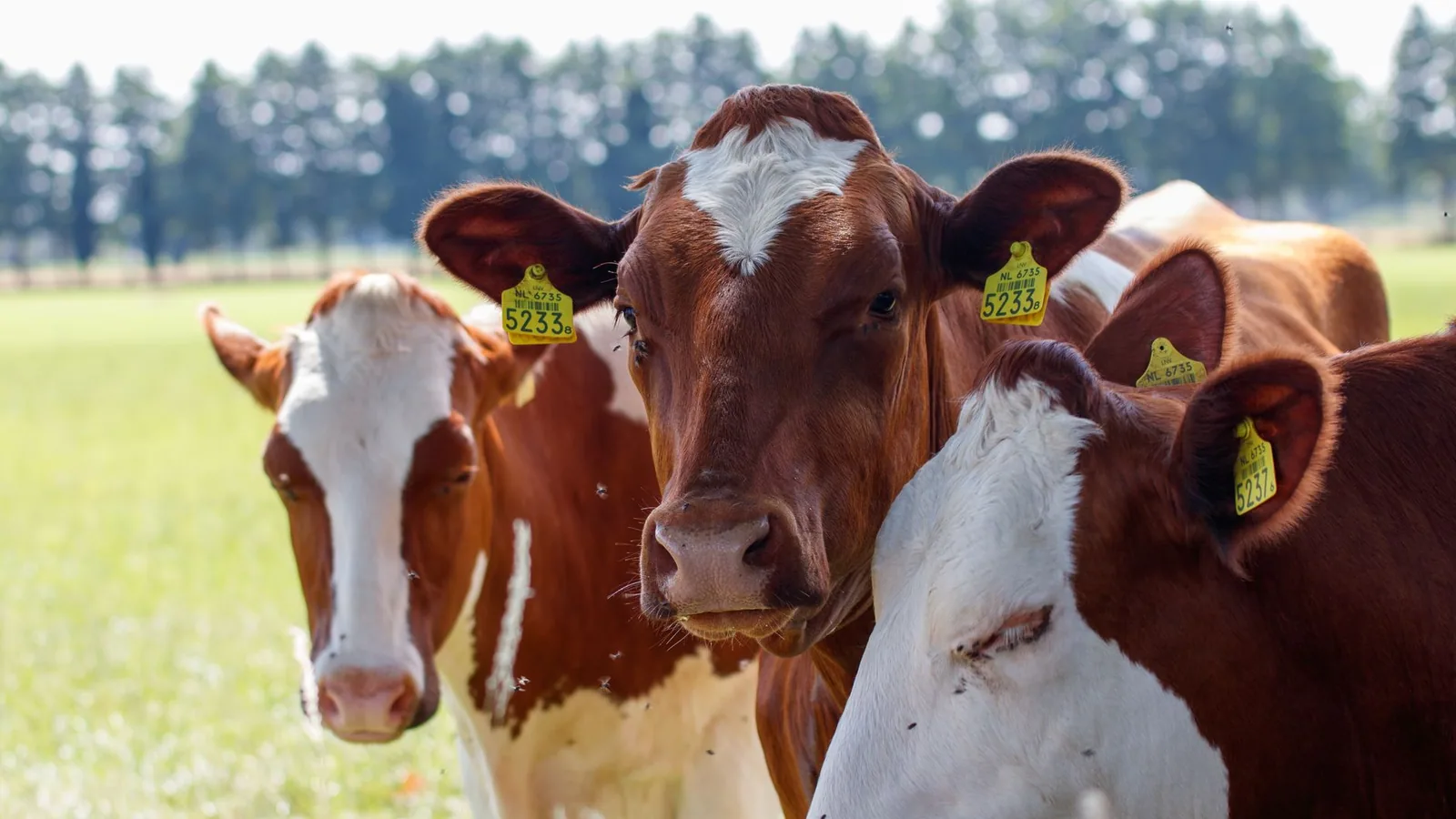
The epidemiology of Cooperia oncophora in calves on pasture is driven by the interaction between environmental conditions, pasture contamination, and host immunity. Calves are the primary age group affected, as they lack acquired immunity. Adult cattle typically harbor low burdens and serve as a source of pasture contamination.
Pasture Contamination and Larval Survival
The primary source of infection for calves is the ingestion of L3 larvae from contaminated pasture. The survival and availability of L3 larvae on pasture are influenced by temperature, humidity, and solar radiation. L3 larvae can survive for extended periods, particularly in temperate climates. Studies have demonstrated high levels of L3 overwinter survival for multiple cattle gastrointestinal nematode species, including C. oncophora, on western Canadian pastures as revealed by ITS2 rDNA metabarcoding [1]. This overwintering capability ensures that pastures become infective early in the grazing season, exposing naive calves to significant larval challenges. The persistence of infective larvae on pasture has been documented in various regions, including Maine dairy cattle pastures [2] and California high Sierra pastures [3].
Seasonal Patterns
Seasonal patterns of fecal egg counts (FECs) and nematode species composition have been characterized in different geographic regions. In Scottish dairy calves, distinct seasonal peaks in FECs have been observed, with C. oncophora often dominating early in the grazing season [4]. In Australian pasture-based dairy herds, FECs also exhibit seasonal variation, influenced by rainfall and temperature [5]. In Atlantic temperate environments, the dynamics of infestation of cattle and pasture by gastrointestinal nematodes, including C. oncophora, follow a predictable pattern: low larval availability in winter, a rise in spring, a peak in summer, and a decline in autumn [6]. In the Red River Delta of Vietnam, the epidemiology of nematode infections in cattle is influenced by the monsoon season, with peak transmission occurring during the rainy months [7].
Hypobiosis
Hypobiosis, or arrested larval development, is a key epidemiological feature of some trichostrongylid nematodes, particularly O. ostertagi. While C. oncophora is less prone to hypobiosis than O. ostertagi, it can still undergo arrested development under certain conditions. Studies in the Pyrenees (Spain) have documented monthly fluctuations of worm burdens and hypobiosis of gastrointestinal nematodes in calves, with C. oncophora showing some degree of arrested development during the winter months [8]. In cow-calf production systems in the American Midwest, hypobiosis of O. ostertagi is a major factor in the epidemiology of ostertagiasis, but C. oncophora burdens also contribute to overall parasite transmission [9].
Host Immunity
Calves gradually acquire immunity to C. oncophora after repeated exposure. However, immunity develops slowly and is often incomplete, particularly in the first grazing season. The effect of preventive anthelmintic treatment on acquired resistance to gastrointestinal nematodes has been studied. Frequent or intensive anthelmintic treatment can interfere with the development of natural immunity, leaving calves more susceptible to infection later in the season [10]. This has important implications for designing control programs that balance parasite suppression with the development of protective immunity.
Wildlife Reservoirs
C. oncophora can infect a range of ruminant hosts, including wildlife. Reindeer have been shown to serve as hosts for nematode parasites of sheep and cattle, including C. oncophora [11]. This cross-species transmission potential complicates control efforts in areas where cattle graze on shared pastures with wild ruminants. In South Africa, helminths of dairy calves on dry-land Kikuyu grass pastures included C. oncophora, with wildlife acting as potential reservoirs [12].
Clinical Signs and Pathology
Infections with C. oncophora are often subclinical, particularly in older animals. In naive calves with high burdens, clinical signs can include diarrhea, reduced appetite, weight loss, and poor growth performance. The primary pathological effect is enteritis, characterized by villous atrophy, crypt hyperplasia, and infiltration of inflammatory cells in the small intestinal mucosa. These changes lead to malabsorption and protein-losing enteropathy. The effect on liveweight gain can be substantial, even in the absence of overt clinical signs. Studies have demonstrated that using anthelmintics with incomplete efficacy against resistant C. oncophora results in reduced liveweight gain in calves [13]. This highlights the economic impact of subclinical infections and the importance of effective control.
Diagnosis
Fecal Egg Counts
The standard diagnostic method for detecting C. oncophora infection is the fecal egg count (FEC) using a modified McMaster technique or a FLOTAC method. Eggs are oval, thin-shelled, and contain a morula. They are morphologically indistinguishable from eggs of other trichostrongylid nematodes, such as O. ostertagi and Cooperia punctata. Therefore, FECs provide a quantitative measure of total nematode egg output but do not allow species-specific identification.
Larval Culture
To differentiate C. oncophora from other trichostrongylids, fecal samples can be cultured to obtain third-stage larvae (L3). L3 larvae are identified based on morphological features, including the length of the sheath tail and the number of intestinal cells. C. oncophora L3 larvae have a characteristic long, whip-like tail and 16 intestinal cells.
Molecular Diagnostics
Molecular methods, particularly ITS2 rDNA metabarcoding, have revolutionized the identification and quantification of gastrointestinal nematode species in mixed infections. This technique allows for the simultaneous detection and relative quantification of multiple nematode species from fecal samples or pasture larval extracts. ITS2 rDNA metabarcoding has been used to demonstrate high levels of L3 overwinter survival for C. oncophora and other species on pasture [1]. This approach provides a more accurate and detailed picture of the parasite community than traditional morphological identification.
Serological Assays
A multiplex fluorescence immunological assay has been developed for the simultaneous detection of antibodies against C. oncophora, Dictyocaulus viviparus, and Fasciola hepatica in cattle [14]. This assay uses recombinant antigens and allows for the detection of exposure to these parasites in a single serum sample. Serological assays are useful for herd-level surveillance and for assessing exposure history, but they do not differentiate between current and past infections.
Diagnostic Decision Tree
The following Mermaid diagram illustrates a diagnostic decision tree for investigating C. oncophora infection in calves on pasture.
graph TD
 A[Calves with poor growth or diarrhea on pasture] --> B{Collect fecal samples}
 B --> C["'Perform Fecal Egg Count (FEC')"]
 C --> D{FEC > 200 epg?}
 D -->|Yes| E[Perform Larval Culture or ITS2 rDNA Metabarcoding]
 D -->|No| F["Consider other causes: viral, bacterial, nutritional"]
 E --> G{Cooperia oncophora dominant?}
 G -->|Yes| H["'Assess anthelmintic efficacy: Fecal Egg Count Reduction Test (FECRT')"]
 G -->|No| I[Identify other nematode species and manage accordingly]
 H --> J{FECRT < 90%?}
 J -->|Yes| K[Anthelmintic resistance suspected. Confirm with molecular testing for resistance alleles.]
 J -->|No| L[Effective anthelmintic. Implement strategic control program.]
 K --> M[Switch to anthelmintic class with different mode of action. Implement integrated control measures.]
Treatment and Anthelmintic Control
Anthelmintic Classes
Several anthelmintic classes are available for the control of C. oncophora in cattle. These include:
- Macrocyclic Lactones (MLs): Ivermectin, doramectin, eprinomectin, and moxidectin. These drugs act on glutamate-gated chloride channels, causing paralysis and death of the nematode.
- Benzimidazoles (BZs): Fenbendazole, oxfendazole, and albendazole. These drugs bind to beta-tubulin, inhibiting microtubule polymerization.
- Imidazothiazoles: Levamisole. This drug acts as a nicotinic acetylcholine receptor agonist, causing spastic paralysis.
- Amino-Acetonitrile Derivatives (AADs): Monepantel. This drug acts on a specific nicotinic acetylcholine receptor subunit (Hco-MPTL-1) found only in nematodes.
- Spiroindoles: Derquantel. This drug acts as a nicotinic acetylcholine receptor antagonist.
Anthelmintic Resistance
Anthelmintic resistance in C. oncophora is a growing global concern. Resistance to macrocyclic lactones, particularly ivermectin, is widespread. The use of anthelmintics with incomplete efficacy against resistant C. oncophora has been shown to negatively affect liveweight gain in calves [13]. This underscores the need for resistance surveillance and the implementation of resistance management strategies.
Fecal Egg Count Reduction Test (FECRT)
The FECRT is the standard method for detecting anthelmintic resistance in the field. Fecal samples are collected from a group of animals before and after treatment. A reduction in FEC of less than 90% (or less than 95% for some guidelines) indicates the presence of resistance. The FECRT should be performed for each anthelmintic class used on the farm.
Integrated Control Strategies
Effective control of C. oncophora in calves on pasture requires an integrated approach that combines strategic anthelmintic use with pasture management.
- Strategic Anthelmintic Treatment: Treatment should be timed to reduce pasture contamination and to protect calves during periods of high larval challenge. A common strategy is to treat calves at turnout and then again 3 to 6 weeks later. The effect of repeated moves to clean pasture on the build-up of gastrointestinal nematode infections has been studied, and this practice can reduce the need for anthelmintic treatment [15].
- Pasture Management: Rotational grazing, moving calves to clean pasture (pasture that has not been grazed by cattle in the previous 12 months), and mixed grazing with sheep or other species can reduce larval contamination. The natural seeding of marshland pastures with bovine gastrointestinal parasites has been documented, highlighting the importance of avoiding high-risk pastures [16].
- Refugia-Based Strategies: Maintaining a population of parasites in refugia (unexposed to anthelmintics) is critical for slowing the development of resistance. This can be achieved by leaving a portion of the herd untreated or by treating only when FECs exceed a threshold.
- Monitoring and Surveillance: Regular FEC monitoring, combined with FECRT and molecular diagnostics, allows for early detection of resistance and adjustment of control strategies.
Conclusion
Cooperia oncophora remains a significant gastrointestinal nematode of calves on pasture, with the potential to cause substantial production losses. Its epidemiology is shaped by environmental factors, pasture management, and host immunity. The emergence of anthelmintic resistance, particularly to macrocyclic lactones, necessitates a shift toward integrated control strategies that combine strategic treatment with pasture management and refugia-based approaches. Molecular diagnostics, including ITS2 rDNA metabarcoding and multiplex serological assays, provide powerful tools for species-specific identification and surveillance. A comprehensive understanding of the epidemiology and resistance dynamics of C. oncophora is essential for designing sustainable control programs that maintain calf health and productivity.
References
[1] Wang T, Avramenko RW, Redman EM, et al. High levels of third-stage larvae (L3) overwinter survival for multiple cattle gastrointestinal nematode species on western Canadian pastures as revealed by ITS2 rDNA metabarcoding. Parasit Vectors. 2020. URL: https://pubmed.ncbi.nlm.nih.gov/32912326/
[2] Gibbs HC. Persistence on pasture of the infective larvae of nematodes parasitizing Maine dairy cattle. Am J Vet Res. 1980. URL: https://pubmed.ncbi.nlm.nih.gov/7224300/
[3] Baker NF, Fisk RA, Rimbey CW. Seasonal occurrence of infective nematode larvae in California high Sierra pastures grazed by cattle. Am J Vet Res. 1984. URL: https://pubmed.ncbi.nlm.nih.gov/24049905/
[4] Campbell P, McIntyre J, O'Neill K, et al. Seasonal patterns of faecal egg counts and gastrointestinal nematode species composition in Scottish dairy calves. Vet Parasitol. 2025. URL: https://pubmed.ncbi.nlm.nih.gov/40795759/
[5] Loughnan T, Mansell P, Playford M, et al. Faecal egg counts in Australian pasture-based dairy herds. Vet Parasitol Reg Stud Reports. 2024. URL: https://pubmed.ncbi.nlm.nih.gov/38772650/
[6] Nogareda C, Mezo M, Uriarte J, et al. Dynamics of infestation of cattle and pasture by gastrointestinal nematodes in an Atlantic temperate environment. J Vet Med B Infect Dis Vet Public Health. 2006. URL: https://pubmed.ncbi.nlm.nih.gov/17062122/
[7] Holland WG, Luong TT, Nguyen LA, et al. The epidemiology of nematode and fluke infections in cattle in the Red River Delta in Vietnam. Vet Parasitol. 2000. URL: https://pubmed.ncbi.nlm.nih.gov/11035232/
[8] Almería S, Llorente MM, Uriarte J. Monthly fluctuations of worm burdens and hypobiosis of gastrointestinal nematodes of calves in extensive management systems in the Pyrenees (Spain). Vet Parasitol. 1996. URL: https://pubmed.ncbi.nlm.nih.gov/9017870/
[9] Stromberg BE, Corwin RM. Epizootiology of Ostertagia ostertagi in cow-calf production systems in the American Midwest. Vet Parasitol. 1993. URL: https://pubmed.ncbi.nlm.nih.gov/8484220/
[10] Claerebout E, Dorny P, Vercruysse J, et al. Effects of preventive anthelmintic treatment on acquired resistance to gastrointestinal nematodes in naturally infected cattle. Vet Parasitol. 1998. URL: https://pubmed.ncbi.nlm.nih.gov/9650866/
[11] Hrabok JT, Oksanen A, Nieminen M, et al. Reindeer as hosts for nematode parasites of sheep and cattle. Vet Parasitol. 2006. URL: https://pubmed.ncbi.nlm.nih.gov/16386848/
[12] Horak IG, Evans U, Purnell RE. Parasites of domestic and wild animals in South Africa. XLV. Helminths of dairy calves on dry-land Kikuyu grass pastures in the Eastern Cape Province. Onderstepoort J Vet Res. 2004. URL: https://pubmed.ncbi.nlm.nih.gov/15732456/
[13] Candy PM, Waghorn TS, Miller CM, et al. The effect on liveweight gain of using anthelmintics with incomplete efficacy against resistant Cooperia oncophora in cattle. Vet Parasitol. 2018. URL: https://pubmed.ncbi.nlm.nih.gov/29426477/
[14] Karanikola SN, Krücken J, Ramünke S, et al. Development of a multiplex fluorescence immunological assay for the simultaneous detection of antibodies against Cooperia oncophora, Dictyocaulus viviparus and Fasciola hepatica in cattle. Parasit Vectors. 2015. URL: https://pubmed.ncbi.nlm.nih.gov/26084663/
[15] Eysker M, van der Aar WM, Boersema JH, et al. The effect of repeated moves to clean pasture on the build up of gastrointestinal nematode infections in calves. Vet Parasitol. 1998. URL: https://pubmed.ncbi.nlm.nih.gov/9653993/
[16] Smith HJ. On the natural seeding of marshland pastures with bovine gastrointestinal parasites. Can J Comp Med. 1974. URL: https://pubmed.ncbi.nlm.nih.gov/4274825/
[17] Agneessens J, Dorny P, Hollanders W, et al. Epidemiological observations on gastrointestinal nematode infections in grazing cow-calf pairs in Belgium. Vet Parasitol. 1997. URL: https://pubmed.ncbi.nlm.nih.gov/9187031/
[18] Gibbs HC. Ostertagiasis in the cow and weaned calf in the northeastern USA. Vet Parasitol. 1993. URL: https://pubmed.ncbi.nlm.nih.gov/8484217/
[19] Charles TP, Baker NF. Seasonal prevalence of gastrointestinal nematodes of beef calves grazed on irrigated pastures in the lower Sacramento Valley of California. Am J Vet Res. 1988. URL: https://pubmed.ncbi.nlm.nih.gov/3377319/
Disclaimer: This article is for educational and informational purposes only. It is not intended to substitute for professional veterinary advice, diagnosis, treatment, or regulatory guidance. Always consult a licensed veterinarian or qualified specialist regarding animal health, disease diagnosis, and therapeutic decisions.