
Coccidiosis in Calves: Pathogenesis, Economic Impact, and Advances in Molecular Diagnostics
Introduction
Bovine coccidiosis is a protozoan enteric disease caused by apicomplexan parasites of the genus Eimeria. The disease represents a major health and economic burden in calf rearing operations worldwide. While subclinical infections are common and often go undetected, clinical outbreaks of hemorrhagic diarrhea, dehydration, and growth retardation can result in substantial mortality and morbidity in young stock [1, 2, 3]. The global prevalence of Eimeria spp. in cattle has been systematically reviewed, with infection rates varying widely by geographic region, management system, and diagnostic methodology [1]. This article provides an exhaustive review of the pathogenesis, economic consequences, and recent advances in molecular diagnostics for bovine coccidiosis, with a focus on the application of modern nucleic acid amplification techniques.
Etiology and Eimeria Species Diversity
Bovine coccidiosis is caused by several species of Eimeria, which are host-specific obligate intracellular parasites. The most pathogenic species in cattle are Eimeria bovis and Eimeria zuernii, which are responsible for the majority of clinical outbreaks [1, 2, 4]. Other species such as Eimeria alabamensis, Eimeria auburnensis, Eimeria ellipsoidalis, and Eimeria canadensis are generally considered less pathogenic but can contribute to mixed infections and subclinical disease [1, 60]. A systematic review and meta-analysis of global Eimeria spp. prevalence in cattle identified E. bovis and E. zuernii as the dominant species across all continents, with pooled prevalence estimates exceeding 40% in some regions [1]. Molecular characterization studies in Greece, Thailand, and Indonesia have confirmed the predominance of these pathogenic species in dairy and beef operations [2, 4, 60].
The life cycle of Eimeria is monoxenous and involves both asexual (merogony) and sexual (gametogony) phases within the intestinal epithelium. Sporulated oocysts are ingested by the calf, and sporozoites are released in the small intestine. Sporozoites invade enterocytes and undergo merogony, producing merozoites that infect adjacent cells. After several generations of asexual replication, gametogony produces macrogametes and microgametes. Fertilization results in the formation of unsporulated oocysts, which are shed in the feces. Sporulation occurs in the external environment under favorable conditions of temperature, humidity, and oxygen [5, 48].
Pathogenesis and Host-Parasite Interactions
The pathogenesis of bovine coccidiosis is driven by the destruction of intestinal epithelial cells during merogony and gametogony. E. bovis primarily parasitizes the ileum, cecum, and colon, while E. zuernii targets the cecum and colon. The massive release of merozoites from first-generation meronts (macromeronts) of E. bovis causes extensive epithelial destruction, leading to hemorrhage, protein-losing enteropathy, and malabsorption [6, 63]. Histopathological examination reveals villous atrophy, crypt hyperplasia, and infiltration of inflammatory cells, predominantly neutrophils and macrophages.
The host immune response to Eimeria infection involves both innate and adaptive components. Intestinal mononuclear phagocytes, including dendritic cells and macrophages, play a critical role in antigen presentation and cytokine production. The transcriptomic response of the intestinal mucosa to Cryptosporidium parvum infection in neonatal calves has been characterized, revealing alterations in epithelial barrier function and transcellular transport systems [7]. While these studies focus on Cryptosporidium, similar mechanisms are likely involved in Eimeria infections, as both are apicomplexan parasites that invade intestinal epithelial cells. Coinfections with Eimeria and other enteric pathogens, including Cryptosporidium parvum and bovine coronavirus, are common and can exacerbate clinical disease [8, 9, 10]. A study in Japan identified Eimeria and Cryptosporidium as the primary protozoan pathogens associated with diarrhea in calves [9]. Metagenomic analysis has further revealed the complex interplay between intestinal protozoan parasites and the gut microbiome, with protozoal infections altering bacterial community composition and diversity.
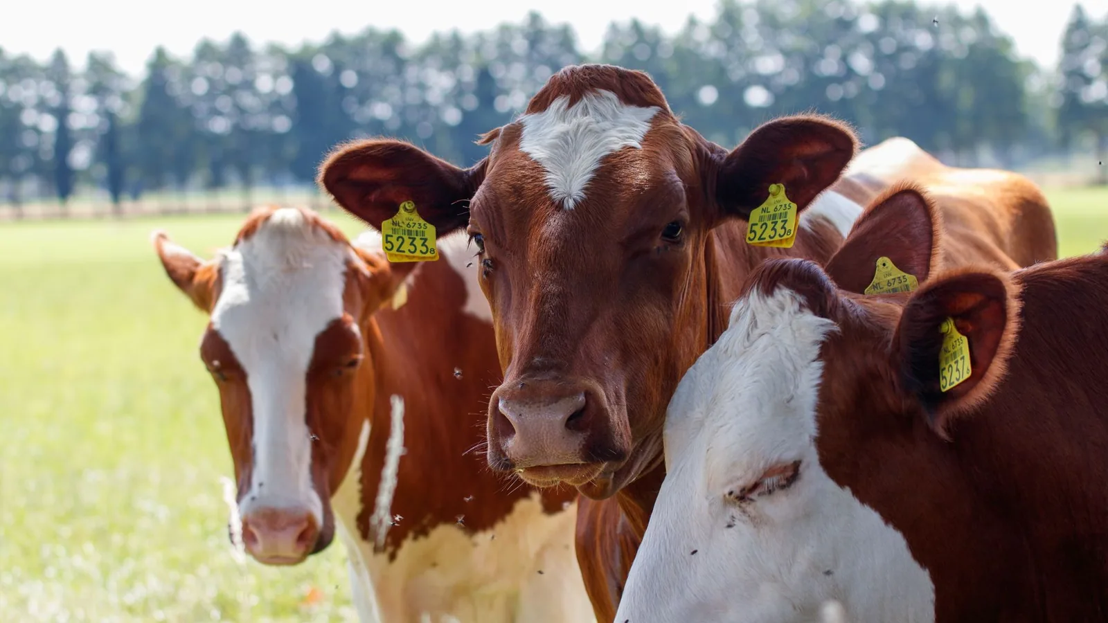
Clinical signs of coccidiosis typically appear in calves between 3 weeks and 6 months of age. The prepatent period ranges from 15 to 21 days for E. bovis and 12 to 17 days for E. zuernii. Acute disease is characterized by profuse watery diarrhea, often with frank blood and mucus, tenesmus, dehydration, anorexia, and fever [6, 36]. Subclinical infections result in reduced feed conversion efficiency and weight gain, which are economically significant but often overlooked. A retrospective evaluation of acid-base imbalances and clinicopathologic alterations in hospitalized calves with Eimeria-associated diarrhea identified metabolic acidosis, electrolyte disturbances, and elevated acute-phase proteins as prognostic indicators [6].
Economic Impact
The economic impact of bovine coccidiosis is substantial and multifaceted. Direct losses include mortality, treatment costs, and reduced weight gain. Indirect losses arise from decreased feed efficiency, increased susceptibility to secondary infections, and the cost of preventive measures such as anticoccidial feed additives and hygiene protocols [11, 48]. A scoping review of antimicrobial drugs used in the prevention and control of protozoal and bacterial calf diarrhea highlighted the widespread use of anticoccidial agents, including ionophores (monensin, narasin) and triazines (toltrazuril, diclazuril), in calf rearing operations [11, 38].
The global prevalence of Eimeria spp. in cattle, as determined by systematic review, indicates that a large proportion of calves are exposed to pathogenic species during the first months of life [1]. In endemic herds, subclinical infections can result in growth depression of 10% to 20% compared to uninfected cohorts. The cost of treatment, including fluid therapy, anticoccidial drugs, and supportive care, can be significant, particularly in outbreaks affecting large numbers of calves. The development of anticoccidial resistance, while less documented in cattle than in poultry, is a growing concern that may increase future economic losses [38, 48].
Management strategies to mitigate economic losses include early detection through regular fecal monitoring, implementation of biosecurity measures, and strategic use of anticoccidial drugs. Supplementation with narasin or monensin has been shown to reduce oocyst shedding and improve growth performance in naturally infected calves. Probiotic supplementation with Enterococcus faecium has also been investigated for its effects on performance and health in preweaning calves, with some studies reporting reduced diarrhea incidence. The use of direct-fed microbials and fermented milk products represents a non-antibiotic approach to disease prevention.
Advances in Molecular Diagnostics
Traditional diagnosis of bovine coccidiosis relies on microscopic examination of fecal samples for the presence of oocysts. While this method is inexpensive and widely available, it has several limitations, including low sensitivity for low-level infections, inability to differentiate between pathogenic and non-pathogenic species based on morphology alone, and the requirement for skilled microscopists [2, 4]. The development of molecular diagnostic assays has addressed many of these limitations, providing high sensitivity, specificity, and the ability to identify and quantify individual Eimeria species.
Quantitative PCR (qPCR)
Quantitative PCR (qPCR) assays targeting the internal transcribed spacer 1 (ITS-1) region of the ribosomal RNA gene have been developed for the detection and quantification of Eimeria species in bovine fecal samples. The ITS-1 region is highly conserved within species but exhibits sufficient interspecific variation to allow for species-specific primer design [2, 4]. Multiplex qPCR assays can simultaneously detect multiple Eimeria species, as well as other enteric pathogens such as Cryptosporidium spp. and Giardia duodenalis [12, 13, 14]. The use of probe-based qPCR (e.g., TaqMan) provides quantitative data on oocyst shedding intensity, which is valuable for assessing infection severity and monitoring treatment efficacy.
A study in Greece used qPCR to investigate Eimeria spp. infection in weaned dairy calves and identified associated risk factors, including age, housing density, and hygiene practices [2]. Molecular characterization of Eimeria species in dairy cattle in Indonesia using qPCR confirmed the presence of E. bovis and E. zuernii as the dominant pathogenic species [4]. The high sensitivity of qPCR allows for the detection of infections in subclinically shedding animals, which are important reservoirs for environmental contamination.
Loop-Mediated Isothermal Amplification (LAMP)
Loop-mediated isothermal amplification (LAMP) is an isothermal nucleic acid amplification technique that offers several advantages over qPCR for field-based diagnostics. LAMP does not require thermal cycling equipment, can be performed at a constant temperature (typically 60-65 degrees Celsius), and produces results in under one hour. A colorimetric LAMP test for the detection of Cryptosporidium in calf feces has been developed, demonstrating high sensitivity and specificity compared to microscopy and qPCR. While this assay targets Cryptosporidium, the same approach can be adapted for Eimeria species detection. The development of a "Rapid-Crypto Colorimetric LAMP Test" for cryptosporidiosis in newborn calves illustrates the potential for similar point-of-care tests for bovine coccidiosis.
LAMP assays for Eimeria species typically target the ITS-1 region or the 18S rRNA gene. The use of colorimetric indicators, such as hydroxynaphthol blue or phenol red, allows for visual interpretation of results without the need for specialized equipment. This makes LAMP particularly suitable for on-farm use in resource-limited settings. The sensitivity of LAMP is comparable to that of qPCR, with detection limits as low as 10 oocysts per gram of feces.
Deep Learning and Automated Detection
Recent advances in computational biology have led to the development of deep learning-based tools for the automated detection of protozoan oocysts in fecal samples. A deep learning-based tool for rapid and automated detection of Cryptosporidium oocysts has been described, using convolutional neural networks to analyze microscopic images [15]. This approach can be extended to Eimeria oocyst detection, providing a high-throughput screening method for epidemiological surveys. The integration of deep learning with digital microscopy has the potential to reduce diagnostic turnaround time and eliminate inter-observer variability.
Genotyping and Molecular Epidemiology
Molecular genotyping of Eimeria species provides valuable information for epidemiological studies and the tracking of infection sources. Sequence analysis of the ITS-1 region and the cytochrome c oxidase subunit I (COI) gene allows for the identification of species and subtypes [2, 4, 16]. Phylogenetic analysis of Eimeria isolates from different geographic regions can reveal patterns of transmission and the introduction of new strains. A study in India used molecular characterization to identify Eimeria species infecting buffaloes, providing insights into the genetic diversity of these parasites in subtropical regions [16].
The application of next-generation sequencing (NGS) and metagenomics to fecal samples allows for the simultaneous detection and characterization of all enteric pathogens, including Eimeria, Cryptosporidium, Giardia, and bacteria [10, 62]. Metagenomic analysis has revealed the relationship between intestinal protozoan parasites and the gut microbiome, highlighting the complex interactions that influence disease outcome. While NGS is not yet practical for routine diagnostic use, it is a powerful tool for research and outbreak investigations.
Diagnostic Workflow
The following Mermaid diagram illustrates a diagnostic workflow for bovine coccidiosis, integrating traditional and molecular methods.
flowchart TD
 A[Fecal Sample Collection] --> B{Clinical Signs Present?}
 B -->|Yes| C[Microscopic Examination]
 B -->|No| D[Regular Screening]
 C --> E{Oocysts Detected?}
 E -->|Yes| F[Species Identification]
 E -->|No| G[Consider Other Pathogens]
 F --> H[Quantification via qPCR]
 H --> I[Treatment Decision]
 I --> J[Anticoccidial Therapy]
 J --> K[Follow-up Fecal Exam]
 K --> L{Resolution?}
 L -->|Yes| M[Return to Normal Management]
 L -->|No| N[Re-evaluate Diagnosis]
 N --> O[LAMP or qPCR for Confirmation]
 O --> P[Adjust Treatment Protocol]
 D --> Q[Pooled Fecal qPCR]
 Q --> R{High Oocyst Count?}
 R -->|Yes| S[Targeted Treatment]
 R -->|No| T[Continue Monitoring]
Management and Control Strategies
Effective management of bovine coccidiosis requires an integrated approach combining hygiene, biosecurity, and strategic anticoccidial use. Oocysts are highly resistant to environmental conditions and can survive for months in contaminated bedding and soil [5]. Disinfectants based on ammonia, chlorine, or steam cleaning are effective for oocyst inactivation, but many common disinfectants have limited efficacy [5]. The viability of sporulated oocysts in water and the efficacy of disinfectants in tropical climates have been studied, emphasizing the need for region-specific protocols [5].
Anticoccidial drugs are used both prophylactically and therapeutically. Ionophores such as monensin and narasin are commonly included in calf starter feeds to prevent coccidiosis. Toltrazuril and diclazuril are triazine derivatives that are effective against both asexual and sexual stages of Eimeria. These drugs are typically administered as a single oral dose at the time of peak exposure, usually around 2 to 3 weeks of age. The development of resistance to anticoccidials is a concern, and rotational use of different drug classes is recommended [11, 38].
Vaccination against bovine coccidiosis is not widely practiced, but research into live attenuated vaccines is ongoing. The use of colostrum-derived immunity is important for neonatal protection, and studies have shown that total and pathogen-specific serum immunoglobulin G concentrations are associated with health and growth in neonatal beef calves [54, 59]. Melatonin treatment at dry-off has been shown to reduce postpartum shedding of coccidia in primiparous dairy cows and their calves, suggesting a potential role for hormonal modulation of immunity [17].
Future Directions
The future of bovine coccidiosis diagnostics lies in the development of rapid, point-of-care molecular assays that can be used on-farm. LAMP and recombinase polymerase amplification (RPA) are promising technologies for this purpose. The integration of microfluidics and lab-on-a-chip devices could further simplify sample processing and reduce the time to result. The use of biological foundation models for predicting host-pathogen interactions and drug resistance is an emerging area of research that may inform the development of new anticoccidial agents.
The application of artificial intelligence and machine learning to epidemiological data can improve risk assessment and early warning systems for coccidiosis outbreaks. Deep learning-based image analysis for automated oocyst detection and quantification is likely to become more widely adopted as the technology matures [15]. The combination of molecular diagnostics with genomic surveillance will provide a more comprehensive understanding of Eimeria population dynamics and the emergence of drug-resistant strains.
Conclusion
Bovine coccidiosis remains a significant challenge for the cattle industry, causing substantial economic losses through mortality, morbidity, and reduced growth performance. The pathogenesis of the disease is driven by the destruction of intestinal epithelial cells by pathogenic Eimeria species, particularly E. bovis and E. zuernii. Advances in molecular diagnostics, including qPCR, LAMP, and deep learning-based image analysis, have greatly improved the sensitivity and specificity of detection, enabling early intervention and targeted treatment. The integration of these diagnostic tools with comprehensive management strategies, including hygiene, biosecurity, and strategic anticoccidial use, is essential for effective control of the disease. Future developments in point-of-care molecular assays and genomic surveillance will further enhance our ability to diagnose, monitor, and manage bovine coccidiosis.
References
[1] Shamsi L, Pouryousef A, Mohammadi MR, et al. Eimeria spp. in Cattle: A Global Systematic Review and Meta-Analysis. Vet Med Sci. 2026. URL: https://pubmed.ncbi.nlm.nih.gov/42113544/
[2] Arsenopoulos KV, Chrysanthopoulos S, Papadopoulos E. Molecular Investigation of Eimeria spp. Infection in Weaned Dairy Calves in Thessaly, Greece, and Associated Risk Factors. Int J Mol Sci. 2026. URL: https://pubmed.ncbi.nlm.nih.gov/41898762/
[3] Vilatuña EJ, Cantón G, Ovelar MF, et al. Bovine coccidiosis: Retrospective study in Central Argentina. Vet Parasitol Reg Stud Reports. 2026. URL: https://pubmed.ncbi.nlm.nih.gov/41651633/
[4] Hastutiek P, Suwanti LT, Suprihati E, et al. Bovine coccidiosis and molecular characterization of pathogenic Eimeria species in dairy cattle on Grati-Pasuruan, East Java, Indonesia. Open Vet J. 2025. URL: https://pubmed.ncbi.nlm.nih.gov/40453855/
[5] Cruvinel LB, de Paula LGF, Dos Santos JCF, et al. Viability time of sporulated oocysts of bovine Eimeria spp. in water and efficacy of disinfectants in a region with tropical climate. Vet Res Commun. 2024. URL: https://pubmed.ncbi.nlm.nih.gov/39196493/
[6] Urgibl-Bauer A, Lorch A, Badura D, et al. Retrospective evaluation of acid-base imbalances, clinicopathologic alterations, and prognostic factors in hospitalized calves with Eimeria-associated diarrhea. Front Vet Sci. 2024. URL: https://pubmed.ncbi.nlm.nih.gov/39834917/
[7] Veshkini A, Kühn C, Dengler F, et al. Cryptosporidium parvum infection alters the intestinal mucosa transcriptome in neonatal calves: impacts on epithelial barriers and transcellular transport systems. Front Cell Infect Microbiol. 2024. URL: https://pubmed.ncbi.nlm.nih.gov/39703373/
[8] Varegg MS, Stokstad M, Bartley PM, et al. Cryptosporidium parvum and bovine coronavirus in naturally and experimentally exposed calves: clinical outcome and pathogen shedding. Vet Res. 2026. URL: https://pubmed.ncbi.nlm.nih.gov/41933416/
[9] Kabir MHB, Murakoshi F, Fukuda Y, et al. Identification of Cryptosporidium and Eimeria associated with diarrhea in calves in Japan (2020-2022). Parasitol Res. 2026. URL: https://pubmed.ncbi.nlm.nih.gov/41667631/
[10] Zhao JQ, Fan YY, Lei YD, et al. Molecular characterization of common zoonotic protozoan parasites and bacteria causing diarrhea in dairy calves in Ningxia Hui Autonomous Region, China. Parasite. 2024. URL: https://pubmed.ncbi.nlm.nih.gov/39353100/
[11] Bernal-Córdoba C, Branco-Lopes R, Alonso-López Y, et al. Antimicrobial drugs used in the prevention and control of protozoal and bacterial calf diarrhea: A scoping review. Prev Vet Med. 2025. URL: https://pubmed.ncbi.nlm.nih.gov/40319541/
[12] Louro M, Linhares JCT, Pinto CA, et al. Integrated epidemiological and molecular analysis of Cryptosporidium spp. and Giardia duodenalis isolates in dairy calves from Terceira Island, Azores. Parasitol Res. 2025. URL: https://pubmed.ncbi.nlm.nih.gov/41350959/
[13] Saleh FER, Abdullah HHAM, Aboelsoued D. Coprological and molecular prevalence of Cryptosporidium and Giardia in cattle and irrigation water from Beni-Suef Governorate, Egypt. Sci Rep. 2025. URL: https://pubmed.ncbi.nlm.nih.gov/40707581/
[15] Şahinduran Ş, Kırbaş İ, Çifci A. A deep learning-based tool for rapid and automated detection of Cryptosporidium oocysts: A new approach for veterinary diagnostics and epizootiological surveys. Exp Parasitol. 2026. URL: https://pubmed.ncbi.nlm.nih.gov/41529739/
[16] Das M, Masharing N, Makri MM, et al. Molecular diagnosis and phylogenetic insights of Eimeria species infecting buffaloes (Bubalus bubalis) in Meghalaya's subtropical hilly region, India. Vet Parasitol Reg Stud Reports. 2024. URL: https://pubmed.ncbi.nlm.nih.gov/39326960/
[17] López-Gatius F, Ganau S, Mora-García M, et al. Melatonin Treatment at Dry-off Reduces Postpartum Shedding of Coccidia in Primiparous Dairy Cows and Their Calves. Animals (Basel). 2024. URL: https://pubmed.ncbi.nlm.nih.gov/39682499/
[18] Yu Q, Chen S, Zhang X, et al. Genetic Characterization and Zoonotic Potential of Cryptosporidium spp. and Giardia duodenalis in Cattle From Northeast China. Transbound Emerg Dis. 2025. URL: https://pubmed.ncbi.nlm.nih.gov/42169685/
[19] Gareh A, Elbarbary NK, Abd El-Halim MO, et al. Cryptosporidiosis at the human-ruminant interface in Aswan, Egypt: a one health epidemiological study using microscopy, immunofluorescence, and PCR. BMC Vet Res. 2026. URL: https://pubmed.ncbi.nlm.nih.gov/42152050/
[20] Riggs MW, Schaefer DA. Calf Clinical Model of Cryptosporidiosis for Efficacy Evaluation of Therapeutics. Methods Mol Biol. 2026. URL: https://pubmed.ncbi.nlm.nih.gov/41144210/
[21] Sonzogni-Desautels K, Chen JF, Mead JR, et al. Mouse Models for Use in Cryptosporidium Infection Studies, Quantification of Parasite Burden Using Flow Cytometry, qPCR and Histopathology, and Confocal Imaging of Oocysts. Methods Mol Biol. 2026. URL: https://pubmed.ncbi.nlm.nih.gov/41144209/
[22] de Siqueira LN, de Souza DCT, Mamani RCC, et al. In Vitro Action of Papaya (Carica Papaya) Latex and Pure Papain Against Eimeria Bovis Oocysts. Acta Parasitol. 2025. URL: https://pubmed.ncbi.nlm.nih.gov/41100022/
[23] Toral FLB, Souza MV, de Moraes MM, et al. Coinfection affects the phenotypic but not genetic resistance of cattle to common parasites. Genet Sel Evol. 2025. URL: https://pubmed.ncbi.nlm.nih.gov/41057794/
[24] Sarfaraz MZ, Abbas S, Zaman MA, et al. Phytochemical profiling and anticoccidial activity of Syzygium cumini and Trachyspermum ammi extracts against Eimeria zuernii: Integrated in vitro, in vivo, and in silico approaches. Vet Parasitol. 2025. URL: https://pubmed.ncbi.nlm.nih.gov/40945468/
[25] Mo Z, Xu B, Quan J, et al. Prevalence and distribution of Cryptosporidium spp. in cattle in central and western Inner Mongolia, China. BMC Microbiol. 2025. URL: https://pubmed.ncbi.nlm.nih.gov/40615937/
[26] Siama A, Kalmobe J, Simonet Poueme Namegni R, et al. Prevalence, distribution, and risk factors of Cryptosporidium spp. infection among calves in the Far-North Region of Cameroon. J Vet Sci. 2025. URL: https://pubmed.ncbi.nlm.nih.gov/40461430/
[27] Ramzy G, Mousa W, Gaidan OK, et al. Molecular characterization and associated risk factors of zoonotic cryptosporidiosis in bovine calves and humans in Menoufia governorate, Egypt. Open Vet J. 2025. URL: https://pubmed.ncbi.nlm.nih.gov/40092185/
[28] Premathilaka C, Kodithuwakku S, Midekessa G, et al. Bovine fecal extracellular vesicles: A novel noninvasive tool for understanding gut physiology and pathophysiology in calves. J Dairy Sci. 2025. URL: https://pubmed.ncbi.nlm.nih.gov/39892598/
[29] Ao Y, Gong X, Li J, et al. Characterization of NFDQ1 in Cryptosporidium parvum. Parasit Vectors. 2024. URL: https://pubmed.ncbi.nlm.nih.gov/39462401/
[30] Wang X, Yan A, Wang B, et al. Prevalence and molecular characterization of Cryptosporidium spp. in pre-weaned diarrheic dairy calves and their bedding materials in northern China. Parasitol Res. 2024. URL: https://pubmed.ncbi.nlm.nih.gov/39432112/
[31] Manjunatha UH, Lakshminarayana SB, Jumani RS, et al. Cryptosporidium PI(4)K inhibitor EDI048 is a gut-restricted parasiticidal agent to treat paediatric enteric cryptosporidiosis. Nat Microbiol. 2024. URL: https://pubmed.ncbi.nlm.nih.gov/39379634/
[32] Sayar E, Keles I. Investigation of the diagnostic and prognostic importance of Tumor Necrosis Factor-alfa (TNF-α), Procalcitonin (PCT), Interleukin-6 (IL-6) and Haptoglobin (HP) in calves with neonatal diarrhea. Vet Immunol Immunopathol. 2024. URL: https://pubmed.ncbi.nlm.nih.gov/39368395/
[33] Tamrat H, Tekle Y, Hailemelekot M, et al. Prevalence and associated risk factors of Cryptosporidium infection in calves and hospitalized humans in Libo Kemkem, North Western Ethiopia. Vet Med Sci. 2024. URL: https://pubmed.ncbi.nlm.nih.gov/39285771/
[34] Bergholm J, Tessema TS, Blomström AL, et al. Detection and molecular characterization of major enteric pathogens in calves in central Ethiopia. BMC Vet Res. 2024. URL: https://pubmed.ncbi.nlm.nih.gov/39227796/
[35] Karimi GR, Paykari HM, Abdi-Goudarzi M, et al. Microscopic, Molecular and Antigen Detection and Isolation of Cryptosporidium parvum Parasites in Diarrheal Disease of Calves in Iran. Arch Razi Inst. 2024. URL: https://pubmed.ncbi.nlm.nih.gov/39192950/
Disclaimer: This article is for educational and informational purposes only. It is not intended to substitute for professional veterinary advice, diagnosis, treatment, or regulatory guidance. Always consult a licensed veterinarian or qualified specialist regarding animal health, disease diagnosis, and therapeutic decisions.