Besnoitia besnoiti: A Comprehensive Reference on Bovine Besnoitiosis
By Dr. Zubair Khalid, DVM, MS, PhD ·
Key Takeaways
- Besnoitia besnoiti is an obligate intracellular apicomplexan parasite causing bovine besnoitiosis, a chronic disease characterized by systemic infection, fever, edema, and tissue cysts in skin and mucous membranes, leading to significant economic losses.
- Transmission is primarily mediated by mechanical vectors like biting flies and direct contact with infected tissues, with potential for vertical transmission, though less frequent than in neosporosis.
- The parasite exhibits a complex pathogenesis involving cellular invasion, immune modulation, and the formation of bradyzoite cysts that induce chronic granulomatous inflammation, leading to characteristic scleroderma and infertility in bulls.
- Diagnosis relies on a combination of clinical signs (scleral and skin cysts), serological tests such as competitive ELISA (cELISA) with high sensitivity and specificity, and molecular detection via PCR.
- Currently, no registered chemotherapeutic agents are approved for bovine besnoitiosis; control strategies focus on biosecurity measures, vector control, and culling of infected animals, with no vaccine available.
Introduction
Besnoitia besnoiti is an obligate intracellular apicomplexan parasite belonging to the family Sarcocystidae, closely related to Toxoplasma gondii and Neospora caninum [<a href="#ref-1">1</a>, <a href="#ref-2">2</a>, <a href="#ref-3">3</a>]. The parasite causes bovine besnoitiosis, a chronic and debilitating disease of cattle characterized by systemic infection, fever, edema, and the formation of thick-walled tissue cysts in the skin, mucous membranes, and internal organs [<a href="#ref-4">4</a>, <a href="#ref-5">5</a>, <a href="#ref-6">6</a>]. The disease is endemic in parts of Europe, Africa, Asia, and South America, and is considered an emerging threat in several regions [<a href="#ref-7">7</a>, <a href="#ref-8">8</a>, <a href="#ref-9">9</a>]. Economic losses arise from reduced milk production, impaired fertility, skin damage, and increased culling rates [<a href="#ref-10">10</a>, <a href="#ref-11">11</a>]. This article provides a detailed, citation-grounded review of the biology, epidemiology, pathogenesis, diagnosis, and control of B. besnoiti.
Taxonomy and Phylogeny
B. besnoiti is classified within the phylum Apicomplexa, class Conoidasida, order Eucoccidiorida, family Sarcocystidae, subfamily Toxoplasmatinae [<a href="#ref-1">1</a>, <a href="#ref-12">12</a>]. The genus Besnoitia includes several species infecting mammals and reptiles, with B. besnoiti being the primary agent of bovine besnoitiosis [<a href="#ref-13">13</a>, <a href="#ref-14">14</a>]. Phylogenetic analyses based on ribosomal RNA and internal transcribed spacer (ITS-1) sequences place B. besnoiti in a clade with B. tarandi and B. caprae, distinct from the T. gondii, N. caninum clade [<a href="#ref-12">12</a>, <a href="#ref-14">14</a>]. The parasite exists in three main life-cycle stages: tachyzoites (rapidly dividing), bradyzoites (slowly dividing within cysts), and sporozoites (within oocysts shed by the definitive host) [<a href="#ref-12">12</a>].
Life Cycle and Transmission
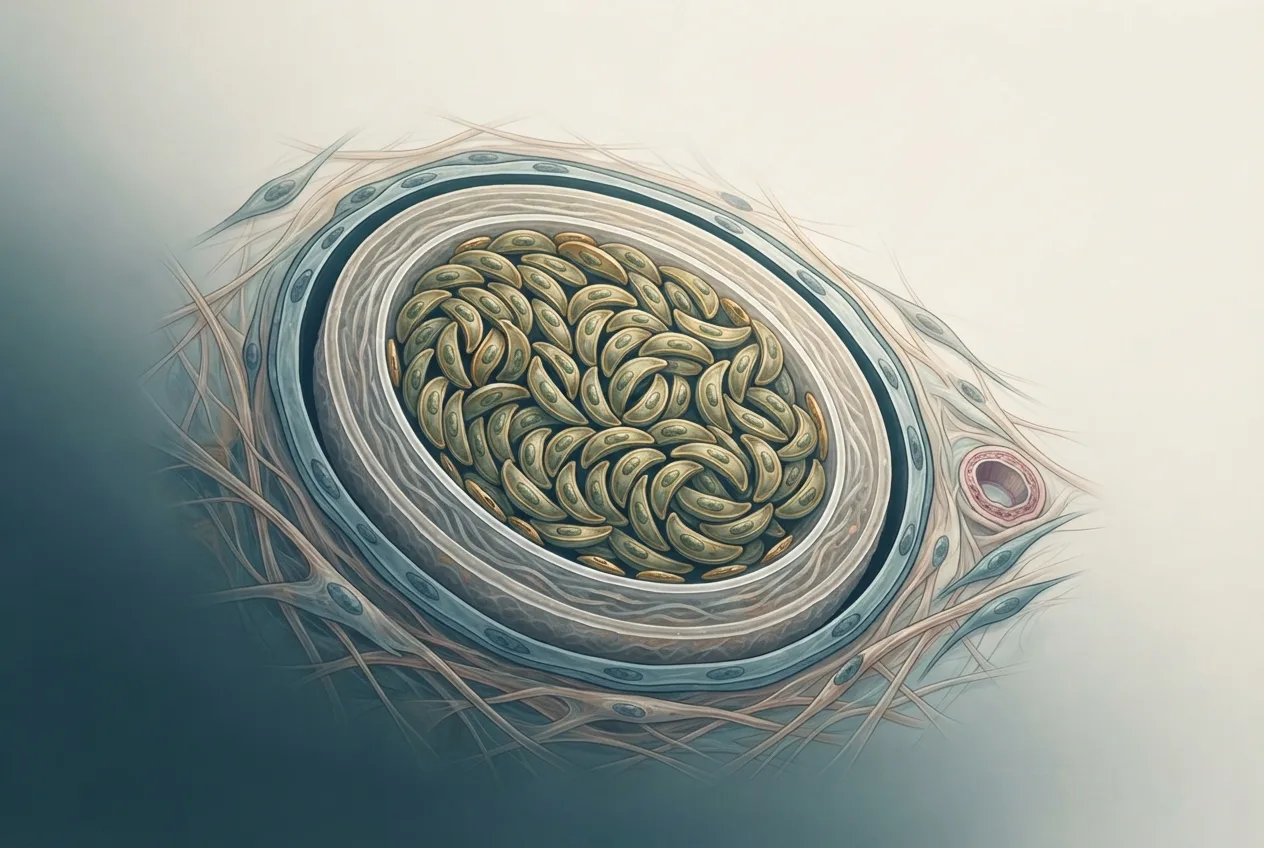
The life cycle of B. besnoiti involves a definitive host (likely felids, though not definitively proven for B. besnoiti) and an intermediate host (cattle and other ruminants) [<a href="#ref-14">14</a>, <a href="#ref-15">15</a>]. Tachyzoites are the proliferative stage in the intermediate host, replicating within endothelial cells, fibroblasts, and macrophages [<a href="#ref-3">3</a>, <a href="#ref-6">6</a>, <a href="#ref-16">16</a>]. Following immune pressure, tachyzoites convert to bradyzoites that form large, whitish, spherical cysts (up to 500 µm in diameter) in connective tissues, particularly the skin, sclera, and nasal mucosa [<a href="#ref-12">12</a>, <a href="#ref-17">17</a>]. Cysts contain hundreds to thousands of bradyzoites and persist for years [<a href="#ref-12">12</a>]. Transmission occurs via mechanical vectors (e.g., biting flies such as Stomoxys calcitrans), direct contact with infected tissues, and possibly through contaminated fomites [<a href="#ref-8">8</a>, <a href="#ref-9">9</a>, <a href="#ref-18">18</a>]. Vertical transmission (transplacental) has been reported but appears less frequent than in neosporosis [<a href="#ref-10">10</a>, <a href="#ref-19">19</a>]. The definitive host, if confirmed as felids, would shed oocysts in feces, but this has not been experimentally demonstrated for B. besnoiti [<a href="#ref-14">14</a>].
graph TD
 A["Definitive host (felid?)"] -->|"Oocysts in feces"| B["Environment"]
 B -->|"Ingestion"| C["Intermediate host (cattle)"]
 C -->|"Tachyzoites"| D["Acute phase: fever, edema"]
 D -->|"Immune response"| E["Bradyzoite cysts in skin, mucosa"]
 E -->|"Mechanical vector (flies)"| F["New intermediate host"]
 E -->|"Direct contact"| F
 F -->|"Tachyzoites"| D
 E -->|"Predation"| A
Epidemiology
Bovine besnoitiosis is endemic in sub-Saharan Africa, southern Europe (Portugal, Spain, France, Italy, Greece), and parts of the Middle East and Asia [<a href="#ref-4">4</a>, <a href="#ref-7">7</a>, <a href="#ref-15">15</a>, <a href="#ref-20">20</a>]. Seroprevalence studies report variable rates: in France, a retrospective study in the Auvergne Rhône-Alpes region found herd-level seropositivity exceeding 50% in some areas [<a href="#ref-4">4</a>]. In Egypt, seroprevalence in Assiut Governorate was 18.7% by ELISA [<a href="#ref-20">20</a>], while in greater Cairo and Beni Suef, antibodies were detected in 12.3% of cattle [<a href="#ref-21">21</a>]. In Saudi Arabia, 8.9% of cattle and 4.2% of sheep were seropositive [<a href="#ref-22">22</a>]. In Malaysia, 6.7% of cattle and 2.5% of goats showed antibodies [<a href="#ref-23">23</a>]. In Turkey, seroprevalence in the Oğuzlar region was 11.4% [<a href="#ref-24">24</a>]. In Iraq, molecular detection (PCR) in Mosul revealed 14.3% of cattle positive [<a href="#ref-25">25</a>]. In Brazil, seroprevalence in horses intended for slaughter was 1.2% [<a href="#ref-26">26</a>]. In Iran, besnoitiosis is considered an emerging threat to livestock and wildlife [<a href="#ref-7">7</a>]. In Europe, outbreaks have been reported in Belgium [<a href="#ref-9">9</a>] and Sicily [<a href="#ref-18">18</a>, <a href="#ref-27">27</a>], and expert elicitation identified climate change, increased vector abundance, and animal movement as key drivers of emergence [<a href="#ref-8">8</a>]. Wild mesocarnivores (foxes, wolves) in Spain have been found shedding Besnoitia-like oocysts, suggesting a potential sylvatic cycle [<a href="#ref-14">14</a>]. European wild lagomorphs appear not to be involved [<a href="#ref-28">28</a>].
Pathogenesis and Host-Parasite Interactions
Cellular Invasion and Replication
B. besnoiti tachyzoites actively invade host cells using a gliding motility mechanism dependent on actin-myosin motors and secretion of microneme and rhoptry proteins [<a href="#ref-2">2</a>, <a href="#ref-3">3</a>]. Calcium signaling plays a critical role in host cell invasion and egress [<a href="#ref-2">2</a>]. Once inside, the parasite resides within a parasitophorous vacuole and acquires nutrients from the host cell. Cholesterol acquisition is essential for tachyzoite replication; the parasite hijacks host Niemann-Pick type C protein 1 (NPC1) to obtain cholesterol from late endosomes/lysosomes [<a href="#ref-3">3</a>]. The scavenger receptor SR-BI is also involved in lipid uptake, and copper chelators such as BLT-1 inhibit replication by blocking SR-BI [<a href="#ref-29">29</a>].
Immune Evasion and Modulation
B. besnoiti modulates host immune responses to establish chronic infection. Transcriptomic analysis of infected fibroblasts reveals upregulation of genes associated with fibrosis and cancer progression, including collagen deposition and transforming growth factor beta (TGF-β) signaling [<a href="#ref-6">6</a>]. In monocyte-derived macrophages, early infection induces type I interferon responses and apoptosis-related transcriptional changes [<a href="#ref-16">16</a>]. The parasite also triggers neutrophil extracellular trap (NET) formation (NETosis) in bovine polymorphonuclear neutrophils (PMNs) [<a href="#ref-30">30</a>, <a href="#ref-31">31</a>, <a href="#ref-32">32</a>, <a href="#ref-33">33</a>]. NETosis is mediated by the CAMKK/AMPK pathway [<a href="#ref-32">32</a>] and P2X1 purinergic receptor signaling [<a href="#ref-33">33</a>]. Extracellular vesicles released by tachyzoites and infected host cells modulate PMN responses, potentially contributing to inflammation and tissue damage [<a href="#ref-34">34</a>]. The metabolic signature of NETs includes increased production of reactive oxygen species and citrullinated histones [<a href="#ref-30">30</a>].
Cyst Formation and Chronic Pathology
Bradyzoite cysts induce a chronic granulomatous inflammatory response. In the skin, cysts cause fibrosis, hyperkeratosis, and alopecia, leading to the characteristic "elephant skin" appearance [<a href="#ref-5">5</a>, <a href="#ref-17">17</a>]. Mass spectrometry imaging of bovine skin cysts reveals distinct lipid and metabolite distributions, including accumulation of sphingolipids and cholesterol esters, which may stabilize the cyst wall [<a href="#ref-17">17</a>]. In the testis, infection leads to degeneration of seminiferous tubules, fibrosis, and impaired spermatogenesis, contributing to male infertility [<a href="#ref-11">11</a>]. Molecular biomarkers of testicular disease progression include upregulation of inflammatory cytokines and matrix metalloproteinases [<a href="#ref-11">11</a>].
Clinical Signs
Bovine besnoitiosis presents in acute and chronic phases. The acute phase, occurring 6-10 days post-infection, is characterized by fever (40-41°C), anorexia, depression, subcutaneous edema (particularly the ventral abdomen, limbs, and head), lymphadenopathy, and nasal and ocular discharge [<a href="#ref-5">5</a>, <a href="#ref-18">18</a>]. Tachyzoites are detectable in blood and tissues during this phase. The chronic phase develops weeks to months later, marked by the appearance of scleroderma (thickened, wrinkled skin), alopecia, and multiple firm, whitish cysts (1-5 mm) visible on the sclera, conjunctiva, nasal mucosa, and skin [<a href="#ref-4">4</a>, <a href="#ref-5">5</a>]. Cysts may also be found in the trachea, esophagus, and tendons. Affected animals often lose condition, suffer from photophobia, and may develop lameness due to tendon cyst formation [<a href="#ref-10">10</a>]. In bulls, orchitis and epididymitis lead to temporary or permanent infertility [<a href="#ref-11">11</a>]. In dairy cows, milk production drops significantly [<a href="#ref-10">10</a>].
Diagnosis
Clinical and Postmortem Examination
Presumptive diagnosis is based on the presence of characteristic scleral and skin cysts in endemic areas [<a href="#ref-5">5</a>, <a href="#ref-18">18</a>]. Postmortem examination reveals cysts in subcutaneous tissue, fascia, and serosal surfaces.
Serological Tests
Several serological assays are available. A competitive ELISA (cELISA) targeting tachyzoite antigens has been validated and shows high sensitivity and specificity [<a href="#ref-35">35</a>]. Indirect fluorescent antibody tests (IFAT) and in-house ELISAs are also used [<a href="#ref-5">5</a>, <a href="#ref-19">19</a>, <a href="#ref-21">21</a>]. A Bayesian latent class model comparing three tests (cELISA, IFAT, and Western blot) estimated sensitivities of 92-98% and specificities of 95-99% for the cELISA [<a href="#ref-5">5</a>].
Molecular Detection
PCR targeting the ITS-1 region or the 18S rRNA gene is used for detection of parasite DNA in blood, skin biopsies, and semen [<a href="#ref-12">12</a>, <a href="#ref-25">25</a>]. Real-time PCR assays offer higher sensitivity for quantifying parasite load [<a href="#ref-25">25</a>].
Histopathology and Cytology
Tissue biopsies stained with hematoxylin and eosin reveal large, thick-walled cysts containing bradyzoites. Immunohistochemistry using polyclonal or monoclonal antibodies against B. besnoiti antigens can confirm the diagnosis [<a href="#ref-12">12</a>].
Table 1: Diagnostic methods for bovine besnoitiosis
| Method | Target | Sensitivity | Specificity | Reference |
|---|---|---|---|---|
| Clinical inspection | Scleral/skin cysts | Moderate | High (in endemic areas) | [<a href="#ref-5">5</a>] |
| Competitive ELISA | Anti-B. besnoiti antibodies | 92-98% | 95-99% | [<a href="#ref-5">5</a>, <a href="#ref-35">35</a>] |
| IFAT | Anti-B. besnoiti antibodies | 85-90% | 90-95% | [<a href="#ref-5">5</a>] |
| Western blot | Anti-B. besnoiti antibodies | 90-95% | 95-98% | [<a href="#ref-5">5</a>] |
| PCR (ITS-1) | Parasite DNA | High | High | [<a href="#ref-12">12</a>, <a href="#ref-25">25</a>] |
| Histopathology | Tissue cysts | Moderate | High | [<a href="#ref-12">12</a>] |
Treatment and Control
No registered chemotherapeutic agent is specifically approved for bovine besnoitiosis. Experimental treatments include bumped kinase inhibitors (BKIs) such as BKI-1708, which interferes with cytokinesis and drives bradyzoite conversion in B. besnoiti and related apicomplexans [<a href="#ref-1">1</a>]. Copper chelators like BLT-1 inhibit tachyzoite replication by targeting SR-BI [<a href="#ref-29">29</a>]. However, these compounds are not yet commercially available. Control relies on biosecurity measures: preventing introduction of infected animals, vector control (e.g., insecticide treatment of barns and animals), and culling of chronically infected animals [<a href="#ref-8">8</a>, <a href="#ref-9">9</a>]. In endemic herds, serological screening and segregation of seropositive animals can reduce transmission [<a href="#ref-10">10</a>, <a href="#ref-19">19</a>]. No vaccine is currently available.
Frequently Asked Questions
What is the definitive host of Besnoitia besnoiti?
The definitive host is suspected to be felids, but this has not been experimentally confirmed for B. besnoiti [<a href="#ref-14">14</a>, <a href="#ref-15">15</a>]. Oocysts morphologically consistent with Besnoitia have been found in feces of wild mesocarnivores in Spain [<a href="#ref-14">14</a>].
How is bovine besnoitiosis transmitted?
Transmission occurs primarily through mechanical vectors such as biting flies, direct contact with infected tissues (e.g., during dehorning or tattooing), and possibly through contaminated needles or fomites [<a href="#ref-8">8</a>, <a href="#ref-9">9</a>, <a href="#ref-18">18</a>]. Vertical transmission is possible but less common [<a href="#ref-10">10</a>, <a href="#ref-19">19</a>].
What are the main clinical signs of bovine besnoitiosis?
Acute signs include fever, subcutaneous edema, and lymphadenopathy. Chronic signs include scleroderma, alopecia, and characteristic cysts on the sclera, conjunctiva, and skin [<a href="#ref-4">4</a>, <a href="#ref-5">5</a>]. Infertility in bulls and reduced milk yield in cows are common [<a href="#ref-10">10</a>, <a href="#ref-11">11</a>].
How is bovine besnoitiosis diagnosed?
Diagnosis is based on clinical examination (scleral cysts), serology (cELISA, IFAT), PCR on blood or tissue, and histopathology of skin biopsies [<a href="#ref-5">5</a>, <a href="#ref-12">12</a>, <a href="#ref-25">25</a>, <a href="#ref-35">35</a>].
Is there a treatment for bovine besnoitiosis?
No registered treatment exists. Experimental compounds such as bumped kinase inhibitors and copper chelators have shown activity in vitro but are not yet available for field use [<a href="#ref-1">1</a>, <a href="#ref-29">29</a>]. Control focuses on biosecurity and vector management [<a href="#ref-8">8</a>, <a href="#ref-9">9</a>].
Can Besnoitia besnoiti infect humans?
B. besnoiti is not considered a human pathogen. No cases of human infection have been reported. The parasite is host-specific to cattle and other ruminants [<a href="#ref-15">15</a>].
What is the economic impact of bovine besnoitiosis?
The disease causes significant economic losses due to reduced milk production, weight loss, infertility, skin damage, and increased culling rates [<a href="#ref-10">10</a>]. In endemic herds, seropositive cows produce less milk and have longer calving intervals [<a href="#ref-10">10</a>].