Histomonas meleagridis (Blackhead): A Comprehensive Veterinary Reference on Etiology, Pathogenesis, Diagnosis, and Control
By Dr. Zubair Khalid, DVM, MS, PhD ·
Key Takeaways
- Histomonas meleagridis is an obligate anaerobic protozoan, the causative agent of histomonosis (blackhead disease), primarily affecting turkeys and characterized by necrotizing inflammation of the ceca and liver.
- Transmission is complex, involving direct cloacal contact, ingestion of Heterakis gallinarum eggs (the primary biological vector), paratenic hosts like earthworms, and potential mechanical transmission by insects.
- Diagnosis relies on gross and histopathological findings of cecal cores and liver necrosis, alongside microscopic identification of trophozoites and highly sensitive PCR assays for parasite DNA in various sample types, including blood.
- Control strategies emphasize stringent biosecurity, preventing contact with known susceptible species, effective control of the Heterakis gallinarum vector through anthelmintics, and maintaining hygienic environments.
- While commercial vaccines are limited, experimental live attenuated vaccines have demonstrated protective efficacy in turkeys and chickens, and research into plant extracts and antimicrobial peptides shows promise for novel therapeutic approaches.
Introduction and Taxonomic Classification
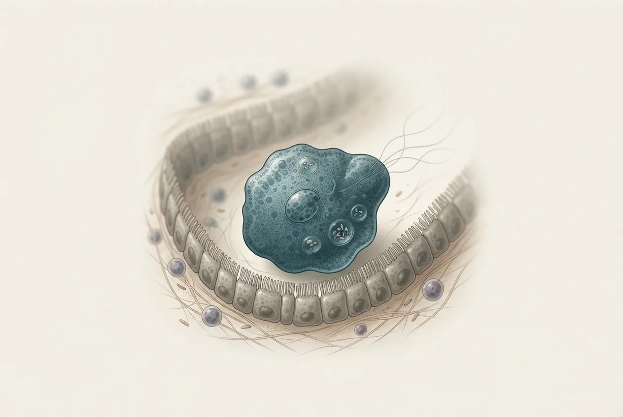
Histomonas meleagridis is a flagellated protozoan parasite belonging to the order Trichomonadida, family Dientamoebidae [<a href="#ref-1">1</a>]. It is the etiologic agent of histomonosis, commonly known as blackhead disease, a highly pathogenic infection primarily affecting turkeys (Meleagris gallopavo) and to a lesser extent chickens, pheasants, peafowl, and other galliform birds [<a href="#ref-2">2</a>, <a href="#ref-3">3</a>, <a href="#ref-4">4</a>]. The organism was first described by Theobald Smith in 1895 and later characterized in detail by Ernst Tyzzer and Everett Lund, whose foundational work established the parasite's life cycle and pathology [<a href="#ref-1">1</a>]. H. meleagridis exists in two morphological forms: a flagellated trophozoite found in the cecal lumen and a non-flagellated invasive form that penetrates tissues [<a href="#ref-1">1</a>, <a href="#ref-5">5</a>]. The parasite is obligately anaerobic and relies on hydrogenosome-based energy metabolism, a feature shared with other trichomonads [<a href="#ref-6">6</a>, <a href="#ref-7">7</a>].
Morphology and Ultrastructure
The trophozoite stage of H. meleagridis is pleomorphic, typically measuring 8 to 15 µm in diameter, and possesses a single nucleus, a prominent axostyle, and one to four flagella depending on the life stage [<a href="#ref-1">1</a>, <a href="#ref-5">5</a>]. The invasive amoeboid form lacks external flagella and exhibits pseudopodial movement, facilitating tissue penetration [<a href="#ref-1">1</a>]. Ultrastructural studies have revealed hydrogenosomes, which are double-membrane organelles involved in pyruvate metabolism and oxygen reduction [<a href="#ref-6">6</a>]. A flavodiiron protein (FDP) has been biochemically characterized in H. meleagridis; this enzyme reduces superoxide radicals, with superoxide serving as a reaction intermediate, and is thought to protect the parasite from oxidative stress within the host [<a href="#ref-6">6</a>]. The surfaceome of clonal strains varies with pathogenicity, with strain-dependent profiles of surface proteins identified via comparative proteomics. Immunoprecipitation studies have further identified host-specific targets of H. meleagridis antigens, revealing differences in antigen recognition between turkeys and chickens [<a href="#ref-8">8</a>].
Life Cycle and Transmission
The life cycle of H. meleagridis is complex and involves both direct and vector-mediated transmission. The primary biological vector is the cecal nematode Heterakis gallinarum, which ingests H. meleagridis trophozoites from infected cecal contents [<a href="#ref-1">1</a>]. The protozoan then invades the nematode's reproductive tissues and is incorporated into H. gallinarum eggs, where it can survive for extended periods in the environment [<a href="#ref-1">1</a>]. Earthworms can act as paratenic hosts, ingesting H. gallinarum eggs and thereby concentrating infective stages for birds that consume them [<a href="#ref-1">1</a>]. Additionally, direct cloacal transmission between birds can occur via the ingestion of fresh cecal droppings, a route that may be particularly important in crowded poultry houses [<a href="#ref-1">1</a>].
Recent molecular evidence has demonstrated the presence of H. meleagridis DNA within Ascaridia galli from chickens in Thailand, suggesting that other nematode species may also serve as vectors [<a href="#ref-9">9</a>]. Insect vectors have been implicated; a survey of dipteran insects around broiler breeder pullet farms identified several species, including house flies and darkling beetles, that carried H. meleagridis DNA, indicating potential mechanical transmission. The parasite can also be transmitted via contaminated fomites, feed, and water.
graph TD
 A["Infected bird: cecal droppings"] -->|"Direct cloacal transmission"| B["Naive bird"]
 A -->|"Ingestion by Heterakis gallinarum"| C["H. gallinarum adult"]
 C -->|"Parasite invades nematode eggs"| D["Eggs shed in feces"]
 D -->|"Earthworm ingestion"| E["Paratenic host: earthworm"]
 E -->|"Bird consumes earthworm"| B
 D -->|"Bird ingests eggs directly"| B
 A -->|"Dipteran vectors"| F["Mechanical transmission via flies/beetles"]
 F --> B
 B -->|"Trophozoites in ceca"| A
Pathogenesis and Pathology
Histomonosis is characterized by necrotizing inflammation of the ceca and liver, with the severity of lesions varying by host species [<a href="#ref-1">1</a>, <a href="#ref-2">2</a>]. In turkeys, the disease is acute and often fatal, whereas chickens typically exhibit milder or subclinical infections [<a href="#ref-2">2</a>, <a href="#ref-10">10</a>]. After ingestion, trophozoites colonize the cecal lumen and subsequently invade the cecal mucosa, causing typhlitis [<a href="#ref-1">1</a>]. The ceca become thickened, ulcerated, and filled with caseous cores composed of necrotic debris and inflammatory cells [<a href="#ref-1">1</a>, <a href="#ref-2">2</a>]. From the ceca, the parasite migrates via the portal circulation to the liver, where it induces focal to coalescing necrotic hepatitis [<a href="#ref-1">1</a>]. Hepatic lesions appear as circular, depressed, yellowish-green areas of necrosis, often described as "target-like" [<a href="#ref-1">1</a>, <a href="#ref-2">2</a>].
Histopathological examination reveals extensive infiltration of heterophils, macrophages, and lymphocytes in both cecal and hepatic tissues [<a href="#ref-2">2</a>, <a href="#ref-11">11</a>]. In chickens, the inflammatory response is less severe, and the parasite may be cleared more effectively, although subclinical infections can still cause production losses [<a href="#ref-2">2</a>, <a href="#ref-10">10</a>]. Concurrent infections with other pathogens can exacerbate pathology. Co-infection with Eimeria species and Escherichia coli in turkey poults resulted in disrupted gut microbiota, suppressed inflammation, and impaired bone health [<a href="#ref-11">11</a>]. Similarly, co-infection with Salmonella has been investigated, though one study found that Salmonella co-infection did not worsen H. meleagridis infection in turkeys [<a href="#ref-12">12</a>, <a href="#ref-13">13</a>]. Concurrent infection with hemorrhagic enteritis virus has been reported in a turkey flock with recurrent blackhead disease [<a href="#ref-14">14</a>]. Additionally, dual infection with Pentatrichomonas hominis has been documented in a blackhead outbreak. In wild turkeys, histomonosis has been found alongside lymphoproliferative disease virus [<a href="#ref-3">3</a>] and other trichomonad species such as Tetratrichomonas, Tritrichomonas, and Simplicimonas [<a href="#ref-4">4</a>].
Clinical Signs and Host Susceptibility
Clinical signs of histomonosis are most pronounced in turkeys and include depression, anorexia, drooping wings, ruffled feathers, sulfur-yellow diarrhea, and cyanosis of the head (the "blackhead" sign, though this is not always present) [<a href="#ref-1">1</a>]. Morbidity and mortality can reach 80-100% in untreated turkey flocks [<a href="#ref-1">1</a>]. In chickens, clinical signs are often absent or limited to mild diarrhea and reduced growth, but severe outbreaks have been reported in backyard flocks [<a href="#ref-2">2</a>]. Peafowl and pheasants are also susceptible, with cecal smear cytology being a useful diagnostic tool in these species [<a href="#ref-5">5</a>, <a href="#ref-15">15</a>]. A new species closely related to H. meleagridis has been identified in turkeys and pheasants in Hungary, suggesting that the genus may be more diverse than previously recognized [<a href="#ref-15">15</a>].
Diagnosis
Diagnosis of histomonosis relies on a combination of clinical history, gross pathology, histopathology, and molecular methods. Microscopic examination of cecal scrapings or liver impressions can reveal the characteristic trophozoites, which are motile and exhibit a jerky, progressive movement in wet mounts [<a href="#ref-5">5</a>]. Staining with Giemsa or trichrome can enhance visualization [<a href="#ref-5">5</a>]. Evans Blue Dye has been validated as an objective quantitative tool for lesion scoring in H. meleagridis and Eimeria infections, providing a reproducible metric for cecal and hepatic damage [<a href="#ref-16">16</a>].
Molecular diagnostics have become increasingly important. PCR assays targeting the 18S rRNA gene or internal transcribed spacer (ITS) regions are highly sensitive and specific for detecting H. meleagridis DNA in cecal contents, liver tissue, and even blood samples [<a href="#ref-17">17</a>]. Early detection of histomoniasis in blood samples by PCR and sequencing has been demonstrated, offering a non-lethal diagnostic option [<a href="#ref-17">17</a>]. Quantitative real-time PCR can also be used to assess parasite load [<a href="#ref-17">17</a>]. Sequencing of PCR amplicons allows differentiation from related trichomonads [<a href="#ref-4">4</a>]. Serum biochemistry changes, including elevated liver enzymes and altered protein profiles, have been described in challenged turkeys and may support diagnosis [<a href="#ref-18">18</a>].
A diagnostic decision tree is presented below.
graph TD
 A["Suspected histomonosis"] --> B{"Clinical signs?"}
 B -->|"Yes: depression, yellow diarrhea, cyanosis"| C["Postmortem examination"]
 B -->|"No: subclinical"| D["Fecal or blood PCR"]
 C --> E{"Gross lesions?"}
 E -->|"Cecal cores + liver necrosis"| F["Microscopy: cecal smear / liver impression"]
 E -->|"No lesions"| D
 F --> G{"Trophozoites observed?"}
 G -->|"Yes"| H["Confirm by PCR/sequencing"]
 G -->|"No"| D
 H --> I["Definitive diagnosis"]
 D --> I
Molecular Biology and Genomics
Proteomic and metabolomic approaches have advanced the understanding of H. meleagridis biology. Tandem mass tag-based quantitative proteomics has been used to compare virulent and attenuated chicken-origin strains, identifying differentially expressed proteins associated with pathogenicity. The metabolic profile of H. meleagridis in Dwyer's media, with and without rice starch, has been characterized using NMR-based metabolomics, revealing shifts in energy metabolism and amino acid utilization [<a href="#ref-7">7</a>]. In vivo metabolomics of chickens co-infected with ascarids and H. meleagridis showed alterations in gut metabolite profiles, including changes in short-chain fatty acids and bile acids [<a href="#ref-19">19</a>]. MicroRNA expression profiling of chicken liver at different times post-infection identified differentially expressed miRNAs that may regulate immune and metabolic pathways [<a href="#ref-20">20</a>]. The surfaceome analysis of clonal strains with different pathogenicity revealed strain-dependent surface protein profiles, which may influence host immune recognition and virulence.
Immunology and Host Response
The host immune response to H. meleagridis involves both innate and adaptive components. In chickens, tissue cytokine responses have been studied in lines selected for high or low humoral antibody responses, with supplemental Limosilactobacillus reuteri modulating the response [<a href="#ref-21">21</a>]. Pro-inflammatory cytokines such as IL-1β, IL-6, and IFN-γ are upregulated in the ceca and liver during infection [<a href="#ref-21">21</a>]. The role of the gut microbiota is critical; infection disrupts the cecal microbial community, with reductions in beneficial bacteria and increases in potentially pathogenic taxa [<a href="#ref-10">10</a>, <a href="#ref-11">11</a>]. In turkeys, co-infection with Eimeria and E. coli exacerbated dysbiosis and suppressed inflammatory signaling [<a href="#ref-11">11</a>]. The microbiota disruption may contribute to the severity of histomonosis by impairing colonization resistance and immune regulation [<a href="#ref-10">10</a>].
Treatment and Control
No commercial vaccines or therapeutic drugs are currently approved for histomonosis in many countries, following the ban of nitroimidazoles (e.g., dimetridazole) in food-producing animals [<a href="#ref-1">1</a>]. Research into alternative control measures is ongoing.
Vaccines: A live clonal monoxenic H. meleagridis vaccine has been shown to provide long-term protection in turkeys [<a href="#ref-22">22</a>]. An attenuated chicken-origin vaccine has also been evaluated for prevention of histomonosis in chickens [<a href="#ref-23">23</a>]. These vaccines rely on the administration of live, attenuated organisms that colonize the ceca and induce protective immunity without causing severe disease [<a href="#ref-22">22</a>, <a href="#ref-23">23</a>].
Plant extracts and natural products: Several plant extracts have demonstrated inhibitory effects against H. meleagridis in vitro and in vivo in chickens, including extracts from Artemisia annua, Allium sativum, and other botanicals [<a href="#ref-24">24</a>]. These compounds may act by disrupting the parasite's membrane integrity or metabolic pathways [<a href="#ref-24">24</a>].
Antimicrobial peptides: Cell-free culture media of Xenorhabdus budapestensis and X. szentirmaii contain antimicrobial peptides that exhibit anti-protozoal activity against H. meleagridis and Leishmania donovani [<a href="#ref-25">25</a>]. These peptides represent a potential novel class of therapeutics.
Management and biosecurity: Control relies on strict biosecurity, including preventing contact between turkeys and chickens or other galliforms, controlling Heterakis gallinarum populations through anthelmintic treatment, and maintaining clean litter and housing. Fenbendazole resistance in H. gallinarum has been documented on a broiler breeder farm, complicating vector control. Dietary factors may influence disease progression; feeding wheat-based diets to turkey poults altered the course of infection compared to corn-based diets [<a href="#ref-26">26</a>]. Retrospective investigations of recurring histomonosis on turkey farms have identified risk factors such as previous outbreaks, presence of H. gallinarum, and poor hygiene. A hypotheses-generating case-series study identified conditions favoring histomonosis, including high stocking density and inadequate downtime between flocks.
Epidemiology and Risk Factors
H. meleagridis has a worldwide distribution, with prevalence varying by region and management system. In backyard chickens from Batna Province, Algeria, a prevalence of 12.5% was reported based on histopathological examination [<a href="#ref-2">2</a>]. In wild turkeys in Alabama, USA, histomonosis was detected in conjunction with lymphoproliferative disease virus [<a href="#ref-3">3</a>]. Molecular surveys in Hungary confirmed endemicity of H. meleagridis and a closely related new species in turkeys and pheasants [<a href="#ref-15">15</a>]. The introduction of histomonosis into German turkey flocks has been linked to contaminated equipment, litter, and the movement of infected birds. The role of insect vectors, particularly dipterans, in spreading the parasite within and between farms is increasingly recognized.
Frequently Asked Questions
What is the primary host of Histomonas meleagridis?
The turkey (Meleagris gallopavo) is the most susceptible host, developing severe and often fatal histomonosis, while chickens typically exhibit milder infections [<a href="#ref-1">1</a>, <a href="#ref-2">2</a>].
How is Histomonas meleagridis transmitted?
Transmission occurs via ingestion of Heterakis gallinarum eggs containing the parasite, consumption of earthworms that have ingested such eggs, direct cloacal contact with infected cecal droppings, and mechanical carriage by insects such as flies and beetles [<a href="#ref-1">1</a>, <a href="#ref-27">27</a>].
What are the characteristic gross lesions of blackhead disease?
The hallmark lesions are severe typhlitis with caseous cecal cores and focal necrotic hepatitis, with yellowish-green target-like areas of necrosis on the liver surface [<a href="#ref-1">1</a>, <a href="#ref-2">2</a>].
Can histomonosis be diagnosed by PCR from blood samples?
Yes, PCR and sequencing of blood samples have been validated for early detection of histomoniasis, providing a non-lethal diagnostic method [<a href="#ref-17">17</a>].
Are there any effective vaccines against Histomonas meleagridis?
Live attenuated vaccines, including a clonal monoxenic vaccine for turkeys and an attenuated chicken-origin vaccine, have shown protective efficacy in experimental settings [<a href="#ref-22">22</a>, <a href="#ref-23">23</a>].
What control measures are recommended for histomonosis?
Control relies on strict biosecurity, separation of turkeys from other poultry, anthelmintic treatment to control Heterakis gallinarum, and management practices that reduce environmental contamination.
Does dietary composition affect the progression of histomonosis?
Dietary wheat has been shown to influence the progression of H. meleagridis infection in turkey poults, suggesting that feed formulation may be a modifiable risk factor [<a href="#ref-26">26</a>].
Can Histomonas meleagridis infect wild birds?
Yes, wild turkeys, pheasants, and peafowl are susceptible, and the parasite has been detected in wild populations in the United States and Europe [<a href="#ref-3">3</a>, <a href="#ref-4">4</a>, <a href="#ref-15">15</a>].
Related Clinical & Scientific Guides
- Tick-Borne Diseases in Dogs: Pathogens, Clinical Signs, Diagnosis, and Prevention
- Toxoplasmosis in Cats and the Risk of Brain Infection in Humans
- Dog Heartworm and Tick-Borne Disease Prevention
- Histomonas meleagridis: Blackhead Disease in Turkeys, Hepatic and Cecal Pathology, Diagnosis, and Control