Dermanyssus gallinae (Red Mite)
By Dr. Zubair Khalid, DVM, MS, PhD ·
Key Takeaways
- Dermanyssus gallinae (poultry red mite) is a significant haematophagous ectoparasite of poultry, causing substantial economic losses through reduced egg production, increased mortality, and acting as a vector for numerous avian pathogens including Salmonella spp., Newcastle disease virus, and fowl poxvirus.
- The mite exhibits a rapid life cycle (7-9 days under optimal conditions of 25-30°C and high humidity) and a remarkable ability to survive for up to 13 months without a blood meal, complicating eradication efforts and facilitating persistence in empty poultry houses.
- Diagnosis relies on visual inspection, particularly at night, and collection from mite harborage sites, with definitive identification achieved through morphological keys or molecular methods like PCR targeting the cox1 gene.
- Control is challenging due to widespread acaricide resistance; strategies include chemical treatments (carbamates, organophosphates, pyrethroids, amidines, systemic fluralaner) with careful monitoring for residues, and non-chemical methods such as entomopathogenic fungi, predatory mites, and environmental manipulation (heat, humidity, lighting).
- The mite's complex bacterial community and potential role as a reservoir for emerging arboviruses, as indicated by the identification of a novel quaranjavirus, highlight its importance as a biosecurity threat and an area for ongoing research into novel control targets.
Introduction
Dermanyssus gallinae, commonly known as the poultry red mite (PRM), is a haematophagous ectoparasite of birds and the most economically important mite infesting egg-laying hens worldwide [<a href="#ref-1">1</a>, <a href="#ref-2">2</a>]. The mite belongs to the order Mesostigmata, family Dermanyssidae, and exhibits a short life cycle that can be completed in as few as 7 days under optimal conditions [<a href="#ref-1">1</a>, <a href="#ref-3">3</a>]. PRM infestations cause reduced welfare, decreased egg production, increased mortality, and act as vectors for several avian pathogens [<a href="#ref-1">1</a>, <a href="#ref-4">4</a>]. Prevalence in European countries, including Serbia, can reach 80-90% in layer flocks [<a href="#ref-1">1</a>]. In England, surveys have similarly documented widespread infestation [<a href="#ref-5">5</a>]. The mite's ability to survive without a blood meal for up to 13 months and to infest new flocks rapidly complicates eradication efforts [<a href="#ref-1">1</a>]. This article provides a detailed, citation-grounded overview of the biology, epidemiology, vector role, diagnostic approaches, and control strategies for D. gallinae, with emphasis on veterinary and molecular perspectives. Cross-references to related parasites such as Ornithonyssus sylviarum (Northern Fowl Mite) Infestation in Poultry and broader Ectoparasites of Poultry are provided where relevant.
Taxonomy and Morphology
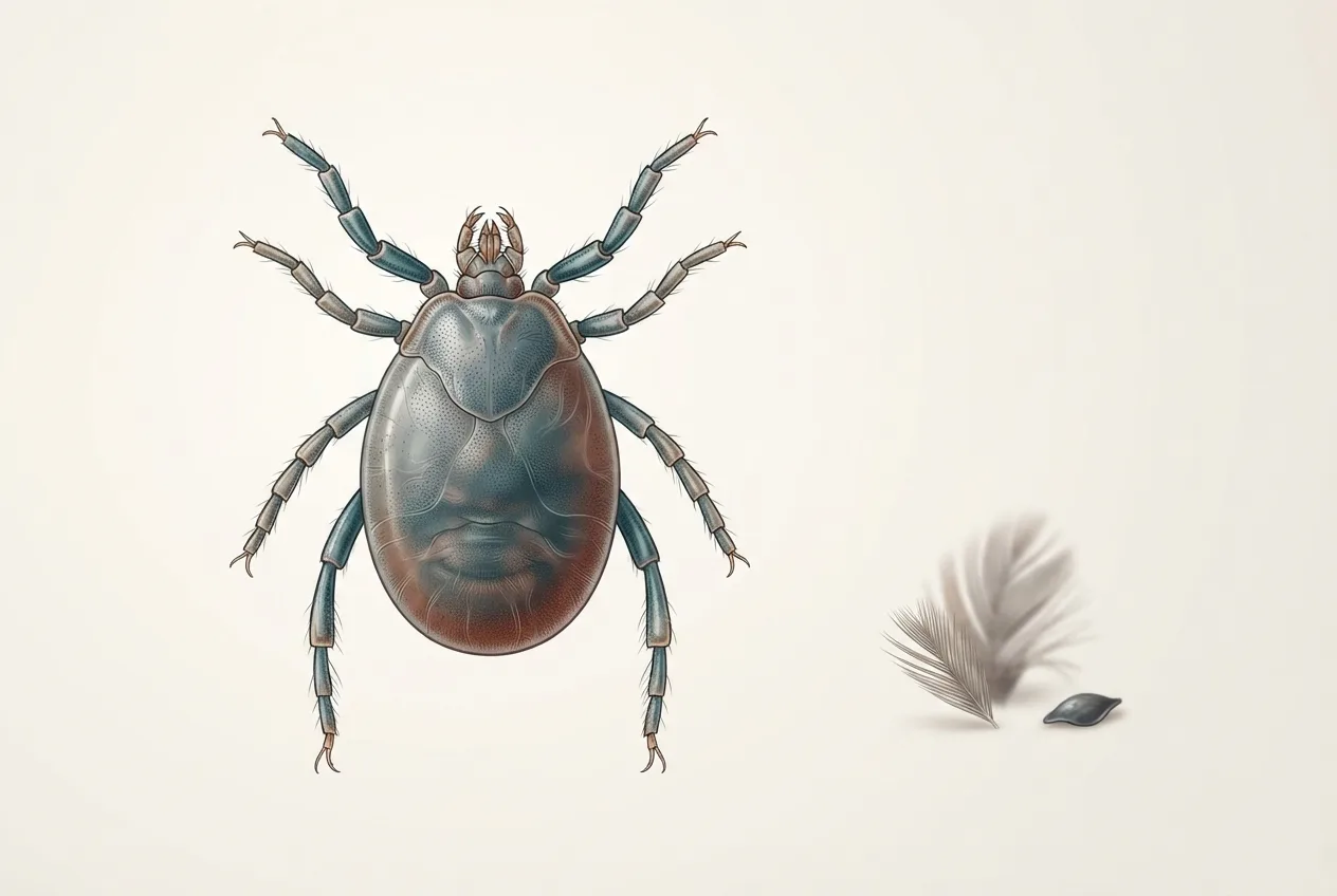
D. gallinae (De Geer, 1778) is a mesostigmatid mite in the family Dermanyssidae [<a href="#ref-6">6</a>]. Adults are approximately 0.7-1.0 mm in length, with an ellipsoid, dorsoventrally flattened idiosoma [<a href="#ref-2">2</a>]. Unfed mites appear greyish-white; engorged individuals turn red due to ingested blood, giving the species its common name [<a href="#ref-2">2</a>]. Key morphological characters that allow differentiation from other bird-infesting mites (e.g., Ornithonyssus sylviarum) include the shape of the dorsal shield, the presence and position of setae, and the structure of the chelicerae [<a href="#ref-7">7</a>]. Scanning electron microscopy of foreleg tarsal sense organs has revealed a complex array of olfactory and mechanoreceptive sensilla that likely mediate host detection and feeding behavior [<a href="#ref-8">8</a>]. A detailed morphological gallery for identification has been published [<a href="#ref-7">7</a>].
Life Cycle
The life cycle of D. gallinae comprises five postembryonic stages: egg, larva, protonymph, deutonymph, and adult [<a href="#ref-1">1</a>, <a href="#ref-2">2</a>]. Embryogenesis within the egg is a critical developmental window during which the mite relies on glucose metabolism via glycolysis and oxidative phosphorylation, with glycogen synthase kinase 3 (GSK3) playing a key regulatory role [<a href="#ref-9">9</a>]. RNA interference targeting Dg-GSK3 significantly reduces female fecundity and egg hatching, highlighting this enzyme as a potential target for novel control strategies [<a href="#ref-9">9</a>].
Under optimal conditions (25-30 °C, high relative humidity), the entire cycle from egg to egg-laying female can be completed in 7-9 days [<a href="#ref-3">3</a>, <a href="#ref-10">10</a>]. The mite typically feeds at night, leaving the host during daylight [<a href="#ref-11">11</a>]. However, under high infestation pressure or with artificial inoculation of protonymphs, mites may remain on the host and feed during daytime as well [<a href="#ref-11">11</a>]. In vitro feeding systems have been developed to support laboratory studies, using either artificial membranes or in vivo devices that attach to live chickens [<a href="#ref-12">12</a>, <a href="#ref-13">13</a>]. A refined in vivo method achieved engorgement rates of 86.8% for adult females and oviposition rates of 98.5%, with a mean of 5.35 eggs per mite [<a href="#ref-12">12</a>]. Darkness significantly promotes feeding rates, engorgement weight, and oviposition, leading to population growth rates 2.4-fold higher under prolonged darkness compared to a 12:12 h light:dark cycle [<a href="#ref-10">10</a>]. Temperature also profoundly affects development; optimal ranges are reported between 20-30 °C, with development ceasing below 10 °C or above 37 °C [<a href="#ref-14">14</a>].
The following table summarizes the duration of each life stage under optimal conditions (data from [<a href="#ref-1">1</a>, <a href="#ref-3">3</a>, <a href="#ref-14">14</a>]):
| Stage | Duration (days) | Key Features |
|---|---|---|
| Egg | 2-3 | Laid in crevices; requires high humidity |
| Larva | 1-2 | Six-legged; does not feed |
| Protonymph | 1-2 | Eight-legged; first blood meal required for molting |
| Deutonymph | 1-2 | Eight-legged; blood meal required |
| Adult female | 7-10 (reproductive lifespan) | Blood meal every 2-3 days; lays up to 5-8 eggs per oviposition |
The ability to survive starvation for extended periods (up to 13 months) allows mites to persist in empty poultry houses between flocks [<a href="#ref-1">1</a>].
Epidemiology and Host Range
D. gallinae has a broad host range that includes domestic fowl, wild birds, and occasionally mammals, including humans [<a href="#ref-2">2</a>, <a href="#ref-15">15</a>]. Phylogenetic studies have identified multiple lineages, including lineage L1 (a cryptic species within D. gallinae sensu lato) that has been associated with urban infestations from pigeon nests and linked to human dermatitis [<a href="#ref-15">15</a>]. Migratory birds and human-mediated transport (e.g., racing pigeon exhibitions) contribute to the intercontinental spread of both D. gallinae and O. sylviarum [<a href="#ref-15">15</a>]. Prevalence in commercial layer farms in Europe is consistently high; surveys in England found 67% of farms infested [<a href="#ref-5">5</a>]. In China, molecular characterization of PRM populations has confirmed widespread presence and high genetic diversity [<a href="#ref-10">10</a>, <a href="#ref-16">16</a>].
Pathogenesis, Vector Capacity, and Economic Impact
Infestation with D. gallinae causes direct damage through blood loss, leading to anemia, decreased egg production, and increased mortality, particularly in young birds [<a href="#ref-1">1</a>, <a href="#ref-4">4</a>]. Immunological effects include altered humoral and cell-mediated responses, reduced serum protein levels, and increased heterophil:lymphocyte ratios [<a href="#ref-4">4</a>]. Egg quality declines with reduced shell thickness and yolk color [<a href="#ref-4">4</a>].
Beyond direct effects, D. gallinae is a competent vector for multiple pathogens. The mite can transmit Salmonella spp., Chlamydia spp., Escherichia coli, Staphylococcus spp., Pasteurella multocida, Newcastle disease virus, and fowl poxvirus [<a href="#ref-1">1</a>]. It also carries antimicrobial resistance genes via its bacterial flora [<a href="#ref-1">1</a>]. The protozoan parasite Atoxoplasma (now Lankesterella) can be transmitted through the mite [<a href="#ref-17">17</a>]. Metagenomic studies of the D. gallinae virome identified a novel quaranjavirus, provisionally named Red Mite Quaranjavirus 1, indicating that the mite may serve as a reservoir for emerging arboviruses [<a href="#ref-18">18</a>]. The bacterial community of the mite changes across developmental stages and midgut compartments, reflecting dynamic host-microbe interactions [<a href="#ref-19">19</a>]. The ability to carry and transmit such many pathogens makes PRM a significant biosecurity threat in poultry production [<a href="#ref-6">6</a>].
Economic losses from D. gallinae infestations are substantial, encompassing reduced egg output, increased feed conversion ratios, mortality, control costs, and carcass condemnation [<a href="#ref-1">1</a>, <a href="#ref-2">2</a>].
Diagnosis
Diagnosis of D. gallinae infestation relies primarily on visual inspection of birds and poultry house structures, especially at night when mites are actively feeding [<a href="#ref-2">2</a>, <a href="#ref-20">20</a>]. Mites can be collected from cracks, perches, and nest boxes using adhesive tape, vacuum samplers, or simple manual collection [<a href="#ref-7">7</a>]. Morphological identification is definitive using keys based on dorsal shield setation and cheliceral morphology [<a href="#ref-7">7</a>]. Molecular diagnostics, particularly PCR targeting the mitochondrial cytochrome c oxidase subunit I (cox1) gene and the 28S rRNA gene, allow species confirmation and lineage discrimination [<a href="#ref-15">15</a>]. Quantitative PCR can quantify mite infestation levels and detect mites in environmental samples [<a href="#ref-18">18</a>]. Transcriptomic and proteomic analyses have been applied to investigate mite biology and identify potential vaccine or acaricide targets [<a href="#ref-18">18</a>, <a href="#ref-21">21</a>]. For field diagnosis, an integrated approach combining visual inspection with molecular testing is recommended.
Control Strategies
Control of D. gallinae is challenging due to the mite’s cryptic behavior, rapid reproduction, and increasing acaricide resistance [<a href="#ref-1">1</a>, <a href="#ref-2">2</a>, <a href="#ref-22">22</a>]. Control measures are broadly categorized as chemical and non-chemical (alternative) methods.
Chemical Control
Acaricides remain the most common intervention, including carbamates, organophosphates, pyrethroids, and amidines [<a href="#ref-1">1</a>, <a href="#ref-23">23</a>]. However, resistance to many compounds is widespread [<a href="#ref-2">2</a>, <a href="#ref-22">22</a>]. Permethrin-impregnated plastic strips have shown efficacy in loose-housing systems [<a href="#ref-24">24</a>]. Systemic acaricides such as fluralaner, administered via drinking water, offer a novel approach with a safety margin of approximately 3-fold in breeder chickens and no adverse effects on reproductive performance [<a href="#ref-25">25</a>]. Ivermectin has been evaluated alone and in combination with allicin, a garlic-derived compound, showing enhanced acaricidal activity in in vivo trials [<a href="#ref-26">26</a>, <a href="#ref-27">27</a>]. A critical concern is acaricide residue accumulation in tissues and eggs. A study of 45 laying hens found that 82.2% were contaminated with carbaryl (a carbamate banned in the EU) and 8.8% with permethrin, with some samples exceeding maximum residue limits [<a href="#ref-23">23</a>]. This underscores the need for careful regulation and monitoring of acaricide use.
Non-Chemical and Alternative Control
Alternative methods include biological control using entomopathogenic fungi, predatory mites, essential oils, temperature extremes, lighting manipulation, and vaccination [<a href="#ref-1">1</a>, <a href="#ref-2">2</a>, <a href="#ref-22">22</a>].
Biological control: The entomopathogenic fungus Aspergillus oryzae (strain Dg-1) isolated from dead D. gallinae significantly increased adult mite mortality (up to 24.8% vs. 15.2% in controls) [<a href="#ref-28">28</a>]. Beauveria bassiana strain JEF-410 induces pathogenesis through upregulation of tryptophan metabolism and secondary metabolite biosynthesis (e.g., beauvericin) in the fungus, while the mite mounts cuticle-strengthening and immune responses [<a href="#ref-21">21</a>]. The predatory mite Cheyletus malaccensis feeds on D. gallinae and can complete its life cycle on a diet of PRM, making it a potential biocontrol agent [<a href="#ref-29">29</a>].
Physical and environmental control: Prolonged darkness promotes population growth, so lighting regimens that include periods of light during the night (intermittent or continuous light) can reduce feeding activity [<a href="#ref-10">10</a>, <a href="#ref-30">30</a>]. High temperatures (>45 °C) and low humidity are lethal [<a href="#ref-14">14</a>]. Simple biosecurity measures such as cleaning, disinfection, and housing design modifications reduce mite harborage [<a href="#ref-22">22</a>].
Vaccine development: Vaccination against D. gallinae is an area of active research. The GSK3 protein has been evaluated as a candidate antigen; immunization of chickens with recombinant Dg-GSK3 significantly reduced female fecundity (2.56 vs. 3.49 eggs per mite) and oviposition rate [<a href="#ref-9">9</a>]. However, developing effective arthropod vaccines is complex due to redundant immune evasion mechanisms [<a href="#ref-2">2</a>].
The decision tree below summarizes the diagnostic and control workflow for D. gallinae infestations.
flowchart TD
 A["Clinical suspicion: anemia, drop in egg production, restlessness"] --> B["Visual inspection at night or use of traps"]
 B --> C{" Mites present? "}
 C -->|"Yes"| D["Morphological identification using key characters"]
 C -->|"No"| E["Monitor with sticky traps or PCR from dust samples"]
 D --> F["Confirmed D. gallinae"]
 F --> G{" Infestation level? "}
 G -->|"Mild"| H["Improve hygiene, apply inert dusts e.g., diatomaceous earth"]
 G -->|"Moderate"| I["Apply chemical acaricide with rotation; consider systemic fluralaner"]
 G -->|"Severe"| J["Combine chemical treatment with environmental control: heat treatment, predatory mites, fungal biopesticides"]
 I --> K["Monitor for acaricide resistance and residues"]
 J --> K
 K --> L{" Control achieved? "}
 L -->|"Yes"| M["Continue surveillance and biosecurity"]
 L -->|"No"| N["Adopt alternative control methods; consider vaccine if available"]
Frequently Asked Questions
What is the optimal temperature for Dermanyssus gallinae development?
The optimal temperature range for development is 20-30°C, with the fastest life cycle (7-9 days) occurring at 25-30°C and high relative humidity [<a href="#ref-1">1</a>, <a href="#ref-3">3</a>, <a href="#ref-14">14</a>]. Below 10°C or above 37°C, development ceases [<a href="#ref-14">14</a>].
Can Dermanyssus gallinae transmit pathogens to poultry?
Yes. D. gallinae is a vector for several bacterial and viral avian pathogens, including Salmonella spp., Chlamydia spp., Escherichia coli, Pasteurella multocida, Newcastle disease virus, and fowl poxvirus [<a href="#ref-1">1</a>]. It also carries antimicrobial resistance genes and can transmit the protozoan Atoxoplasma [<a href="#ref-1">1</a>, <a href="#ref-17">17</a>]. Metagenomic studies have identified a novel quaranjavirus in the mite [<a href="#ref-18">18</a>].
What non-chemical methods are effective against red mites?
Effective non-chemical methods include biological control with entomopathogenic fungi (e.g., Beauveria bassiana, Aspergillus oryzae) and predatory mites (e.g., Cheyletus malaccensis) [<a href="#ref-21">21</a>, <a href="#ref-28">28</a>, <a href="#ref-29">29</a>]; environmental manipulation such as high temperature (>45°C) and low humidity [<a href="#ref-14">14</a>]; lighting regimens that reduce darkness (e.g., intermittent lighting) [<a href="#ref-10">10</a>, <a href="#ref-30">30</a>]; and use of essential oils or plant oils [<a href="#ref-1">1</a>, <a href="#ref-2">2</a>]. Vaccination using recombinant proteins such as Dg-GSK3 is under development [<a href="#ref-9">9</a>].
How is Dermanyssus gallinae distinguished from Ornithonyssus sylviarum?
Morphological differentiation relies on dorsal shield shape and setation: D. gallinae has a single dorsal shield with 12-13 pairs of setae, while O. sylviarum has two separate shields and fewer setae [<a href="#ref-7">7</a>]. Molecular confirmation using cox1 and 28S rRNA gene sequencing provides definitive species identification [<a href="#ref-15">15</a>].
Related Clinical & Scientific Guides
- Tick-Borne Diseases in Dogs: Pathogens, Clinical Signs, Diagnosis, and Prevention
- Toxoplasmosis in Cats and the Risk of Brain Infection in Humans
- Dog Heartworm and Tick-Borne Disease Prevention