Smooth ER: Function and Role in the Cell
By Dr. Zubair Khalid, DVM, MS, PhD ·
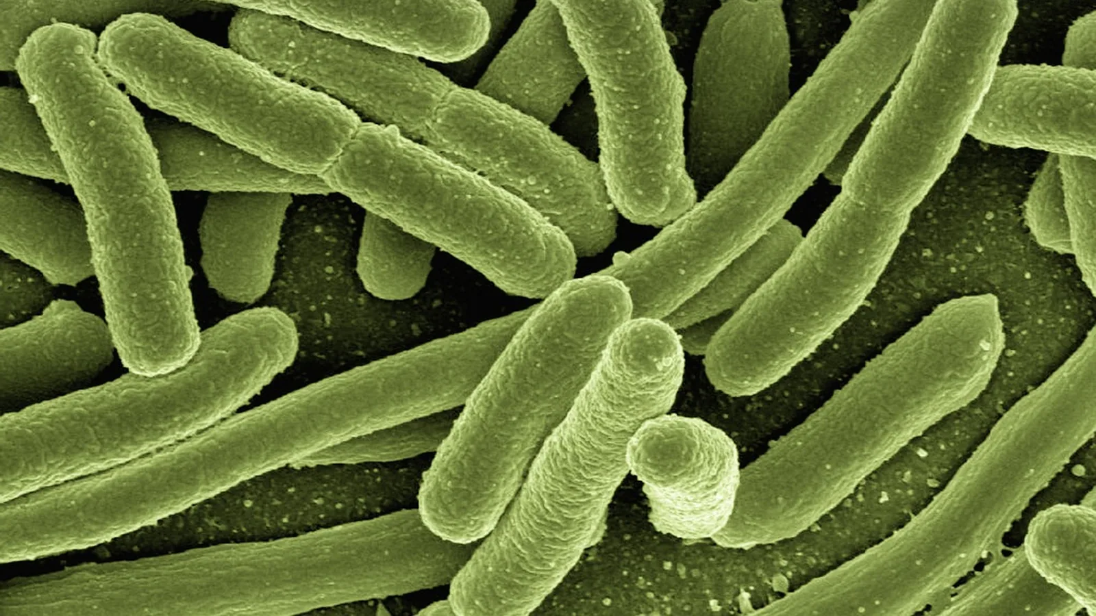
The smooth endoplasmic reticulum (smooth ER) is the ribosome-free region of the endoplasmic reticulum, a continuous membrane network that synthesizes lipids and steroids, detoxifies small molecules, and stores calcium ions. The endoplasmic reticulum (ER) is the largest membrane-bound organelle in the cell and is built from rough ER, smooth ER, and the nuclear envelope [1].
The ER matters because almost every secreted protein, every membrane lipid, and a large share of the cell's calcium traffic passes through it. Two domains of the same continuous membrane handle those jobs with different machinery. Rough ER is studded with ribosomes and runs protein synthesis, folding, and N-linked glycosylation. Smooth ER lacks ribosomes and runs lipid and steroid synthesis, detoxification through cytochrome P450 enzymes, and calcium storage. In striated muscle the smooth ER is specialized into the sarcoplasmic reticulum (SR), which controls contraction by releasing and recapturing calcium [2]. When protein folding in the ER goes wrong, the cell triggers the unfolded protein response (UPR), a signaling program run by three sensors: IRE1, PERK, and ATF6 [3].
What the Endoplasmic Reticulum Is


The ER is a single continuous membrane system. Its lumen, the enclosed interior space, is topologically equivalent to the outside of the cell, which is why proteins inserted into the ER membrane can later appear on the plasma membrane without crossing another lipid bilayer. The organelle is composed of distinct domains, including rough and smooth ER and membrane regions that contact other organelles directly [4].
The ratio of rough to smooth ER shifts with cell type and metabolic demand. Hepatocytes carry abundant smooth and rough ER because the liver handles both protein secretion and lipid and drug metabolism [5]. Cells that secrete large amounts of protein, such as pancreatic acinar cells and antibody-producing plasma cells, are dominated by rough ER. Cells that produce steroids, such as adrenal cortical cells and Leydig cells of the testis, are dominated by smooth ER. The two domains are not fixed compartments. They are zones of one membrane whose protein composition and curvature differ.
Rough ER: the ribosome-studded domain
Rough ER (also written rER or r-ER) gets its name from the electron-dense ribosomes docked on its cytosolic surface. Those ribosomes translate proteins that carry a signal peptide, which directs the ribosome to the ER membrane. The nascent chain enters the lumen, where chaperones fold it and enzymes attach N-linked glycans to asparagine residues. Rough ER is the primary site for synthesis and folding of membrane and secretory proteins, which together make up a large fraction of total protein output in mammalian cells [2]. Rough ER also serves as the entry point for the secretory pathway, and defects in its integrity and function activate the unfolded protein response [6].
Smooth ER: the ribosome-free domain
Smooth ER lacks bound ribosomes, so it appears as a smooth tubular network on electron micrographs rather than as stacked sheets. It is continuous with rough ER, and the boundary between them is a transition zone rather than a wall. The smooth ER is where the cell builds most of its membrane lipids, manufactures steroid hormones, runs oxidative detoxification, and stores calcium. Because it has no ribosomes, it does not synthesize proteins directly.
Summary Table: Rough ER vs Smooth ER
| Feature | Rough ER | Smooth ER |
|---|---|---|
| Ribosomes | Bound to the cytosolic surface | Absent |
| Appearance | Parallel stacked sheets (cisternae) | Tubular network |
| Main job | Protein synthesis, folding, N-linked glycosylation | Lipid and steroid synthesis, detoxification, calcium storage |
| Signature enzymes | Signal peptidase, oligosaccharyltransferase, BiP/GRP78 | Cytochrome P450, enzymes of cholesterol and phospholipid synthesis |
| Calcium role | Minor compared with smooth ER | Major intracellular Ca2+ store |
| Muscle specialization | Not the dominant domain | Becomes the sarcoplasmic reticulum |
| Stress pathway | UPR triggered by misfolded protein load | UPR can also be triggered by lipid and calcium disturbance |
| Typical cell type | Plasma cells, pancreatic acinar cells | Hepatocytes, adrenal cortex, Leydig cells |
Functions of Smooth ER
Lipid and steroid synthesis
Smooth ER is the main site of lipid and steroid synthesis in the cell [1]. Membrane phospholipids and cholesterol are assembled on the cytosolic face of the smooth ER membrane and inserted into the bilayer there. From the ER, lipids move to the Golgi, mitochondria, and plasma membrane by vesicular traffic and by direct membrane contact sites. Cells that produce steroid hormones, including cells of the adrenal cortex, ovary, testis, and placenta, expand their smooth ER to support the conversion of cholesterol into steroid products. Because cholesterol is the precursor for all steroid hormones, steroidogenic cells depend on a large, active smooth ER compartment.
Detoxification and cytochrome P450
The smooth ER of hepatocytes carries cytochrome P450 enzymes that oxidize drugs, alcohol, and metabolic waste so the products can be conjugated and excreted. This is the same enzyme family that performs phase I drug metabolism, and it explains why repeated exposure to a drug or toxin can cause the smooth ER to proliferate. A larger smooth ER compartment means more P450 enzyme and faster clearance. The reaction consumes molecular oxygen and NADPH and generates reactive intermediates, which is why some compounds become more toxic after P450 processing rather than less.
Calcium storage and signaling
The ER is the main intracellular Ca2+ store [7]. Smooth ER membranes carry pumps that move calcium into the lumen against its concentration gradient and channels that release it back into the cytosol on demand. Calcium release from the ER is a central signaling event. In oocytes, calcium storage and release are especially prominent features of the ER, and an appropriate calcium signaling response initiates oocyte development and embryogenesis [1]. In non-muscle cells, the same machinery drives secretion, gene expression changes, and cell movement.
Glycogen metabolism and glucose handling
Hepatocyte smooth ER participates in glucose handling. The ER lumen houses glucose-6-phosphatase, the enzyme that releases free glucose into the blood during fasting. The ER also senses nutritional status and external stimuli and helps regulate glucose and lipid metabolism through UPR signaling pathways [8]. This is one reason the liver is so sensitive to ER stress.
The sarcoplasmic reticulum in muscle
In striated muscle, the smooth ER is called the sarcoplasmic reticulum. It is a specialized membrane system that regulates calcium homeostasis and contraction [2]. When a motor neuron fires, an action potential travels along the muscle cell membrane and reaches deep invaginations called T-tubules. The T-tubule signal opens calcium release channels in the SR, calcium floods the cytosol, and the contractile apparatus shortens. When the signal ends, SR calcium pumps return calcium to the lumen and the muscle relaxes. In adult cardiac myocytes, the SR becomes the dominant membrane network throughout the cell, while ER markers stay enriched in the perinuclear region [2]. Excitation-contraction coupling is localized to the SR, and initiation of secretion is concentrated in the ER [2]. The two compartments perform overlapping but distinct specialized functions.
Small regulatory proteins tune this calcium cycle. Phospholamban and sarcolipin are microproteins, meaning proteins under about 100 amino acids, that regulate calcium handling at the ER and SR [4]. Newly discovered ER microproteins also contribute to calcium regulation, the ER stress response, organelle communication, and protein translocation [4].
How the Unfolded Protein Response Works
The ER monitors the quality of the proteins it folds. When the load of protein synthesis and folding exceeds the processing capacity of the ER, misfolded or unfolded proteins accumulate and trigger ER stress [5]. The cell responds with the unfolded protein response, a transcriptional and translational program that aims to restore homeostasis by reducing oxidative stress, slowing protein synthesis, and limiting calcium leakage [7].
Three sensors sit in the ER membrane and launch the response.
- IRE1 detects unfolded proteins in the lumen, oligomerizes, and splices XBP1 mRNA so it produces a functional transcription factor, XBP1s. The IRE1-XBP1 branch expands the ER's folding and secretory capacity.
- PERK phosphorylates eIF2 alpha, which temporarily slows global translation and selectively increases translation of ATF4, a transcription factor that drives adaptation genes.
- ATF6 moves to the Golgi when it is released from the chaperone BiP, where it is cleaved to release a transcription factor that turns on chaperone genes.
ER stress activates the UPR and thereby influences downstream PERK, IRE1 alpha, and ATF6 pathways [3]. Short-term ER stress lets cells survive and adapt. Severe or sustained ER stress typically provokes cell death through multiple routes [5]. The UPR is therefore a survival-versus-death decision point, not a simple on-off switch.
How Smooth ER Is Studied in the Lab
Researchers identify rough and smooth ER by a mix of imaging, fractionation, and reporter assays.
- Electron microscopy distinguishes rough sheets from smooth tubules by the presence or absence of bound ribosomes. In one study of duck intestinal epithelial cells, viral infection caused severe rough ER dilation, a morphological readout of ER stress [9].
- Subcellular fractionation separates rough and smooth microsomes by density gradient centrifugation. Rough microsomes carry ribosomes and are denser. Smooth microsomes are lighter and enriched for lipid synthesis enzymes and P450.
- Fluorescent markers such as GFP or mCherry fused to ER-resident proteins label the whole network in live cells. Immunocytochemistry with antibodies against ER and SR markers showed that ER and SR have overlapping but distinct distributions in cardiac myocytes [2].
- Calcium imaging with dyes such as Fura-2 or genetically encoded indicators measures ER and SR calcium release in real time.
- UPR reporters such as XBP1 splicing assays, ATF4 translation reporters, and CHOP promoter constructs quantify which UPR branch is active. In C. elegans, neuronal overexpression of the gap junction protein UNC-9 activates the IRE-1-XBP-1 branch, and loss of the early secretory proteins ERGI-2 or ERGI-3 suppresses that response [10].
- Proteomics and ribosome profiling quantify which proteins and which microproteins occupy the ER under different conditions [4].
Comparative and Clinical Relevance
ER composition changes with age and with metabolic state. In aging models ranging from yeast to C. elegans and mammals, ER mass declines in most tissues and ER morphology shifts from rough sheets to tubular ER, with a corresponding shift from protein synthesis toward lipid metabolism [11]. ER-phagy, the selective autophagic turnover of ER, drives this remodeling and appears to be protective, because impairing ER-phagy limits lifespan in yeast [11]. This is a useful reminder that the smooth-to-rough balance is dynamic, not fixed.
ER stress sits at the center of many metabolic and inflammatory conditions. ER stress and metabolic deregulation are functionally intertwined and are both considered contributing factors in liver disease [5]. In human airway smooth muscle, the inflammatory cytokine TNF alpha increases reactive oxygen species, which triggers the IRE1 alpha-XBP1s ER stress pathway and downstream mitochondrial fragmentation [12]. In the liver, hepatocytes are rich in smooth and rough ER and use ER-resident metabolic enzymes to sense nutritional status and external stimuli [5]. ER stress also shapes immune cell production: UPR signaling increases myeloid cell production through the XBP1 pathway and cooperates with the Jak2V617F mutation through the ATF4 pathway in a mouse model of polycythemia vera [13].
Viral and parasitic infections routinely manipulate the ER. African swine fever virus protein pMGF110-9L is an ER-localized membrane protein whose expression reorganizes cellular compartments, causes ER swelling and aggregation, and activates all three UPR branches [14]. Duck enteritis virus infection activates the PERK-eIF2 alpha-ATF4-CHOP and IRE1 alpha-XBP1 axes in intestinal epithelial cells [9]. In plants, phytoplasma infection alters sieve-element ER morphology and triggers release of bZIP signals from the ER, with differential expression of UPR-related genes [6].
The UPR also connects to regulated cell death. Necroptosis, a programmed necrosis pathway driven by RIPK1, RIPK3, and MLKL, is regulated by the UPR during ER stress [3]. This link explains why ER stress shows up in so many disease models even when the primary defect is elsewhere.
Common Mistakes and Limitations
Confusing the two domains as separate organelles. Smooth and rough ER are continuous. A protein can diffuse within the same membrane from a rough sheet into a smooth tubule. The distinction is functional and compositional, not a physical separation.
Assuming smooth ER makes proteins. It does not. Protein synthesis, folding, and N-linked glycosylation happen on rough ER. Smooth ER handles lipids, steroids, detoxification, and calcium.
Treating the sarcoplasmic reticulum as a different organelle. The SR is the smooth ER of muscle cells. It shares the same basic membrane machinery but is specialized for calcium-driven contraction [2].
Believing all calcium storage is in the ER. The ER is the main intracellular store [7], but mitochondria and acidic organelles also hold calcium. ER calcium is the dominant rapidly releasable pool in most cells.
Reading the UPR as purely protective. Short-term UPR helps cells adapt. Chronic UPR drives reactive oxygen species formation, inflammation, and apoptosis, which worsens the stress and spreads damage to other organelles [7].
Overreading morphology. A dilated rough ER on electron microscopy indicates stress but does not identify which UPR branch is active. Branch-specific assays are needed [9].
Individual variation. Cell type, metabolic state, and age all shift the rough-to-smooth ratio [11]. A single image or fractionation result describes one condition, not a universal rule. Individual experimental or clinical cases need expert interpretation.
Quick Review
- Smooth ER is the ribosome-free domain of the ER and handles lipid and steroid synthesis, detoxification, and calcium storage.
- Rough ER carries bound ribosomes and handles protein synthesis, folding, and N-linked glycosylation [2].
- The sarcoplasmic reticulum is the smooth ER of striated muscle and controls contraction through calcium release and reuptake [2].
- Cytochrome P450 enzymes in hepatocyte smooth ER oxidize drugs, alcohol, and metabolic waste.
- The ER is the main intracellular Ca2+ store and a central hub for calcium signaling [1] [7].
- ER stress activates the UPR through three sensors: IRE1, PERK, and ATF6 [3].
- Short-term UPR restores homeostasis. Chronic UPR drives inflammation and cell death [5] [7].
Frequently Asked Questions
What is the main function of smooth ER?
The main function of smooth ER is lipid and steroid synthesis, detoxification of drugs and metabolic waste through cytochrome P450 enzymes, and storage and release of calcium ions. It does not synthesize proteins because it lacks ribosomes.
How does smooth ER differ from rough ER?
Rough ER is studded with ribosomes and handles protein synthesis, folding, and N-linked glycosylation. Smooth ER lacks ribosomes and handles lipid and steroid synthesis, detoxification, and calcium storage. The two domains are continuous parts of one membrane network.
What is the sarcoplasmic reticulum?
The sarcoplasmic reticulum is the specialized smooth ER of striated muscle cells. It stores calcium and releases it on demand to trigger contraction, then pumps it back to allow relaxation [2].
Why does the liver have so much smooth ER?
Hepatocytes carry abundant smooth ER because the liver must synthesize lipids, process drugs and toxins through cytochrome P450 enzymes, and regulate blood glucose. Hepatocytes are rich in smooth and rough ER and use ER-resident enzymes to sense nutritional status [5].
What triggers the unfolded protein response?
The unfolded protein response is triggered when misfolded or unfolded proteins accumulate in the ER lumen faster than the organelle can fold or clear them. The three sensors IRE1, PERK, and ATF6 detect the disturbance and launch adaptive transcriptional and translational changes [3].
Does smooth ER store calcium in all cell types?
Yes. The ER is the main intracellular Ca2+ store in essentially all eukaryotic cells [7]. Muscle cells simply expand and specialize this calcium function into the sarcoplasmic reticulum to support rapid contraction cycles.
Related Articles
- Cell Membrane Cholesterol: Its Role in Membrane Fluidity and Function
- The Role of Chromatin Remodelers in Cell Differentiation: A Review of SWI/SNF and ISWI Functions in Lineage Commitment
- Cell Membrane Function Biology
- Endoplasmic Reticulum Cell Function
- Anticodon Sequence: Definition, Function, and Role in Translation
- 5 Prime Cap: Definition, Function, and Role in mRNA
Sources
- Endoplasmic reticulum in oocytes: spatiotemporal distribution and function.
- Development of a protein synthesis network at the sarco/endoplasmic reticulum in adult cardiac myocytes.
- Necroptosis-related diseases are regulated by the unfolded protein response during endoplasmic reticulum stress.
- Emerging roles for microproteins as critical regulators of endoplasmic reticulum function and cellular homeostasis.
- Regulation mechanism of endoplasmic reticulum stress on metabolic enzymes in liver diseases.
- The sieve-element endoplasmic reticulum: A focal point of phytoplasma-host plant interaction?
- [[Activation of endoplasmic reticulum stress sensors by metabolic disease-associated diets and COVID-19].](https://pubmed.ncbi.nlm.nih.gov/35759643/)
- Role of endoplasmic reticulum stress in hepatic glucose and lipid metabolism and therapeutic strategies for metabolic liver disease.
- DEV infection induces endoplasmic reticulum stress and activates inflammatory responses in duck intestinal epithelial cells.
- ER-to-Golgi transport machinery promotes the excessive cargo-triggered unfolded protein response in C. elegans.
- ER-phagy drives age-onset remodeling of endoplasmic reticulum structure-function and lifespan.
- ROS Scavenging Mitigates TNFα Induced Endoplasmic Reticulum Stress and Mitochondrial Fragmentation in Human Airway Smooth Muscle.
- Unfolded protein response signaling promotes myeloid cell production and cooperates with oncogenic mutation.
- African swine fever virus pMGF110-9L plays a critical role in the integrated stress response and viral pathogenesis.