Lytic vs Lysogenic Cycle of Bacteriophages
By Dr. Zubair Khalid, DVM, MS, PhD ·

The lytic cycle is a bacteriophage reproductive program in which the virus replicates inside a bacterial host and then ruptures the cell to release progeny virions. The lysogenic cycle is a temperate-phage program in which the phage genome persists in the host, often integrated into the bacterial chromosome as a prophage or maintained as an extrachromosomal plasmid, and is copied with the host until induction switches the cell to lytic growth.
The choice between these cycles is one of the clearest examples of a molecular decision with ecological and clinical consequences. A single phage can destroy a bacterial population quickly, or it can become a stable genetic passenger that changes host phenotype, protects the host from related phages, and moves bacterial genes between strains. The classic distinction between the lysogenic and lytic cycles was established in the mid-twentieth century through studies of lysogenic bacteria and phage multiplication [1][2][3]. Modern work has added mechanistic detail from temperate phages of Streptococcus pneumoniae, Staphylococcus aureus, Lactobacillus casei, Salmonella enterica, Bacillus species, and Bacteroides symbionts [4][5][6][7][8]. The same decision also affects bacterial virulence, antimicrobial resistance, immune systems such as CRISPR-Cas, and the safety and efficacy of phage-based biocontrol or therapy [9][10][11].
Lysogenic Cycle Definition and Core Vocabulary
The lysogenic cycle definition is simple at the level of host fate: a bacteriophage enters a bacterial cell, its genome becomes established in a stable form, and the host survives and continues to divide. The phage genome is replicated as part of the host's growth. The host cell is called a lysogen, and the carried phage genome is called a prophage. A prophage can be integrated into the bacterial chromosome, or it can be maintained as a plasmid or phage-plasmid, depending on the phage. For example, Bacillus phage phi18-2 is a temperate virus whose genome can be present in the cytoplasm of lysogenic cells as a linear phage-plasmid rather than an integrated prophage [12]. This exception matters because students often treat integration as a universal rule.
Several terms are needed to read the primary literature accurately.
- A temperate phage is a bacteriophage that can establish lysogeny under some conditions and enter the lytic cycle under other conditions.
- A virulent phage is a bacteriophage that generally completes a lytic cycle and does not establish a stable lysogenic state.
- A prophage is the phage genome carried by a lysogen. It may be integrated or extrachromosomal.
- A lysogen is a bacterial cell or strain that carries a prophage.
- Induction is the process that switches a prophage from the lysogenic state to the lytic cycle.
- Superinfection immunity is the resistance of a lysogen to infection by the same or closely related phages, usually because the prophage expresses a repressor that blocks lytic gene expression.
- Lysogenic conversion is a change in host phenotype caused by genes carried or expressed by a prophage. It is not the same as transduction, which is phage-mediated transfer of bacterial DNA.
The lysogenic cycle is not a dormant state in the ordinary sense. A prophage can express genes, especially the repressor that maintains lysogeny. In phage A2 of Lactobacillus casei, lysogens contain detectable cI and cro-gp25 mRNA, and the CI repressor only partially represses the early lytic promoter [7]. In S. aureus lysogens carrying phage SapYZUs7, integrated omics showed changes in host gene expression and metabolism [9]. The prophage is therefore a regulated genetic element, not simply a silent piece of stored DNA.
Lytic vs Lysogenic Cycle at a Glance
| Feature | Lytic cycle | Lysogenic cycle |
|---|---|---|
| Typical phage type | Virulent phage, or a temperate phage after induction | Temperate phage |
| Host outcome | Lysis and death | Survival and continued growth |
| Phage genome state | Replicating and packaging many copies | Integrated prophage or extrachromosomal plasmid |
| Dominant regulation | Early and late lytic genes active | Repressor blocks lytic promoters |
| Transmission | Horizontal release of virions | Vertical inheritance with host division |
| Immunity to related phage | Not usually a stable state | Superinfection immunity is common |
| Exit route | Completion of lysis | Induction, then lytic cycle |
| Examples in the literature | Lytic infection by verocytotoxigenic phages [13] | P2 [14], P22 [8], Phi11 [6], A2 [7], SapYZUs7 [9] |
Lysogenic Cycle Steps
The lysogenic cycle steps are best learned as a sequence of commitments. The order can vary between phages, and some steps are bypassed in phages that maintain a plasmid prophage. The core logic is still useful.
- Adsorption and genome delivery. The phage binds a specific host surface receptor and injects its nucleic acid. The host range is set by that receptor interaction and by post-entry defenses.
- Genome stabilization inside the cell. Many phage genomes circularize or become protected by phage proteins. The genome must avoid degradation and must prepare for either integration or replication.
- The lysogeny decision. Early phage gene expression and host physiology set the balance between repressor and antirepressor activity. This is the genetic switch. In phage P2, the choice depends on a transcriptional switch [14]. In pneumococcal phage MM1, a regulatory system controls the lytic and lysogenic pathways [4].
- Integration or plasmid maintenance. In integrating phages, a phage-encoded integrase catalyzes site-specific recombination between a phage attachment site and a bacterial attachment site. The integrated prophage is flanked by recombination junctions. In non-integrating temperate phages, the genome is maintained as a plasmid. phi18-2 is an example of a linear phage-plasmid prophage [12].
- Repressor expression and immune establishment. The prophage expresses a repressor that binds operator sites and blocks lytic promoters. In phage A2, Cro binds the genetic switch region [15], while CI represses the early lytic promoter and Cro opposes the lysogenic promoter [7]. The repressor also gives the lysogen superinfection immunity against related phages.
- Vertical propagation. When the bacterium replicates its chromosome, the integrated prophage is duplicated with it. The daughter cells inherit the prophage. In a plasmid prophage, the extrachromosomal element is partitioned to daughter cells.
- Induction. DNA damage, host stress, or specific inducing agents can trigger the lytic cycle. In S. aureus phage Phi11, the transcriptional repressor LexA and co-protease RecA play a crucial role in lytic-lysogenic switching [6]. In phage A2, partial degradation of CI during induction allows Cro accumulation and lytic activation [7]. Classic experiments showed that the lytic cycle can be induced in lysogenic bacteria by agents such as phagolessin A 58 [16].
- Excision and lytic replication. Integrated prophages must excise from the host chromosome before or during lytic replication. The timing is not universal. Wild-type Salmonella phage P22 uses a replication-packaging-excision program and delays excision to just before it would be detrimental to phage production [8].
- Lysis and release. The phage completes virion assembly, produces lysis proteins, and releases progeny. The released virions can infect new susceptible cells.
The lysogenic cycle bacteriophage does not simply choose lysogeny once and stop. The decision can be reversed, and the probability of lysis versus lysogeny can be tuned by host state and phage regulators. In phage A2, Gp25 binds cro-gp25 mRNA between the ribosome binding site and the cro start codon and inhibits Cro translation [7]. During induction, CI is partially degraded and Cro accumulates. A concomitant increase in Gp25 restricts Cro translation, and low but detectable CI late in the lytic cycle promotes reentry of part of the population into lysogeny. The result is that only about 1% of A2 lysogens become lysed in the reported system [7]. That number is a study-specific measurement, not a universal rule.
The Genetic Switch: Repressors, Antirepressors, and RNA-Level Control
The lysogenic cycle vs lytic cycle decision is often drawn as a bistable switch. In the canonical lambda-style model, CI repressor maintains lysogeny and Cro promotes the lytic cycle. At low CI activity, lytic promoters fire. At high CI activity, CI binds operators, blocks lytic transcription, and activates its own expression. Cro accumulates during lytic growth and opposes CI. This textbook abstraction is useful, but real phages modify it.
Phage P2 uses a transcriptional switch to choose between lysogenic and lytic development [14]. Phage MM1 has a molecular and biochemical regulatory system for the lytic and lysogenic cycle [4]. In phage A2, CI represses the early lytic promoter PR, and Cro abolishes expression from the lysogenic promoter PL [7]. Cro specifically binds the genetic switch region of A2 [15]. The A2 system adds an RNA-binding protein, Gp25, which suppresses Cro translation and helps explain why A2 produces stable lysogens despite a switch architecture that might otherwise favor lytic dominance [7]. This is a good example of why students should not memorize a single lambda diagram as the universal mechanism.
Phage Phi11 of S. aureus adds another variation. Gp07 functions as a novel antirepressor and regulates development by enhancing binding of the Cro repressor to its operator. The CI repressor of Phi11 binds the putative operator of Gp07 and regulates its expression. LexA and RecA are involved in lytic-lysogenic switching. The N-terminal Bro-N domain of Gp07 is responsible for enhancing Cro binding to its operator [6]. The authors note that Phi11 prophage induction differs from that of other bacteriophages, which reinforces the point that the switch is modular.
Phage A2 also illustrates post-transcriptional control. In Escherichia coli, the A2 cro gene can produce Cro and a shorter Cro* polypeptide through a -1 ribosome frameshift. Cro* is twelve amino acids shorter than Cro. However, frameshifting was not observed during phage development in L. casei, and wild-type phages and cro-frameshifting negative mutants had the same phenotype. Only the canonical Cro form was needed for viable phage progeny [17]. This result is a caution against assuming that a regulatory mechanism seen in a heterologous host operates in the natural host.
How the Lysogenic Cycle Diagram Is Used in Teaching and Research
A useful lysogenic cycle diagram should separate three spaces: the extracellular environment, the bacterial cytoplasm, and the bacterial chromosome. It should also distinguish the prophage state from the replicating lytic state. A student who can label those spaces can follow most papers on temperate phages.
A strong teaching diagram has these elements.
- Free phage and receptor binding. Show the phage particle outside the cell and the specific host receptor at the surface.
- Genome entry and stabilization. Show the phage DNA inside the cell and indicate whether it circularizes or remains linear.
- The decision node. Label the repressor, the antirepressor, and the promoters that control the switch.
- Integration. Show the bacterial chromosome, the attachment site, the integrase reaction, and the integrated prophage flanked by recombination junctions.
- Alternative plasmid prophage. Add a separate branch for phages such as phi18-2, whose lysogenic genome can be an unintegrated linear phage-plasmid [12].
- Vertical transmission. Show bacterial division carrying the prophage into daughter cells.
- Induction. Show the inducing signal, repressor inactivation, excision, lytic replication, and lysis.
A common labeling error is to draw every prophage as an integrated segment. That is wrong for plasmid prophages. Another error is to draw the lysogenic state as a completely silent genome. Repressor expression and other prophage genes can be active. A third error is to show induction as a single arrow from lysogeny to lysis. In P22, the order of replication, packaging, and excision is tuned, and wild-type P22 delays excision to support lateral transduction [8]. The arrow is real, but the timing is biologically important.
Lysogenic Cycle vs Lytic Cycle: Molecular and Ecological Differences
The lysogenic cycle vs lytic cycle comparison is often reduced to "kill versus not kill." That is accurate at the level of host fate, but it misses the molecular and ecological distinctions.
In the lytic cycle, the phage commits to producing many progeny. Early lytic genes activate replication, middle and late genes build virions, and lysis releases the particles. The host cell dies. In the lysogenic cycle, the phage commits to a stable association. The prophage is usually present at one copy per bacterial chromosome, or at a low and regulated copy number for a plasmid prophage, and it is replicated with the host. The host survives and can continue to divide.
The regulatory difference is the repressor. A lysogen expresses a repressor that blocks lytic promoters and provides immunity to superinfection by related phages. That immunity is specific, not universal. A lysogen can still be infected by unrelated phages. In Phi11, CI regulates Gp07 expression, and Gp07 enhances Cro binding to its operator, showing how repressor and antirepressor activities can be layered [6]. In A2, CI and Cro oppose each other at early promoters, and Gp25 adds translational control [7].
The ecological difference is strategy. Lysis is a rapid horizontal transmission strategy. Lysogeny is a vertical persistence strategy that can also create opportunities for horizontal gene transfer. Prophages can carry bacterial genes, and induction can mobilize host DNA. Wild-type P22 delays prophage excision and thereby engages in lateral transduction, in which large fragments of host DNA are packaged and transferred during the lytic transition [8]. This is a clear example of the lysogenic cycle feeding molecular evolution. Environmental conditions and host physiology can also shift the balance between lytic and lysogenic paths in natural communities [18]. Mathematical models have examined how the lytic and lysogenic paths affect coexistence of bacteria and bacteriophage [19].
The binary comparison also has limits. The hankyphage of Bacteroides symbionts spontaneously produces virions in laboratory conditions even without inducer, but those virions showed no evidence of infectivity. Silencing the master repressor RepCHP increased gene expression, replicative transposition, and virion production, but infection or lysogenization of new host cells remained unsuccessful. Transmission electron microscopy and capsid DNA sequencing revealed abnormal virion morphology and incomplete DNA packaging [5]. This is an incomplete lytic cycle, not a clean switch between two fully functional programs. The lysogenic cycle and lytic cycle are therefore useful categories, not guarantees of a complete infectious outcome.
Lysogenic Cycle Examples
Lysogenic cycle examples span very different phages and hosts. The examples below show the range of mechanisms that can support the same broad lifecycle.
Integrated Prophages
Many temperate phages integrate into the host chromosome. Phage P2 uses a transcriptional switch to control lysogenic versus lytic development [14]. Phage MM1 is a pneumococcal temperate phage with a regulatory system for the lytic and lysogenic cycle [4]. Phage Phi11 of S. aureus has a CI repressor, a Cro repressor, and a Gp07 antirepressor that modulates the switch, with LexA and RecA involved in induction [6]. Phage A2 of L. casei uses CI, Cro, and Gp25 to maintain stable lysogeny and to limit the fraction of lysogens that lyse after induction [7][15].
Extrachromosomal and Phage-Plasmid Lysogeny
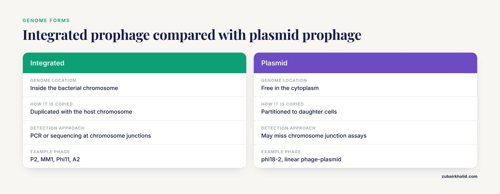
Not every prophage integrates. Bacillus phage phi18-2 is a novel temperate virus whose genome is present in the cytoplasm of lysogenic cells as a linear phage-plasmid [12]. The filamentous phage Cflt from Xanthomonas campestris pv. citri also has a lysogenic cycle [20]. These examples are useful because they break the assumption that lysogeny equals chromosomal integration. They also complicate prophage detection by PCR or sequencing, since an extrachromosomal prophage may not sit at a predictable chromosome junction.
Lysogenic Conversion and Host Fitness
Lysogenic conversion occurs when a prophage changes host phenotype. A new temperate Enterococcus phage, vB_GEC_EFS_2, has potential for lysogenic conversion [10]. S. aureus phage SapYZUs7 carries smaII, which is associated with a single-protein MazF-like antiphage system, and the phage can integrate into the S. aureus genome. Lysogenic S. aureus showed higher phage resistance, a lower growth rate, and inhibited metabolic activity compared with parental strains. prophages carrying smaII are widely distributed in S. aureus and harbor other virulence factor and antimicrobial resistance genes. SapYZUs7 integration increased phagocytosis resistance but decreased adhesion, biofilm formation, and antimicrobial resistance. The combination of SapYZUs7 and antibiotics showed a better bactericidal effect than either agent alone [9]. This example shows that lysogeny can impose costs and benefits at the same time.
Verocytotoxigenic bacteriophages can carry out lytic and lysogenic infection of diverse Escherichia coli and Shigella strains [13]. That broader host range matters for understanding how phage-carried traits might move among enteric bacteria. The hankyphage example adds another dimension: a widespread transposable phage of Bacteroides symbionts can produce virions spontaneously, yet the virions are abnormal and incomplete in laboratory conditions [5]. The lesson is that lysogeny in a natural host does not always translate into a simple plaque-forming laboratory system.
How Lysogeny Is Tested and Observed
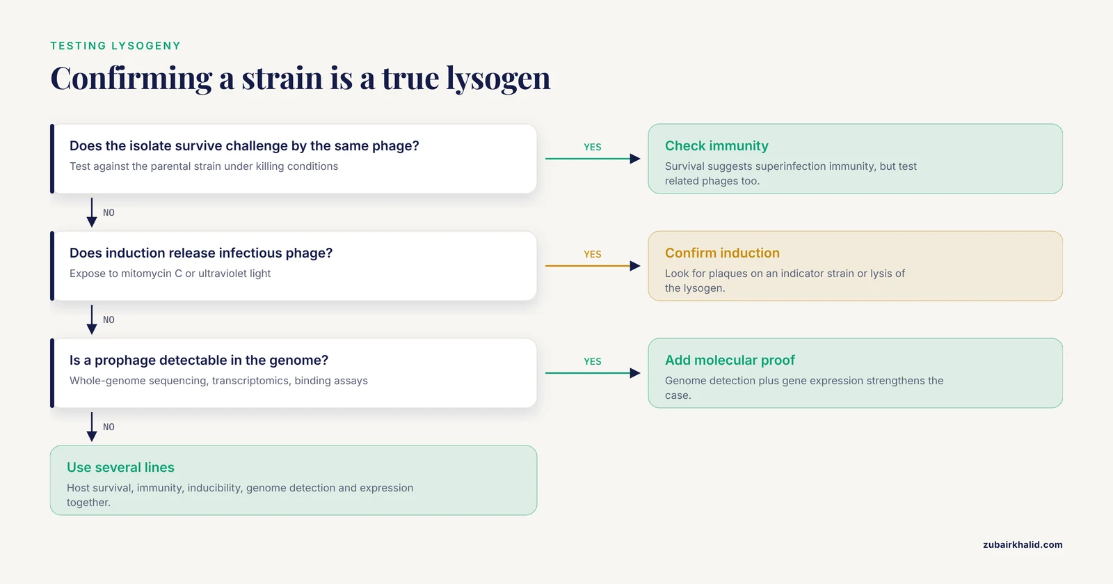
Lysogeny is detected by combining classical microbiology with molecular assays. No single assay answers every question.
Plaque Morphology and Lysogen Isolation
A plaque assay measures phage infection by counting zones of lysis in a bacterial lawn. Virulent phages often produce clear plaques because the infected cells lyse. Temperate phages often produce turbid plaques because some infected cells survive as lysogens and continue to grow within the plaque. Plaque morphology is a useful first clue, not proof. A turbid plaque can have other explanations, and some temperate phages plaque poorly in vitro.
To isolate a lysogen, a researcher picks bacteria from a turbid plaque or from a surviving colony after phage exposure, purifies the strain, and tests whether it releases phage after induction. The isolate is also tested for superinfection immunity. A true lysogen should survive challenge by the same phage under conditions that kill the parental strain. The immunity is usually specific to related phages, so a broader panel is often needed.
Induction and Functional Tests
Lysogens can be exposed to inducing agents such as mitomycin C or ultraviolet light. Induction is detected by the appearance of phage particles, plaques on an indicator strain, or lysis of the lysogen. Classic work showed that the lytic cycle can be induced in lysogenic bacteria by phagolessin A 58 [16]. Modern studies use induction together with molecular assays to determine whether the prophage can complete a productive cycle. The hankyphage example shows why this matters: spontaneous virion production occurred, but the virions were not infectious, and infection or lysogenization attempts failed [5].
Molecular Detection of Prophages
Whole-genome sequencing can identify integrated prophages, prophage boundaries, and prophage-carried genes. In S. aureus, whole-genome analysis showed that SapYZUs7 could integrate into the genome, and prophages carrying smaII were widely distributed and associated with virulence factor and antimicrobial resistance genes [9]. Transcriptomics and metabolomics can show which prophage genes are expressed and how lysogeny changes host physiology [9]. Binding assays and gene expression studies can resolve the genetic switch. In Phi11, Gp07 was amplified and analyzed as a regulator, and its domain requirements were mapped [6]. In A2, Cro binding to the genetic switch region was tested directly [15]. In A2, Gp25 binding to cro-gp25 mRNA was linked to translational repression [7].
Electron microscopy and capsid DNA sequencing can reveal virion morphology and genome packaging defects. These methods were central to showing that hankyphage virions were abnormal and incompletely packaged [5]. Metaviromics and phylogenetic analyses can then ask whether related sequences are present in natural communities, even when laboratory infection fails [5]. Environmental surveys can link lytic and lysogenic cycles to host physiology and environmental conditions [18]. The practical point is that lysogeny is best established with multiple lines of evidence: host survival, immunity, inducibility, genome detection, and gene expression.
Molecular Evolution and Clinical Relevance
Lysogeny is a major force in bacterial evolution. A prophage can be inherited vertically, but it can also move horizontally when induction produces virions. Prophages can carry virulence factors, antimicrobial resistance genes, immune systems, and metabolic genes. In S. aureus, SapYZUs7 lysogeny altered phage resistance, growth, metabolism, phagocytosis resistance, adhesion, biofilm formation, and antimicrobial resistance [9]. Those effects can change how a strain behaves in an infection or on a mucosal surface.
Lateral transduction is a particularly important evolutionary mechanism. Wild-type Salmonella phage P22 delays prophage excision during induction and uses a replication-packaging-excision program. This timing allows P22 to package and transfer large fragments of host DNA, powering host evolution through gene transfer [8]. The old assumption that prophage excision always occurs early after induction came from an experimentally favored P22 mutant. The wild-type program is different. This is a useful case study in how a model system can mislead if the strain background is not considered.
Host immune systems also shape lysogeny. Type III-A CRISPR-Cas systems can restrict temperate phage lytic infections while tolerating lysogenic infections when prophage targets are transcriptionally repressed. However, long-term maintenance of conditionally tolerant type III-A systems can produce fitness costs in S. aureus lysogens. The costs depend on prophage-internal promoter activity and Cas nuclease activity, can be more severe in double lysogens, and can be alleviated by spacer-target mismatches that do not abolish immunity during the lytic cycle [11]. This shows that lysogeny is not a private relationship between phage and host. It is embedded in a larger network of host defense, prophage regulation, and fitness trade-offs.
Clinically, temperate phages can be useful or problematic. They can carry genes that increase virulence or antimicrobial resistance, and they can impose fitness costs on the host. They can also be developed as biocontrol agents. SapYZUs7 was studied as a potential biocontrol agent for S. aureus, and the combination of phage and antibiotics produced a better bactericidal effect than either alone in that system [9]. The takeaway is not that temperate phages are always good or always bad. The takeaway is that the lysogenic cycle must be characterized before a phage is used as a therapeutic or biocontrol tool.
Common Mistakes and Limitations
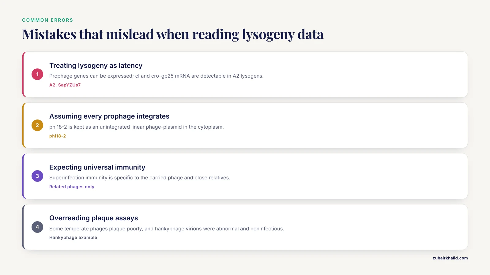
- Treating lysogeny as latency. A prophage is not necessarily silent. Repressor genes and other prophage genes can be expressed. In A2, cI and cro-gp25 mRNAs are detectable in lysogens [7]. In S. aureus, SapYZUs7 lysogeny changes host transcriptome and metabolome profiles [9].
- Assuming every prophage is integrated. phi18-2 is maintained as an unintegrated linear phage-plasmid in the cytoplasm of lysogenic cells [12]. Detection methods that only search for chromosome junctions will miss this form.
- Assuming a lysogen is immune to all phages. Superinfection immunity is usually specific to the carried phage and close relatives. Unrelated phages can still infect the cell.
- Confusing lysogenic conversion with transduction. Lysogenic conversion is a phenotype change caused by prophage genes. Transduction is phage-mediated transfer of bacterial DNA. The two processes can be connected during induction, but they are not the same.
- Treating the switch as a simple on-off circuit. A2 uses Gp25 to control Cro translation [7]. Phi11 uses Gp07 as an antirepressor and involves LexA and RecA [6]. P22 uses a replication-packaging-excision program that differs from the classic excision-first model [8].
- Assuming prophages are harmless passengers. Prophages can carry virulence and antimicrobial resistance genes, and they can impose fitness costs under some conditions [9][11].
- Overinterpreting plaque assays. Some temperate phages do not plaque well in vitro, and some produce abnormal or noninfectious virions in laboratory conditions [5]. Plaque formation is useful evidence, not a universal requirement for proving that a phage is temperate.
- Ignoring host and environmental context. The balance between lysis and lysogeny can shift with host physiology and environmental conditions [18]. A result from one host or one induction condition may not generalize.
For animal infections or environmental samples, individual cases need a veterinarian and laboratory confirmation before any phage-based intervention is considered.
Quick Review
- The lytic cycle kills the host and releases progeny. The lysogenic cycle keeps the host alive and carries the phage genome through host division.
- A prophage is the phage genome in a lysogen. It can be integrated or extrachromosomal.
- Temperate phages can switch from lysogeny to lysis through induction.
- Repressors maintain lysogeny and provide superinfection immunity. Antirepressors and RNA-binding regulators can modify the switch.
- The genetic switch is not identical in all phages. P2, Phi11, A2, MM1, and P22 each add variation [14].
- Lysogeny matters for bacterial evolution, virulence, antimicrobial resistance, CRISPR-Cas fitness costs, and phage therapy or biocontrol [9][8][11].
- Plaque morphology is a clue, not a diagnosis. Lysogeny is best confirmed with immunity tests, induction, genome detection, and gene expression data.
Related Guides
Frequently Asked Questions
What is the lysogenic cycle in simple terms?
The lysogenic cycle is a bacteriophage lifecycle in which the phage genome remains in a surviving bacterial host and is copied as the host divides. The phage does not usually lyse the cell during this phase. The host cell is called a lysogen, and the carried phage genome is called a prophage.
What are the steps of the lysogenic cycle?
The main steps are phage attachment and genome entry, stabilization of the phage genome, the lysogeny decision, integration or plasmid maintenance, repressor expression, vertical transmission with host division, and induction. After induction, the phage excises if needed, replicates, and enters the lytic cycle.
How does the lysogenic cycle differ from the lytic cycle?
The lytic cycle produces many phage particles and lyses the host. The lysogenic cycle keeps the host alive and carries the phage genome through bacterial generations. The lysogenic state is maintained by repressors that block lytic promoters, while induction reverses that repression.
What is a prophage?
A prophage is the form of a temperate phage genome inside a lysogen. It can be integrated into the bacterial chromosome or maintained as an extrachromosomal plasmid. The prophage is replicated along with the host and can be induced to produce virions.
Do all bacteriophages have a lysogenic cycle?
No. Virulent phages generally do not establish a stable lysogenic state. Temperate phages can choose between lysogeny and lysis, but even temperate phages vary in genome form, induction behavior, and host range.
Can a lysogenic phage become lytic?
Yes. A prophage can be induced to enter the lytic cycle by DNA damage, host stress, or specific inducing agents. Induction inactivates or overcomes the repressor, allows lytic gene expression, and leads to phage replication and release.
Sources
- Lysogenic versus lytic cycle of phage multiplication.
- [[Cycle of bacteriophage in a lysogenic bacterium].](https://pubmed.ncbi.nlm.nih.gov/13134614/)
- [[Bacteriophage cycle in lysogenic bacteria].](https://pubmed.ncbi.nlm.nih.gov/14954423/)
- Molecular and biochemical analysis of the system regulating the lytic/lysogenic cycle in the pneumococcal temperate phage MM1.
- Incomplete lytic cycle of a widespread Bacteroides bacteriophage leads to the formation of defective viral particles
- Studies on the gene regulation involved in the lytic-lysogenic switch in Staphylococcus aureus temperate bacteriophage Phi11.
- Modulation of Lactobacillus casei bacteriophage A2 lytic/lysogenic cycles by binding of Gp25 to the early lytic mRNA
- Lateral transduction is inherent to the life cycle of the archetypical Salmonella phage P22
- Temperate bacteriophage SapYZUs7 alters Staphylococcus aureus fitness balance by regulating expression of phage resistance, virulence and antimicrobial resistance gene.
- NEW TEMPERATE ENTEROCOCCUS PHAGE VB_GEC_EFS_2 WITH POTENTIAL OF LYSOGENIC CONVERSION.
- Incomplete prophage tolerance by type III-A CRISPR-Cas systems reduces the fitness of lysogenic hosts
- Bacillus phage phi18-2 is a novel temperate virus with an unintegrated genome present in the cytoplasm of lysogenic cells as a linear phage-plasmid
- Lytic and Lysogenic Infection of DiverseEscherichia coli and Shigella Strains with a Verocytotoxigenic Bacteriophage
- Lysogenic or lytic cycle of bacteriophage P2 : a transcriptional switch
- A2 cro, the lysogenic cycle repressor, specifically binds to the genetic switch region of Lactobacillus casei bacteriophage A2.
- The induction of the lytic cycle in lysogenic bacteria by phagolessin A 58.
- Differential expression of cro, the lysogenic cycle repressor determinant of bacteriophage A2, in Lactobacillus casei and Escherichia coli.
- Linking the lytic and lysogenic bacteriophage cycles to environmental conditions, host physiology and their variability in coastal lagoons.
- Effects of Lysogenic and Lytic Path of Bacteriophage on the Coexistence of Bacteria and Bacteriophage Sads
- The lysogenic cycle of the filamentous phage Cflt from Xanthomonas campestris pv. citri.