Staphylococcus lugdunensis
By Dr. Zubair Khalid, DVM, MS, PhD ·

Staphylococcus lugdunensis is a Gram-positive, coagulase-negative coccus that is part of the normal human skin flora but carries an unusually high virulence potential for a coagulase-negative staphylococcus. It is frequently misidentified or dismissed as a contaminant, yet it causes destructive infections, especially infective endocarditis, that resemble those of Staphylococcus aureus rather than those of typical skin commensals [1][2].
The clinical relevance of S. lugdunensis lies in this paradox: it is biochemically and phylogenetically a coagulase-negative staphylococcus (CoNS), but its disease spectrum, tissue destruction, and mortality approach those of S. aureus. Laboratories that report it as "coagulase-negative staphylococcus, probable contaminant" without species-level identification miss an organism that requires prompt, aggressive treatment [3]. This article covers the microbiology, virulence, laboratory identification, antimicrobial susceptibility, and treatment of S. lugdunensis, with emphasis on the practical decisions a clinical microbiology bench makes when this organism appears in culture.
Classification and Microbiology
Taxonomic Position
S. lugdunensis belongs to the genus Staphylococcus, family Staphylococcaceae, order Bacillales. It is a member of the coagulase-negative staphylococci, a heterogeneous group of more than 40 species that colonize human skin and mucous membranes. The species name derives from Lugdunum, the Latin name of Lyon, France, where the organism was first described in 1988.
Like all staphylococci, S. lugdunensis is a facultatively anaerobic, catalase-positive, Gram-positive coccus that clusters in grape-like arrangements. It is nonmotile, nonspore-forming, and grows readily on routine media such as blood agar under aerobic conditions.
Coagulase-Negative Status
The term "coagulase-negative" refers to the absence of free coagulase, the enzyme that converts fibrinogen to fibrin and causes plasma to clot. S. aureus produces both free coagulase (detected by the tube coagulase test) and bound coagulase or clumping factor (detected by the slide coagulase test). S. lugdunensis produces no free coagulase, although some strains give a positive slide coagulase (clumping factor) test, which places it in the CoNS group [4][2]. This classification has clinical consequences because many laboratories historically treated all CoNS as contaminants, a practice that is unsafe for S. lugdunensis [5][3].
Key Biochemical Characteristics
S. lugdunensis has a distinctive biochemical profile that allows its separation from other CoNS. Two tests are particularly useful:
- Pyrrolidonyl arylamidase (PYR): S. lugdunensis is PYR positive. This enzyme hydrolyzes L-pyrrolidonyl-beta-naphthylamide. Among clinically relevant CoNS, only S. lugdunensis and S. haemolyticus are typically PYR positive.
- Ornithine decarboxylase (ODC): S. lugdunensis is ODC positive. This is an unusual trait among staphylococci and is highly discriminating. S. epidermidis, S. haemolyticus, and most other CoNS are ODC negative.
In a study of 251 CoNS isolates from blood culture bottles, 23 were positive in either or both of the PYR and ODC tests, and the combination of dual positivity with API Staph correctly identified S. lugdunensis [4]. The practical takeaway is that any CoNS isolate that is PYR positive, ODC positive, or both should be worked up to species level rather than reported as "CoNS, not S. aureus."
Other biochemical features include acid production from trehalose, sucrose, and mannose, no acid from mannitol (in contrast to S. aureus), variable urease activity, positive nitrate reduction, and negative esculin hydrolysis.
Colony Morphology
On sheep blood agar after 24 to 48 hours at 35°C, S. lugdunensis forms small (1 to 2 mm), smooth, entire, low-convex colonies that are creamy white to pale yellow. A weak beta-hemolysis may appear after 48 to 72 hours. The colony morphology is not distinctive enough to separate S. lugdunensis from other CoNS, so species-level identification requires biochemical testing, MALDI-TOF mass spectrometry, or molecular methods.
Staphylococcus lugdunensis vs Staphylococcus epidermidis
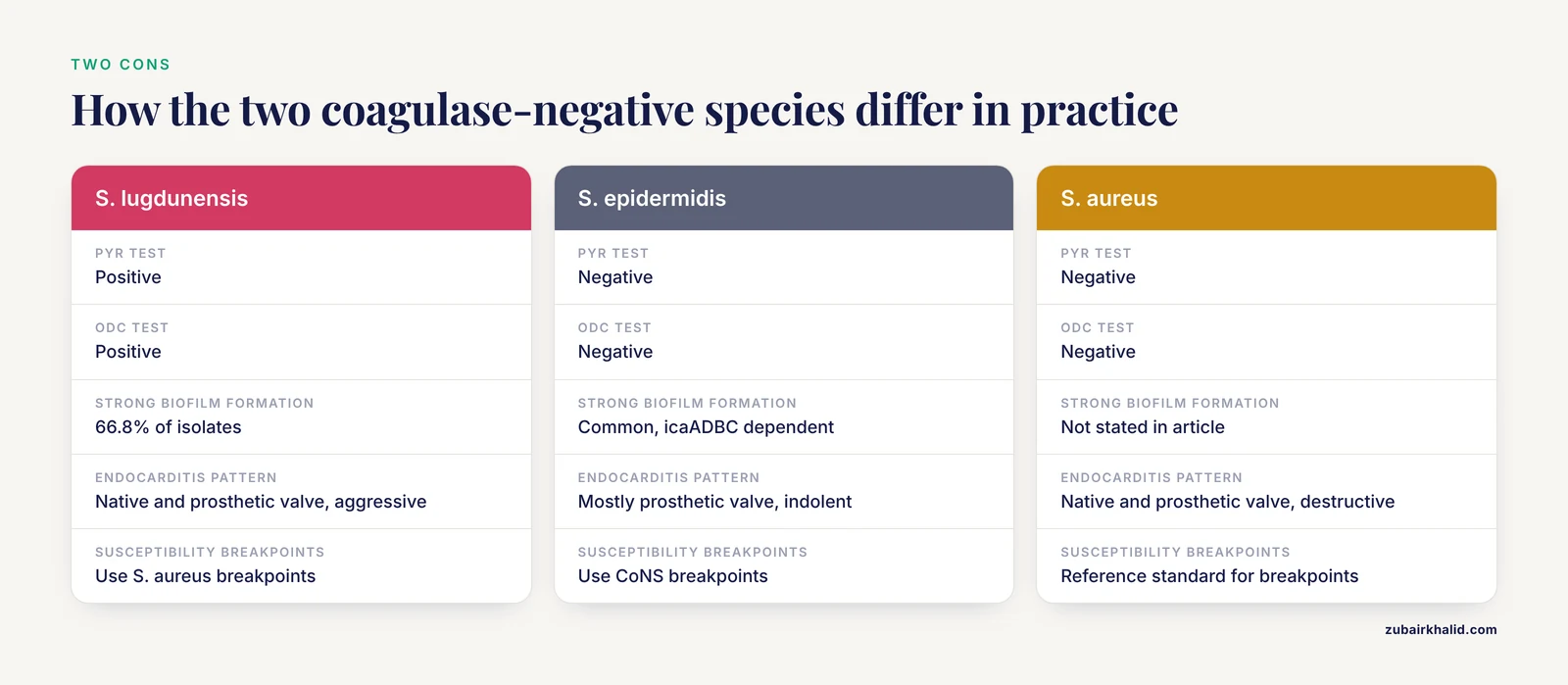
The comparison most often requested at the bench is between S. lugdunensis and S. epidermidis, because both are CoNS and both are common skin commensals. The differences are clinically decisive.
| Feature | Staphylococcus lugdunensis | Staphylococcus epidermidis |
|---|---|---|
| Coagulase | Negative | Negative |
| PYR test | Positive | Negative |
| ODC test | Positive | Negative |
| Clumping factor | Variable (some strains positive) | Negative |
| Strong biofilm formation | 66.8% of isolates [6] | Common (icaADBC dependent) |
| Virulence profile | S. aureus-like, destructive [1][2] | Low, opportunistic |
| Endocarditis | Native and prosthetic valve, aggressive [1][7] | Mostly prosthetic valve, indolent |
| Clinical significance | True pathogen in a high proportion of isolates [2] | Often a contaminant |
| Susceptibility interpretation | Use S. aureus breakpoints [4] | Use CoNS breakpoints |
| Methicillin resistance (mecA) | 14.6% of isolates [8] | Often greater than 60% |
The single most important message in this table is that S. lugdunensis must not be managed like S. epidermidis. The microbiology laboratory should perform full species identification on any clinically significant CoNS isolate, and when S. lugdunensis is identified, the susceptibility report should follow the S. aureus interpretive criteria [4].
Virulence Factors
Genomic Relationship with Staphylococcus aureus
S. lugdunensis shares more than 70% of its genome with S. aureus [9]. This genomic similarity explains why the two species produce overlapping clinical syndromes. Whole-genome sequencing of seven clinical strains identified four new prophages (named φSL2 to φSL4, belonging to the Siphoviridae family) and five plasmids (pVISLISI_1 to pVISLISI_5), three of which are homologous to known plasmids including one from S. aureus [9]. Mobile genetic elements are the main vehicles for virulence and resistance genes in staphylococci, and their presence in S. lugdunensis suggests that horizontal gene transfer contributes to its pathogenic potential.
Metalloprotease and Tissue Destruction
The VISLISI prospective study, conducted at the University Hospital of Strasbourg from November 2013 to March 2016, collected 347 samples positive for S. lugdunensis, of which 129 (37.2%) came from confirmed infections [2]. A novel secreted metalloprotease was identified and purified from 50 of 81 included patients (61.7%), and it was most frequently associated with deep infections and prosthetic joint infections [2]. Peptide sequencing showed that this protease shares 62.42% identity with ShpI, a metalloprotease from Streptococcus pyogenes, a pathogen known for tissue destruction. This enzyme likely contributes to the ability of S. lugdunensis to destroy cardiac valves and other structural tissue [2].
Biofilm Formation
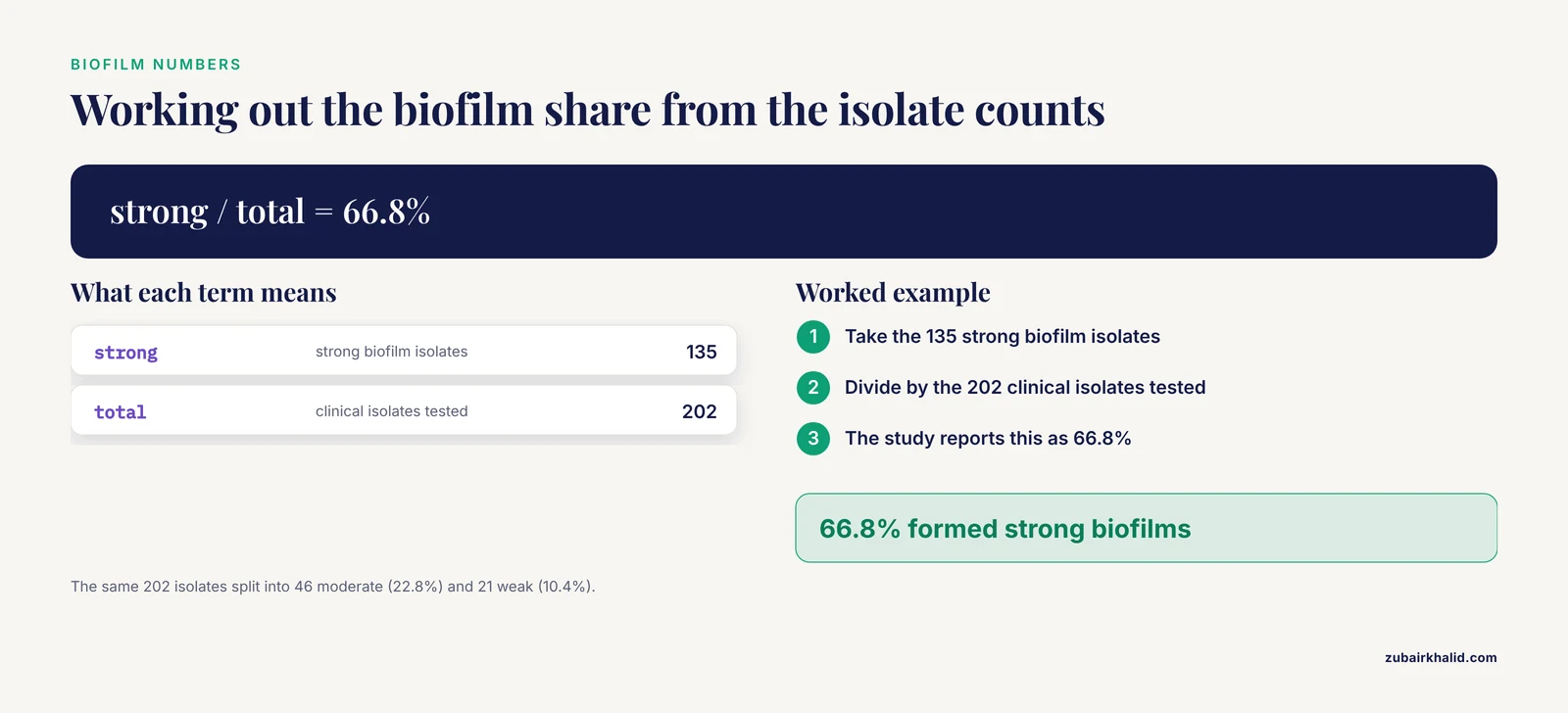
Biofilm formation is a major virulence mechanism in CoNS because it allows bacteria to adhere to medical devices and host tissue, resist antibiotics, and evade immune clearance. Among 202 clinical isolates of S. lugdunensis, 135 (66.8%) formed strong biofilms, 46 (22.8%) moderate biofilms, and 21 (10.4%) weak biofilms [6]. The same study found that isolates from primary bloodstream infections formed significantly stronger biofilms than isolates judged to be contaminants, and that ST3 isolates had higher biofilm-forming capacity than ST6 and ST27 isolates [6]. Biofilm-associated genes were highly conserved across all strains regardless of biofilm strength, meaning that the capacity to form biofilm is intrinsic to the species and the phenotypic expression varies by strain [6].
Clumping Factor and Adhesins
S. lugdunensis produces a clumping factor that is detectable in some strains, which can cause a false-positive slide coagulase test. This is a classic identification pitfall: a laboratory that uses only the slide coagulase test may misidentify S. lugdunensis as S. aureus. The tube coagulase test (free coagulase) remains negative, which is the definitive way to separate the two species. The clumping factor is a cell-wall-associated adhesin that binds fibrinogen, and it contributes to the ability of the organism to adhere to damaged endothelium, which is relevant to endocarditis pathogenesis.
Other Virulence Determinants
A genomic study of 144 S. lugdunensis genomes found that virulence genes, including cap8E (capsular polysaccharide biosynthesis) and esxA (type VII secretion system substrate), were widely distributed [8]. The type VII secretion system is a protein export apparatus that in S. aureus contributes to immune evasion and persistence. The presence of these genes across diverse sequence types indicates that the species carries a core virulence arsenal rather than relying on strain-specific acquisitions.
Clinical Syndromes
Infective Endocarditis
S. lugdunensis is the most feared CoNS in the context of endocarditis. Unlike S. epidermidis, which almost exclusively causes prosthetic valve endocarditis with an indolent course, S. lugdunensis causes both native and prosthetic valve endocarditis with rapid valvular destruction, perivalvular abscess formation, and high mortality [1][7][10].
The clinical picture is S. aureus-like. A case report of prosthetic mitral valve endocarditis in a woman in her 70s described rapid deterioration and death within 5 days of admission despite targeted intravenous antibiotics [1]. Another case documented native mitral valve endocarditis complicated by cerebral embolic infarctions in a peritoneal dialysis patient, requiring surgical valve replacement [7]. An unusual case of tricuspid valve endocarditis occurred in a patient with an unrepaired ventricular septal defect and autoimmune hepatitis [11]. S. lugdunensis has also been reported as the cause of an infected left atrial myxoma with concomitant mitral valve endocarditis and septic emboli [12].
A particularly instructive case involved an elderly male who presented with P wave asystole as the initial manifestation of S. lugdunensis endocarditis, in the absence of fever or classic symptoms [10]. Transesophageal echocardiography revealed severe aortic regurgitation from cusp destruction, a perivalvular abscess, and an acquired Gerbode defect (a left ventricular-to-right atrial communication). The patient required urgent aortic valve replacement, and the Gerbode defect was found to have spontaneously closed at surgery [10]. This case emphasizes that S. lugdunensis endocarditis can present with conduction abnormalities and that structural complications develop quickly.
The modified Duke criteria for infective endocarditis rely on isolating the causative organism. When S. lugdunensis is reported as a contaminant, the diagnosis can be delayed. One case report described an elderly female whose blood cultures grew methicillin-sensitive S. lugdunensis that was initially reported as a contaminant; a transthoracic echocardiogram eventually revealed tricuspid vegetations with moderate regurgitation, and the patient was successfully treated with cefazolin [3].
Bloodstream Infections and Bacteremia
S. lugdunensis is isolated from blood cultures more often than previously recognized. In a laboratory that switched from phenotypic methods to MALDI-TOF MS, S. lugdunensis was identified more commonly than before, and the authors raised the possibility of vertical transmission from mother to infant [13]. The clinical significance of a blood culture isolate is not always clear: in one study of 202 isolates, 36 bacteremia isolates were classified as primary infections and 40 as contaminants [6]. The distinction matters because primary infection isolates formed significantly stronger biofilms, suggesting that biofilm capacity is a marker of true pathogenicity [6].
A nationwide surveillance study in China from 2014 to 2023 collected 11,277 CoNS isolates and found that S. epidermidis and S. lugdunensis increased in bloodstream infections over the study period [14]. This trend, combined with the high per-isolate virulence of S. lugdunensis, means that clinical laboratories should not reflexively dismiss CoNS bacteremia without species identification.
Bone and Joint Infections
In the VISLISI prospective study, bone and prosthetic joint infections were the most frequent site of S. lugdunensis infection, accounting for 28 of 81 cases (34.6%) [2]. The metalloprotease was most frequently associated with deep infections and prosthetic joint infections, with probabilities of 0.97 and 0.91 respectively [2]. S. lugdunensis should be considered a true pathogen when isolated from joint aspirates, bone biopsies, or prosthetic material, and it should not be dismissed as skin contamination.
Skin and Soft Tissue Infections
Skin and soft tissue infections were the second most common presentation in the VISLISI study, accounting for 23 of 81 cases (28.4%) [2]. These include abscesses, wound infections, and postoperative surgical site infections [15]. The organism can cause brain abscess, breast abscess, and spondylodiscitis [15].
Other Infections
The disease spectrum of S. lugdunensis includes meningitis, septic arthritis, peritonitis, and post-surgical wound infections [15]. S. lugdunensis has also been isolated from bovine mastitis in India, where CoNS constituted 64% of staphylococcal isolates from mastitis cases and S. lugdunensis was among the species identified [16]. This veterinary finding is relevant to the One Health perspective but does not change the human clinical approach.
Laboratory Identification
Specimen Handling and Culture
S. lugdunensis grows on standard media, including sheep blood agar, and chocolate agar. Like other staphylococci, it does not grow on MacConkey agar. Blood culture bottles flag positive in standard automated systems. Once Gram stain shows Gram-positive cocci in clusters and the catalase test is positive, the isolate is a Staphylococcus. The next step is the coagulase test.
Coagulase Testing and the PYR/ODC Workup
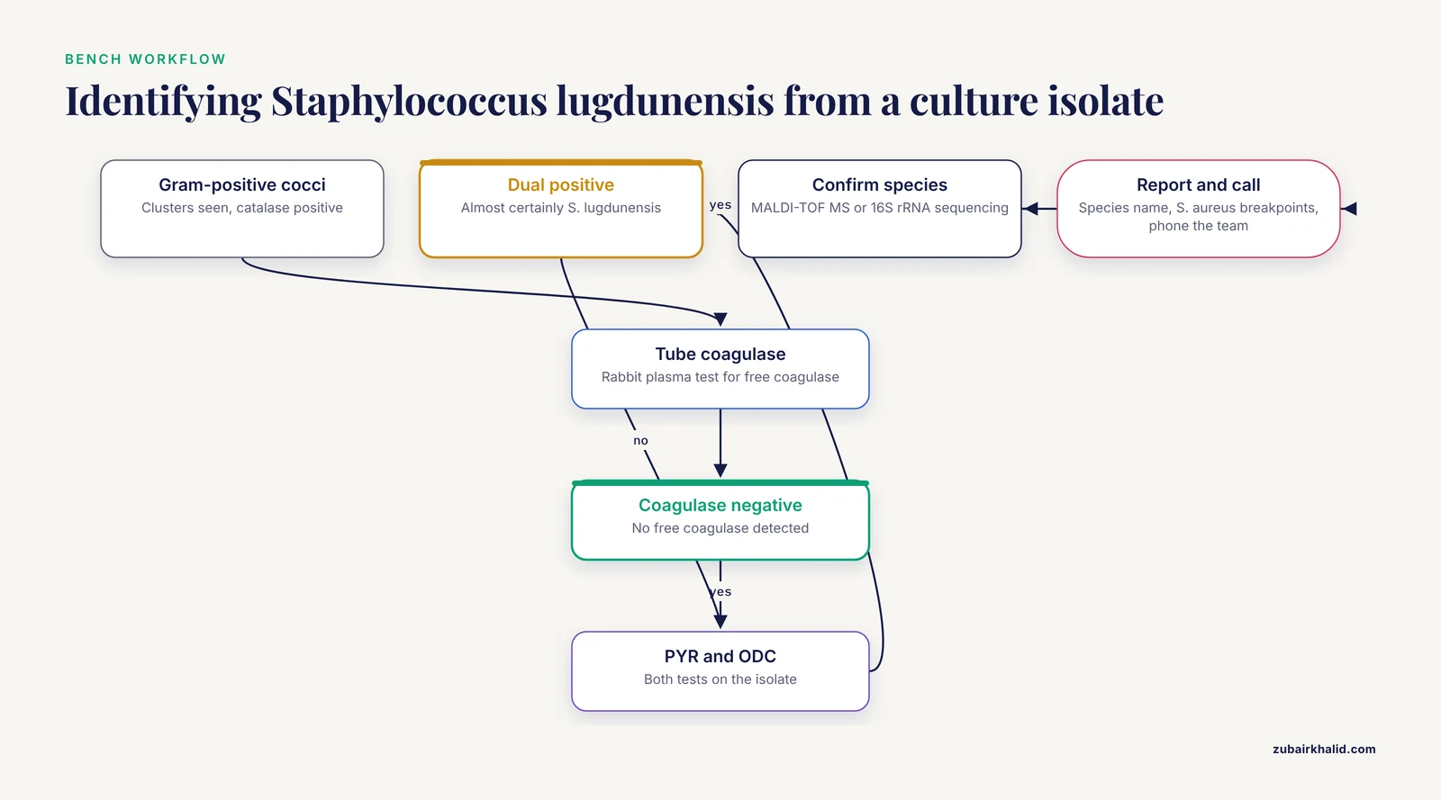
The tube coagulase test uses rabbit plasma and detects free coagulase. S. lugdunensis is tube coagulase negative [4]. The slide coagulase test detects clumping factor and can be positive in some S. lugdunensis strains, which can mislead the bench into calling the isolate S. aureus. Any isolate that is slide coagulase positive but tube coagulase negative should be worked up as a possible S. lugdunensis.
For tube coagulase negative isolates, the PYR and ODC tests are the fastest phenotypic route to S. lugdunensis. PYR tests are available as commercial disks or strips. ODC tests require a Moeller decarboxylase broth with ornithine and a mineral oil overlay, incubated for 24 to 48 hours. An isolate that is PYR positive and ODC positive is almost certainly S. lugdunensis [4]. In the Turkish study, the single S. lugdunensis isolate from eye vitreous fluid was dual positive with ODC and PYR, and API Staph confirmed the identification [4].
MALDI-TOF Mass Spectrometry
Matrix-assisted laser desorption ionization-time of flight mass spectrometry (MALDI-TOF MS) is now the standard method for species-level identification of CoNS in clinical laboratories. In a study comparing phenotypic methods with MALDI-TOF MS, phenotypic methods identified only 14% of CoNS isolates to species level, whereas MALDI-TOF MS identified the large majority [5]. The same study found that 40% of S. lugdunensis strains were associated with infection risk, a rate far higher than for other CoNS [5].
MALDI-TOF MS also revealed the presence of recently characterized staphylococcal species such as S. pettenkoferi, S. condimenti, and S. piscifermentans, which had been previously unrecognized because phenotypic methods could not separate them [5]. For S. lugdunensis specifically, MALDI-TOF MS provides a definitive identification in minutes once a colony is available.
Molecular Methods
16S rRNA gene sequencing is the gold standard for species identification when phenotypic and MALDI-TOF results are discrepant. In a study of CoNS from bovine mastitis, 16S rDNA sequencing confirmed the biochemical identification of seven representative isolates and identified additional species such as S. schleiferi, S. haemolyticus, and S. sciuri [16]. For S. lugdunensis, species-specific PCR targeting the fbl gene (encoding the fibrinogen-binding protein) is also available in reference laboratories.
Reporting and Communication
When S. lugdunensis is identified, the laboratory should:
- Report the species name, not "coagulase-negative staphylococcus."
- Perform antimicrobial susceptibility testing and interpret using S. aureus breakpoints [4].
- Flag the isolate as clinically significant if it is from a sterile site.
- Communicate directly with the clinical team for blood culture isolates, because the organism can be fatal if treated as a contaminant [3].
Antimicrobial Susceptibility and Treatment
Beta-Lactamase Production
S. lugdunensis frequently produces beta-lactamases, which hydrolyze penicillin and aminopenicillins [15]. The blaZ gene was present in 37.5% of 144 genomes in a global genomic study [8]. As a result, penicillin is not reliable for treatment unless the isolate tests penicillin susceptible by the penicillin zone-edge test or beta-lactamase test. Most isolates are susceptible to oxacillin, cefazolin, and other antistaphylococcal beta-lactams.
Methicillin Resistance
Methicillin resistance in staphylococci is mediated by the mecA gene, which encodes an altered penicillin-binding protein (PBP2a) with low affinity for beta-lactams. The mecA gene was present in 14.6% of S. lugdunensis genomes in the global study, and staphylococcal cassette chromosome mec (SCCmec) elements were identified in all mecA-positive isolates, with type V predominant in ST3 and type II in ST6 [8]. Importantly, 76.2% of SCCmec-positive isolates were infection-associated, meaning that methicillin-resistant S. lugdunensis is not merely a colonizer [8].
Ceftaroline
Ceftaroline is a fifth-generation cephalosporin with activity against methicillin-resistant staphylococci. In a US surveillance study of 1,593 clinically significant CoNS isolates, all S. lugdunensis isolates were inhibited at ceftaroline MIC of ≤1 μg/mL, with MIC50/90 values of 0.06 to 0.25 and 0.25 to 0.5 μg/mL across species including S. lugdunensis [17]. Ceftaroline inhibited 99.2% of all CoNS at ≤1 μg/mL, including 98.7% of methicillin-resistant CoNS [17]. These data support ceftaroline as a reliable option for S. lugdunensis, including methicillin-resistant strains.
Combination Therapy for Persistent Bacteremia
A multicenter retrospective study evaluated ceftaroline in combination with daptomycin or vancomycin for methicillin-resistant CoNS bacteremia, including S. lugdunensis [18]. All 12 patients who received combination therapy had sterile blood cultures on the subsequent blood culture after ceftaroline initiation. Patients who received ceftaroline combination within 7 days had a faster time to blood culture sterilization than those who received it after 7 days (6 days, range 3 to 7, versus 17 days, range 12 to 19, p = 0.031) [18]. This supports early escalation to combination therapy when vancomycin or daptomycin alone fails to clear CoNS bacteremia.
Treatment Principles
The consensus approach, supported by the literature, is to treat S. lugdunensis infections with the same urgency and antibiotic regimens used for S. aureus [4][1]. Specific principles include:
- Native valve endocarditis requires intravenous bactericidal therapy, typically an antistaphylococcal beta-lactam (nafcillin, oxacillin, cefazolin) for methicillin-susceptible isolates, or vancomycin plus ceftaroline for methicillin-resistant isolates.
- Prosthetic valve endocarditis often requires surgical intervention in addition to antibiotics, because the organism destroys cardiac structures rapidly [1][10].
- Blood culture isolates should never be dismissed as contaminants without clinical correlation [3].
- The duration of therapy follows the S. aureus endocarditis guidelines (4 to 6 weeks for native valve, 6 weeks or longer for prosthetic valve), because S. lugdunensis behaves like S. aureus [1].
Epidemiology and Genomics
Colonization and Transmission
S. lugdunensis is a normal inhabitant of human skin, particularly in the perineal area, axillae, and lower extremities. The finding of possible vertical transmission from mother to infant suggests that colonization can be acquired early in life [13]. The organism is also found in the environment and in animals, including bovine mastitis [16].
Population Structure
A global genomic study of 144 S. lugdunensis genomes identified 19 sequence types (STs) grouped into five clonal complexes (CCs), with CC3 (notably ST3) and ST27 predominating [8]. A sequence type is a grouping of isolates based on the alleles of seven housekeeping genes, and a clonal complex is a group of related sequence types that share a recent common ancestor. ST3 isolates exhibited higher biofilm formation than ST6 and ST27 isolates [6]. Eighteen antimicrobial resistance genes were identified across the population, with blaZ (37.5%), qacD (34.7%), and mecA (14.6%) being the most frequent [8]. Seven isolates carried five or more resistance genes, and novel plasmids carrying multiple resistance genes were identified in two ST3 isolates [8]. This population structure matters because ST3 appears to combine high biofilm formation with a high prevalence of SCCmec type V, making it the lineage most likely to cause difficult-to-treat infections.
Trends Over Time
In China, the proportion of methicillin-resistant CoNS decreased significantly in both bloodstream and cerebrospinal fluid isolates from 2014 to 2023, while vancomycin susceptibility remained stable [14]. S. lugdunensis increased in bloodstream infections over the same period [14]. The combination of increasing frequency and high per-isolate virulence means that S. lugdunensis will become a more common clinical problem as MALDI-TOF MS becomes universal.
Common Mistakes and Limitations
- Reporting S. lugdunensis as "CoNS, no species identification." This is the most dangerous mistake. Phenotypic methods identify only 14% of CoNS to species level, and S. lugdunensis is exactly the species that must not be missed [5]. Any laboratory using MALDI-TOF MS should reflexively identify all CoNS from sterile sites to species level.
- Misidentifying S. lugdunensis as S. aureus due to a positive slide coagulase test. The clumping factor in some S. lugdunensis strains causes a false-positive slide coagulase result. Always confirm with the tube coagulase test, which is negative in S. lugdunensis.
- Interpreting susceptibility results with CoNS breakpoints. S. lugdunensis should be interpreted using S. aureus breakpoints, particularly for oxacillin and cefoxitin [4].
- Treating S. lugdunensis bacteremia as contamination. In one case, the organism was initially reported as a contaminant, delaying the diagnosis of endocarditis [3]. A substantial proportion of isolates are true pathogens.
- Underestimating the speed of valvular destruction. S. lugdunensis endocarditis can be fatal within 5 days of admission despite appropriate antibiotics [1]. Surgical consultation should occur early.
- Assuming all S. lugdunensis are methicillin susceptible. The mecA gene is present in 14.6% of isolates, and 76.2% of SCCmec-positive isolates are infection-associated [8]. Oxacillin susceptibility must be tested, not assumed.
- Ignoring the possibility of non-endocarditis deep infections. S. lugdunensis causes prosthetic joint infections, spondylodiscitis, brain abscess, and peritonitis [15][2]. A sterile-site isolate should trigger a full workup.
- Relying on phenotypic methods alone for species identification. MALDI-TOF MS identifies approximately 85% of CoNS to species level, compared to 14% for phenotypic methods [5]. Laboratories without MALDI-TOF MS should use the PYR and ODC tests to screen for S. lugdunensis [4].
Individual treatment decisions must be made by the clinical team caring for the patient, and this article does not replace infectious disease consultation.
Quick Review
- S. lugdunensis is a coagulase-negative staphylococcus that behaves clinically like S. aureus and shares more than 70% of its genome with S. aureus [9].
- It is PYR positive and ODC positive, a combination that is nearly unique among CoNS [4].
- It causes native and prosthetic valve endocarditis with rapid valvular destruction, perivalvular abscess, and high mortality [1][10].
- Bone and joint infections are the most frequent clinical presentation, followed by skin and soft tissue infections [2].
- A novel metalloprotease, secreted by 61.7% of clinical isolates, is associated with deep infections and prosthetic joint infections [2].
- Strong biofilm formation is present in 66.8% of isolates, and the mecA gene is present in 14.6% of isolates [8][6].
- Antibiotic susceptibility must be interpreted using S. aureus breakpoints, ceftaroline inhibits all tested isolates at ≤1 μg/mL, and treatment should follow S. aureus endocarditis guidelines [4][1][17].
Related Guides
- Gram Stain Reporting for Blood Cultures: Explains Gram stain reporting practices for positive blood cultures
- Antimicrobial Resistance in Livestock-Associated Staphylococcus aureus: Genomic Epidemiology and One Health Implications: Provides One Health context for staphylococcal antimicrobial resistance and livestock-associated clones
- How to Detect Functional Genes in Long-Read Metagenomes: A Guide to Antibiotic Resistance and Virulence Factor Annotation: Explains detecting resistance and virulence genes in genomic data, complementing S. lugdunensis genomics
- Veterinary Culture and Susceptibility Sampling: Antimicrobial Selection and Treatment Review: Veterinary culture and susceptibility sampling context for animal S. lugdunensis isolates
Frequently Asked Questions
Is Staphylococcus lugdunensis coagulase negative?
Yes. S. lugdunensis is a coagulase-negative staphylococcus, meaning it does not produce free coagulase in the tube coagulase test [4]. However, some strains produce a clumping factor that can cause a positive slide coagulase test, which can lead to misidentification as S. aureus.
How is Staphylococcus lugdunensis different from Staphylococcus epidermidis?
S. lugdunensis is PYR positive and ODC positive, while S. epidermidis is negative for both. S. lugdunensis is far more virulent, causing native valve endocarditis and destructive infections similar to S. aureus, while S. epidermidis is mostly an opportunistic pathogen of prosthetic devices [1][2].
What antibiotics are used for Staphylococcus lugdunensis treatment?
Treatment follows S. aureus guidelines. Methicillin-susceptible isolates are treated with antistaphylococcal beta-lactams such as nafcillin or cefazolin. Methicillin-resistant isolates are treated with vancomycin or daptomycin, often with ceftaroline as combination therapy for persistent bacteremia [17][18].
Does Staphylococcus lugdunensis cause endocarditis?
Yes, and this is its most feared manifestation. S. lugdunensis causes both native and prosthetic valve endocarditis with rapid valvular destruction, perivalvular abscess, embolic stroke, and high mortality [1][7][10]. It can be fatal within days despite appropriate antibiotics [1].
How is Staphylococcus lugdunensis identified in the laboratory?
The organism is identified by MALDI-TOF mass spectrometry, which identifies approximately 85% of coagulase-negative staphylococci to species level [5]. In laboratories without MALDI-TOF, the PYR and ODC tests are the key phenotypic screens, and 16S rRNA gene sequencing is the molecular gold standard [4][16].
Is Staphylococcus lugdunensis a contaminant in blood cultures?
It can be a contaminant, but it must never be assumed to be one. Any S. lugdunensis blood culture isolate should prompt clinical correlation and an evaluation for endocarditis, because the organism causes destructive native and prosthetic valve infections that are fatal if treatment is delayed [1][3].
Sources
- Case Report: Fatal prosthetic valve endocarditis due to Staphylococcus lugdunensis-a wolf in coagulase-negative clothing.
- VISLISI trial, a prospective clinical study allowing identification of a new metalloprotease and putative virulence factor from Staphylococcus lugdunensis.
- The Deadly Contaminant: A Case of Staphylococcus lugdunensis Endocarditis.
- Investigation of Staphylococcus lugdunensis and Selected Coagulase Negative Staphylococci Isolated from Blood Culture bottles and Determination of their Sensitivities to Antibiotics.
- Implementation of Matrix-Assisted Laser Desorption Ionization-Time of Flight Mass Spectrometry in Routine Clinical Laboratories Improves Identification of Coagulase-Negative Staphylococci and Reveals the Pathogenic Role of Staphylococcus lugdunensis.
- Biofilm-forming ability of Staphylococcus lugdunensis clinical isolates associates with sequence type and virulence in a Galleria mellonella model.
- Innocent Until Proven Guilty: Native Valve Staphylococcus lugdunensis Infective Endocarditis Complicated by Embolic Stroke in a Peritoneal Dialysis Patient: A Case Report.
- Global genomic landscape of Staphylococcus lugdunensis: population structure, antimicrobial resistance, and virulence determinants.
- Whole-Genome Sequencing of Seven Strains of Staphylococcus lugdunensis Allows Identification of Mobile Genetic Elements.
- Staphylococcus lugdunensis endocarditis presenting with P wave asystole, valvular destruction and spontaneous closure of an acquired Gerbode defect.
- A rare case of Staphylococcus lugdunensis endocarditis of the tricuspid valve in a patient with unrepaired ventricular septal defect and autoimmune hepatitis.
- Staphylococcus lugdunensis-Infected Left Atrial Myxoma Complicated by Mitral Valve Endocarditis and Septic Emboli: A Case Report.
- Unbiased species-level identification of clinical isolates of coagulase-negative Staphylococci: does it change the perspective on Staphylococcus lugdunensis?
- A decade-long nationwide surveillance of antimicrobial resistance in coagulase-negative staphylococci in China (2014-2023) and characterisation of a rare teicoplanin-resistant Staphylococcus haemolyticus.
- Identification of New Drug Target in Staphylococcus lugdunensis by Subtractive Genomics Analysis and Their Inhibitors through Molecular Docking and Molecular Dynamic Simulation Studies.
- Identification of coagulase negative staphylococcal species from bovine mastitis in India.
- Antimicrobial activity of ceftaroline and comparator agents when tested against numerous species of coagulase-negative Staphylococcus causing infection in US hospitals.
- Ceftaroline combination therapy for methicillin resistant coagulase negative Staphylococcus bacteraemia and endocarditis.