How to Write an SOP for Gram Staining in Microbiology
By Dr. Zubair Khalid, DVM, MS, PhD ·
By the end of this guide you will be able to write a complete, bench-ready Standard Operating Procedure (SOP) for the Gram stain, from the document header to the sign-off page. You will have a section-by-section template format you can copy, a filled-in example showing what each section looks like when completed, a worked calculation for preparing your own reagents, and a quality control (QC) plan you can defend during an inspection.
Everything in this article can be done with tools you already have on the bench: a word processor or document control system, your current staining protocol written down in draft form, the package inserts for your stain reagents, a compound microscope with a 100x oil objective, known quality control organisms, and a signed copy of your laboratory's document control policy.
Why a Written Gram Stain SOP Matters
The Gram stain separates bacteria into Gram-positive (purple, crystal violet retained) and Gram-negative (pink, crystal violet lost, safranin taken up) groups based on cell wall structure. It is usually the first result a physician sees for a culture workup, and it directs everything that follows, from media selection to the empiric therapy conversation. Because it is read by humans and judged by humans, it is one of the most operator-dependent tests in the microbiology laboratory.
That operator dependence is exactly why the SOP exists. A multi-center study of immunologic assays found that when laboratories each ran their own in-house procedures, results diverged enough to complicate any comparison between sites. After the teams adopted harmonized written SOPs, variation between laboratories dropped sharply, with coefficients of variation for stimulated responses falling from 148% to 77% for one antigen and from 126% to 73% for another [1]. The lesson transfers directly to the Gram stain: the single most effective intervention against operator-to-operator variation is a written procedure that specifies every variable, including reagent lots, contact times, and how the reader decides what to call the result.
A laboratory quality management system treats the SOP as a controlled document. It has a unique identifier, a version number, an effective date, named approvers, a defined review cycle, and a revision history. If a surveyor asks "show me how you do a Gram stain," the answer is a document, not a person's memory.
What a Gram Staining SOP Template Format Looks Like
A complete Gram staining SOP template contains twelve sections. Every accredited clinical or research laboratory template uses some version of this structure, and most document control systems enforce it. The sections, in order:
- Header and document control block
- Purpose
- Scope
- Principle (scientific basis)
- Responsibilities
- Definitions and abbreviations
- Reagents, supplies, and equipment
- Safety and waste handling
- Stepwise procedure
- Quality control
- Interpretation, reporting, and troubleshooting
- References, revision history, and approval signatures
If your laboratory already has an SOP for another stain, such as acid-fast or calcofluor white, reuse its structure. Consistency between SOPs in the same manual is itself a quality management practice, because staff stop relearning the document layout and start reading the content.
The Gram Staining SOP Template: Section-by-Section Walkthrough
Section 1. Header and Document Control
Every SOP begins with an identification block. At minimum it carries:
- SOP title: "Gram Stain, Bacterial (Smear)"
- SOP number: use your laboratory's scheme, for example
MIC-STAIN-001 - Version number and effective date
- Supersedes: the version this document replaces
- Department: Bacteriology
- Author, reviewer (usually the technical supervisor), and approver (usually the laboratory director)
- Review due date: most laboratories set annual or biennial review
- Page numbering in the format "Page X of Y", because loose pages are a classic inspection finding
Write this block as a table at the top of page 1. Do not bury the version number in a footer, because printed working copies get separated from their headers.
Section 2. Purpose
One or two sentences. State what the procedure accomplishes and why it exists.
The purpose of this SOP is to describe the preparation, staining, microscopic examination, and reporting of Gram-stained smears from clinical specimens and cultures in the Bacteriology Laboratory.
Section 3. Scope
State which specimen types and situations the SOP covers, and which it does not. A scope statement might read:
This SOP applies to Gram stains prepared from clinical specimens (wound, respiratory, sterile body fluids, urine, genital sources) and from isolated colonies on solid or in liquid media. It does not cover tissue section staining or fungal calcofluor stains, which are addressed in separate SOPs.
Naming what is excluded prevents scope creep and tells a new employee exactly where the boundary lies.
Section 4. Principle
Explain the chemistry in four to six sentences a competent new technologist can follow. The standard account, as described in bacteriology textbooks, runs as follows. Crystal violet, an aniline dye, enters all bacterial cells and binds to their nucleic acids and cell wall components. Gram's iodine, an iodine-potassium iodide complex, acts as a mordant, forming an insoluble crystal violet-iodine complex inside the cell. Decolorization with alcohol or an acetone-alcohol mixture dissolves lipids and extracts the dye complex. Gram-negative bacteria, with their thin peptidoglycan layer and outer membrane rich in lipids, lose the complex rapidly and then take up the pink counterstain safranin. Gram-positive bacteria, with a thick, cross-linked peptidoglycan layer and no outer membrane, retain the crystal violet-iodine complex and stay purple. Write the exact sequence your laboratory uses in the procedure section, and write the reasoning once, here.
Section 5. Responsibilities
Assign each task to a role, not a person's name, because roles survive staff turnover. A typical assignment table:
| Role | Responsibility |
|---|---|
| Medical laboratory scientist (bench) | Performs staining, reads smears, records QC, reports results |
| Technical supervisor | Reviews QC, approves reagent lots, trains new staff, maintains the SOP |
| Laboratory director | Approves the SOP and any revisions, reviews competency documentation |
Section 6. Definitions and Abbreviations
Define every term a new hire will not already know. For this SOP the standard set includes:
- SOP: Standard Operating Procedure, a controlled written document describing how a task is performed.
- Smear: a thin film of specimen or organisms spread on a glass slide.
- Heat fixation: passing an air-dried smear through a flame, or resting it on a warm surface, to adhere organisms to the glass and kill them.
- Mordant: a substance, here Gram's iodine, that fixes a dye in or on a material by forming an insoluble complex.
- Decolorizer: the solvent step that removes the crystal violet-iodine complex from Gram-negative cells.
- QC organism: a characterized reference strain of known staining reaction used to verify reagent performance.
- Gram-variable: organisms that stain both purple and pink in the same field or give inconsistent reactions.
Section 7. Reagents, Supplies, and Equipment
List every item with enough detail that a purchasing agent could reorder it without calling the bench. For each reagent record the expected formulation, storage condition, and stability. The core reagent set is:
- Crystal violet (Hucker or common commercial formulation)
- Gram's iodine
- Decolorizer (95% ethanol, or an acetone-alcohol mixture)
- Safranin counterstain
- Distilled or deionized water
- Immersion oil
- Clean glass slides, marked with a solvent-resistant pencil or slide labels
- Inoculating loops and needles, Bunsen burner or slide warmer, staining rack, bibulous paper or blotting pads
- Compound brightfield microscope with 100x oil objective
- Personal protective equipment: lab coat, gloves, eye protection
The worked calculation later in this article shows how to prepare crystal violet and Gram's iodine from raw chemicals if your laboratory mixes its own stains.
Section 8. Safety
Address the actual hazards of this procedure: fixed and unfixed smears from patient specimens are handled as potentially infectious until heat-fixed, open flames are used during fixation if you do not use a slide warmer, and the stains are chemical hazards. Specify that crystal violet is a potential carcinogen in some animal models, that gloves and a lab coat are worn during staining, that spent stain goes into the chemical waste stream your institution defines, and that specimen smears are handled per your laboratory's biosafety manual. Cite your institution's chemical hygiene plan by its internal document number rather than restating its contents.
Section 9. Stepwise Procedure
This is the heart of the SOP. Write it as numbered steps in the imperative mood, with a defined time or endpoint for every staining step. Do not write "stain briefly". Write the number of seconds. The times below are a widely used representative scheme from standard bacteriology references. Your laboratory validates its own times and writes those validated times into its own SOP.
A. Smear preparation
- Label the frosted end of a clean slide with the specimen or culture identifier using a solvent-resistant marker.
- For colonies, place one to two loopfuls of a 18 to 24 hour colony in one or two drops of sterile saline on the slide and emulsify. For liquid specimens, place one drop of well-mixed specimen directly on the slide. For swabs, roll the swab over an area of roughly 1 by 2 cm.
- Spread to a thin, even film the size of a dime to a quarter. A good smear dries without visible opacity. Too thick a smear is the most common cause of an uninterpretable stain.
- Air dry completely at room temperature. Do not apply heat to speed drying before the film is dry, because wet heat fixation boils the cells and distorts morphology.
- Heat fix by passing the slide, film side up, rapidly through the blue cone of a Bunsen burner flame two to three times, or place it on a slide warmer at 60 to 65 degrees Celsius for several minutes. The slide should feel warm, not hot, on the back of your hand.
- Allow the slide to cool before staining.
B. Staining
- Flood the smear with crystal violet. Hold for 1 minute.
- Drain by tipping the slide. Rinse gently with a gentle stream of distilled water. Do not aim water directly at the film.
- Flood with Gram's iodine. Hold for 1 minute.
- Drain. Rinse gently with distilled water.
- Decolorize with 95% ethanol (or your validated decolorizer) by letting it flow across the tilted slide drop by drop until the solvent coming off the film runs clear, typically 5 to 10 seconds for thin smears. This is the critical, timing-sensitive step.
- Rinse immediately and gently with distilled water to stop decolorization.
- Flood with safranin. Hold for 30 to 60 seconds.
- Rinse gently with distilled water.
- Blot dry gently with bibulous paper or air dry. Do not rub the film.
- Examine under oil with the 100x objective. Record total magnification used, commonly 1000x.
Notice what the numbers in the SOP do. They remove every judgment call except the decolorization endpoint, and even that endpoint has a defined visual criterion ("solvent runs clear"). The FLUCOP project reached the same conclusion from the opposite direction: when laboratories documented exactly which tubes, times, and reagent volumes to use, the between-laboratory differences that had plagued their assays collapsed [1].
Section 10. Quality Control
Microbiology quality control for the Gram stain answers one question with a written record: "are my reagents working today?" The standard approach is to stain control organisms of known reaction alongside or in parallel with your test smears.
Control organisms. Use well-characterized strains with known, stable reactions. Laboratories conventionally use a Gram-positive coccus such as Staphylococcus aureus ATCC 25923 and a Gram-negative bacillus such as Escherichia coli ATCC 25922. Prepare stocks from a reference supplier, follow the supplier's storage and passage instructions, and do not pass controls repeatedly in-house, because repeated subculture can shift staining behavior.
Acceptance criteria. Write them explicitly:
- The S. aureus control stains purple (Gram-positive cocci in clusters).
- The E. coli control stains pink (Gram-negative bacilli).
- Background debris and host cells stain faintly pink and do not obscure morphology.
- If either control fails, do not report patient results. Discard or troubleshoot the reagent in question, repeat QC, and document the corrective action.
Frequency. State the schedule your laboratory's quality plan requires. Common practice is to perform QC with each new lot or shipment of reagent, when transferring a reagent to a new staining setup, and at a defined interval such as with each staining run or weekly. Whatever your schedule, the SOP must name it, and the QC result must be recorded on a form or in the laboratory information system with date, technologist initials, lot numbers, and pass or fail.
Reagent records. Record the date opened or prepared, the lot number, the expiration date, and the storage condition for each staining reagent. Iodine solutions fade with use and age, so your SOP should direct staff to replace iodine when the deep brown color has visibly lightened or at a defined interval, whichever comes first.
Section 11. Interpretation, Reporting, and Troubleshooting
Define how a result is described so two technologists reading the same smear write compatible reports. The standard reporting elements are:
- Gram reaction and morphology of organisms seen, for example "many Gram-positive cocci in clusters", "moderate Gram-negative bacilli"
- Relative quantity, using your laboratory's defined scale (for example: rare, few, moderate, many, with a defined number of organisms per oil immersion field for each tier)
- Presence and relative amount of host cells, for example polymorphonuclear cells and squamous epithelial cells
- For sputum and similar specimens, your laboratory's screening criteria for specimen acceptability, stated here by reference to the relevant SOP
Interpretation rules to write into the SOP:
- Read multiple fields before reporting a quantity. Define how many fields, for example at least 10.
- Report what you see, not what you expect. A Gram-negative diplococcus report says "Gram-negative diplococci", not "Neisseria", unless your system authorizes presumptive identification.
- If organisms are present but the stain is technically uninterpretable, state the reason, for example "smear too thick for evaluation", and request or prepare a new smear.
- Define who may read and release the result, and what the supervisor review requirements are.
Troubleshooting table. Build this into the SOP so corrective action happens at the bench:
| Observation | Likely cause | Corrective action |
|---|---|---|
| Gram-positive control stains pink | Over-decolorization, decolorizer too strong or step too long, thin film washed off | Shorten decolorization, verify decolorizer formulation, check smear thickness, repeat QC |
| Gram-negative control stains purple | Under-decolorization, decolorizer expired or diluted, smear too thick | Verify decolorizer lot and concentration, make thinner smears, extend decolorization endpoint, repeat QC |
| All cells faint and washed out | Old iodine (lost mordant activity), rinse too forceful, film not heat fixed | Replace iodine, gentler rinse, verify fixation step |
| Crystal violet crystals on smear | Stain allowed to dry on slide, dye precipitated | Keep slide wet between steps, filter or replace crystal violet |
| Nothing visible at 100x | Smear absent, film washed off, wrong side examined, focus error | Verify smear placement and fixation, mark film side, recheck microscope |
| Organisms Gram-variable | Old culture, thick smear, inherent variability of the organism | Use fresh 18 to 24 hour culture, make thinner smear, report as Gram-variable when consistent |
Section 12. References, Revision History, and Approval
Close the SOP with three blocks. First, references: the bacteriology textbooks or manuals your procedure is based on, cited by edition and page where applicable. Second, a revision history table with version, date, author, and a one-line summary of what changed. Third, signature lines for author, reviewer, and director, with dates. An SOP without signatures is a draft.
Worked Example: A Gram Staining SOP Template Example, Filled In
The skeleton below doubles as a free gram staining SOP template you can paste directly into your document control system and complete. Every line in brackets gets replaced with your laboratory's specifics.
SOP NUMBER: [MIC-STAIN-001]        VERSION: [2.0]
TITLE: Gram Stain, Bacterial (Smear)
EFFECTIVE DATE: [YYYY-MM-DD]       REVIEW DUE: [YYYY-MM-DD]
SUPERSEDES: [Version 1.0, YYYY-MM-DD]
AUTHOR: [Name, title]              REVIEWED BY: [Name, title]
APPROVED BY: [Name, title, Laboratory Director]

1. PURPOSE
[One or two sentences, imperative.]

2. SCOPE
[Specimen types covered. What is explicitly excluded.]

3. PRINCIPLE
[4-6 sentences on crystal violet, iodine mordant,
decolorization, safranin, and cell wall basis.]

4. RESPONSIBILITIES
Bench technologist: [tasks]
Technical supervisor: [tasks]
Laboratory director: [tasks]

5. DEFINITIONS
[List each term with a one-line definition.]

6. REAGENTS AND EQUIPMENT
| Reagent | Formulation | Storage | Expiry | QC frequency |
| [Crystal violet] | [...] | [...] | [...] | [...] |
| [Gram's iodine] | [...] | [...] | [...] | [...] |
| [Decolorizer] | [...] | [...] | [...] | [...] |
| [Safranin] | [...] | [...] | [...] | [...] |
Equipment: [microscope, racks, burner or warmer, ...]

7. SAFETY
[PPE, infectious material handling, chemical waste stream,
flame or heat source precautions, chemical hygiene plan ref.]

8. PROCEDURE
8.1 Smear preparation: [numbered steps 1-6 as above]
8.2 Staining: [numbered steps with exact times]
8.3 Microscopy: [magnification, fields read]

9. QUALITY CONTROL
Control strains: [S. aureus ATCC 25923, E. coli ATCC 25922]
Expected results: [purple cocci in clusters / pink bacilli]
Frequency: [each new lot / per run / weekly]
Acceptance criteria: [as written above]
Failure action: [stop testing, troubleshoot, repeat, document]

10. INTERPRETATION AND REPORTING
Quantity scale: [rare / few / moderate / many, defined]
Morphology terms: [approved terminology list]
Uninterpretable smears: [criteria and action]

11. TROUBLESHOOTING
[Table of observation / cause / action.]

12. REVISION HISTORY
| Version | Date | Author | Summary of change |

13. APPROVAL
Author signature/date, Reviewer signature/date,
Director signature/date.
When completed, a real line from section 8.2 of your document should read like this:
8.2.1  Flood the smear with crystal violet (Lot CV-24117,
       exp. 2027-03-15). Hold exactly 60 seconds.
8.2.2  Drain by tipping the slide. Rinse gently with
       distilled water for 2-3 seconds, directing the
       stream above the film, not onto it.
That level of specificity, including the lot number convention and the direction of the rinse stream, is the difference between an SOP and a memory aid.
Preparing Your Own Reagents: Worked Calculations
If your laboratory prepares stains from raw chemicals following the classic Hucker modification, write the formulas and the calculations into the SOP so any technologist can reproduce them. Standard Hucker formulations are:
- Crystal violet stock: dissolve 2 g crystal violet (90% dye content) in 20 mL of 95% ethyl alcohol. Separately dissolve 0.8 g ammonium oxalate in 80 mL distilled water. Mix the two solutions, let stand 24 hours, then filter. Store in amber glass at room temperature.
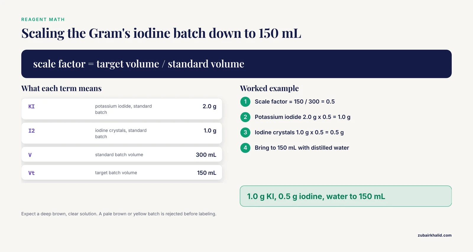
- Gram's iodine: dissolve 2 g potassium iodide in a small volume (about 10 mL) of distilled water, add 1 g iodine crystals, shake until dissolved, then bring the volume to 300 mL with distilled water. Store in amber glass away from light.
- Decolorizer: 95% ethyl alcohol, or a 1:1 mixture of acetone and 95% ethyl alcohol for faster action.
- Safranin working solution: 0.25 g safranin O in 10 mL of 95% ethyl alcohol, brought to 100 mL with distilled water.
Here is the worked calculation for scaling the iodine down to a 150 mL batch, as you would show it in the SOP:
Standard formula (makes 300 mL):
  Potassium iodide  2.0 g
  Iodine crystals   1.0 g
  Distilled water   to 300 mL

Target batch = 150 mL, so scale factor = 150 / 300 = 0.5

  Potassium iodide  2.0 g x 0.5 = 1.0 g
  Iodine crystals   1.0 g x 0.5 = 0.5 g
  Distilled water   to 150 mL

Expected output: a deep brown, clear solution. If the
solution is pale brown or yellow on the day of preparation,
the iodine has not fully dissolved, and the batch is
rejected before labeling. Record lot, preparation date,
preparer initials, and a 3-month expiry on the label.
Showing the expected output matters. A technologist who has never seen good Gram's iodine cannot recognize bad Gram's iodine, and the SOP is the right place to teach the difference.
Quality Control Records: What a Completed QC Entry Looks Like
The QC section of the SOP becomes real on a record form. Here is what a passing daily entry looks like in a plain-text version of your form, so staff know the expected output before they ever fill one in:
GRAM STAIN QC RECORD                    Date: 2026-11-11
Technologist: J. Rivera        Initials: JR
Reagent lots in use:
  Crystal violet  CV-24117  exp 2027-03-15
  Gram's iodine   IO-2609   exp 2027-02-01
  Decolorizer     DC-2544   exp 2027-06-30
  Safranin        SA-2380   exp 2027-05-12

Control slide results:
  S. aureus ATCC 25923: purple cocci in clusters ... PASS
  E. coli  ATCC 25922:  pink bacilli ............... PASS
Background: acceptable, host cells faint pink ..... PASS

Overall QC result: ACCEPTABLE   Patient testing may proceed.
Supervisor review (weekly): ______ date ______
If the Gram-positive control had read pink, the record would show FAIL in that line, Overall QC result: UNACCEPTABLE, a line stating "Patient staining held", and a corrective action note referencing the troubleshooting table. Reviewing blank versus failing forms during training is one of the fastest ways to teach new staff what the quality system expects.
Building Gram Stain QC into Your Laboratory Quality Management Program
The Gram stain QC record should not live in isolation. In a working laboratory quality management system it connects to four other elements:
- Document control. The SOP itself is versioned, approved, and periodically reviewed. Staff confirm in writing that they have read the current version before performing the procedure.
- Competency assessment. Each technologist's Gram stain reading is assessed against known material on a defined schedule, typically at initial training and then periodically, with the method and frequency defined by your laboratory's competency policy.
- Proficiency testing. External or inter-laboratory challenges verify that your written procedure produces correct and comparable results. The FLUCOP experience illustrates the pattern well: the project teams ran proficiency tests before and after introducing harmonized SOPs, and the post-harmonization tests demonstrated both reduced background noise in the assay and statistically significant reduction in between-laboratory variation, confirming that the written procedures had done their job [1]. Your laboratory can apply the same principle internally by having two technologists read the same set of smears blind and comparing their interpretations.
- Corrective action. Every QC failure generates a documented investigation: what failed, why, what was fixed, and how re-acceptance was demonstrated. The troubleshooting table in section 11 is the first stop of that investigation.
Auditors rarely ask "did you do QC today." They ask "show me the trail from your QC failure in March to the corrective action you took." The SOP, the QC record, and the corrective action file together are that trail.
Common Mistakes and Limitations
These are the errors that appear most often when laboratories write or run Gram stain SOPs.
- Writing "stain until decolorized" without an endpoint. An SOP that leaves the decolorization endpoint undefined produces one answer from a senior technologist and a different answer from a new hire. Define the visual endpoint in words.
- Omitting reagent lot and expiry recording. If a staining problem appears, the first question is always which lot was in use. A SOP that does not require lot recording makes that question unanswerable.
- Skipping QC after a new reagent lot. New lots can differ in dye concentration and solvent strength. Staining patient material on the lot's first day without control organisms is a reportable-quality-event waiting to happen.
- Letting the smear be the last thing considered. Most unreadable stains are unreadable because the smear was too thick, not because the reagents failed. State smear quality criteria in the procedure, not just in troubleshooting.
- Reporting interpretation beyond the stain. The Gram stain supports presumptive description, not definitive identification. An SOP that lets the report drift toward genus-level calls without a defined rule will generate inconsistency.
- Treating the SOP as static. If the bench starts doing something differently, either the practice is wrong or the SOP is outdated. Both require action, and neither is solved by silence.
- Assuming the stain is the only diagnostic layer. Molecular and point-of-care methods continue to expand, and a recent review of point-of-care nucleic acid amplification platforms highlights how much of diagnostics is moving toward rapid, decentralized testing [2]. Stain-based microscopy remains foundational on the bacteriology bench, but your SOP and your test menu should acknowledge where molecular confirmation sits in your workflow.
One limitation applies to this article as a whole. Individual laboratories operate under different accreditation requirements, and every patient case requires judgment by qualified laboratory and clinical staff. Use this guide to draft your SOP, then reconcile it with your own quality manual and director's expectations.
Implementation Checklist and Next Steps
Work through these steps in order when you adopt the template:
- Photograph or copy your current bench practice exactly as performed today, inconsistencies included.
- Fill in the template against actual practice, deciding deliberately which inconsistencies to fix rather than letting the document lie.
- Validate your chosen contact times with control organisms across at least one full reagent lot cycle before freezing the numbers.
- Route the draft through reviewer and director signatures in your document control system.
- Train all staff on the new version and collect read-and-understood sign-offs.
- Run the QC and competency elements for one full cycle, then schedule the SOP's first periodic review.
- When you next face an inspection or an inter-laboratory comparison, pull the trail: SOP, training records, QC records, and corrective actions. That trail is what your template was built to produce.
Frequently Asked Questions
What is a Gram staining SOP?
A Gram staining SOP is a controlled written document that specifies exactly how your laboratory prepares, stains, reads, reports, and quality-controls Gram-stained smears. It exists so that any qualified technologist following the document produces the same result.
What should a gram staining sop template include?
A complete template includes document control, purpose, scope, principle, responsibilities, definitions, reagents and equipment, safety, a stepwise procedure with exact times, quality control with named control organisms and acceptance criteria, interpretation and reporting rules, a troubleshooting table, and revision history with signatures. Anything less leaves a decision to memory.
How often should Gram stain reagents be quality controlled?
Follow the schedule your laboratory's quality plan defines. Common practice is QC with each new reagent lot, when the staining setup changes, and at a recurring interval such as every staining run or weekly, with results recorded on a dated, signed form.
Why do my Gram-positive organisms stain Gram-negative?
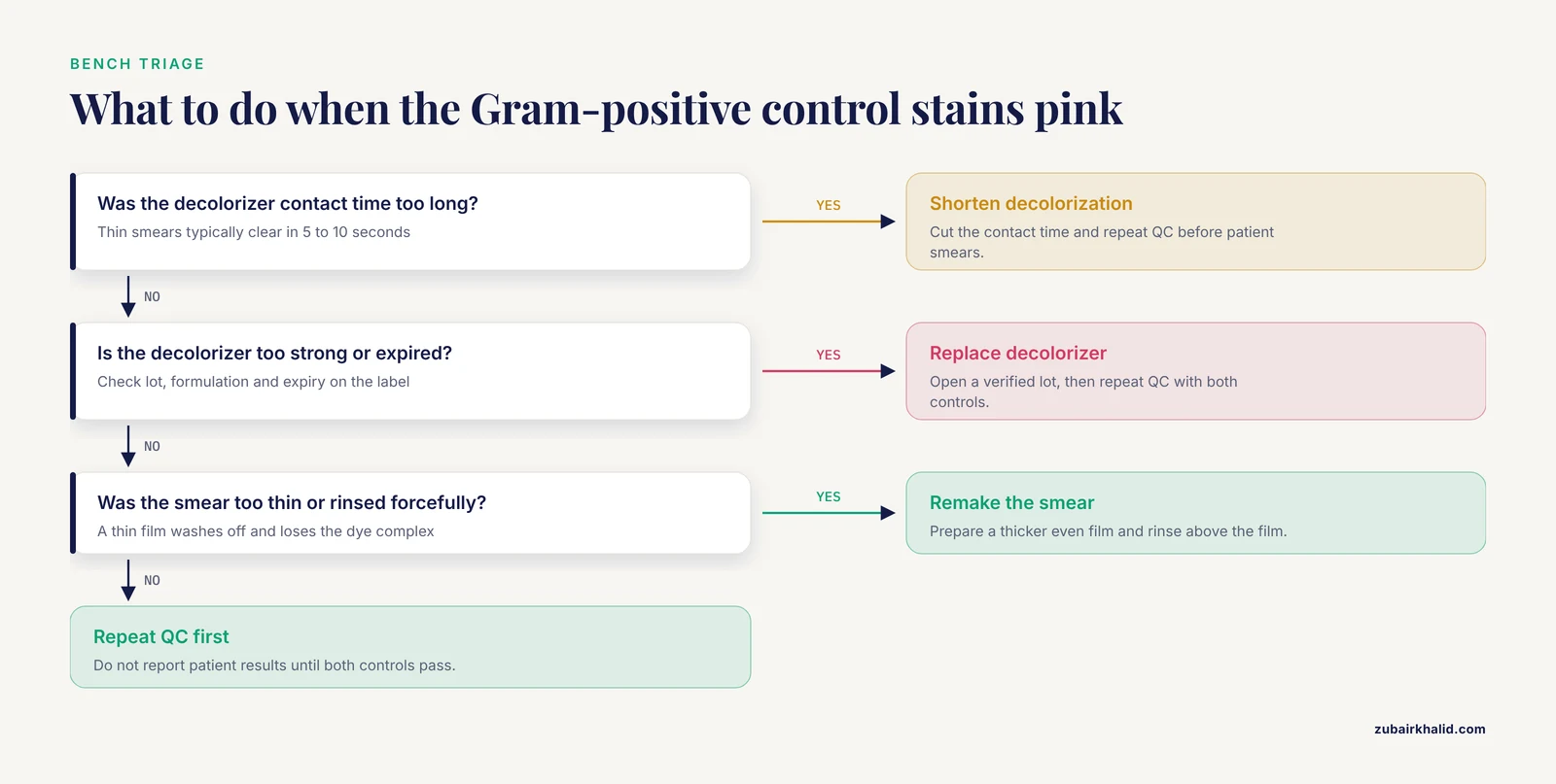
The most common cause is over-decolorization, meaning the decolorizer contact time was too long, the decolorizer is too strong, or the smear was too thin. Old crystal violet and forceful rinsing produce the same picture. Fix the step, then repeat QC with control organisms before trusting patient smears.
What is the difference between an SOP and a work instruction?
An SOP defines the full controlled procedure, its responsibilities, its QC, and its documentation. A work instruction is a shorter, job-focused document that may sit inside an SOP and describe one task in detail. Many laboratories make the stepwise staining sequence a work instruction that lives at the bench under the SOP.
How do I validate a new lot of Gram stain reagents?
Stain your Gram-positive and Gram-negative control organisms with the new lot before it enters patient use, compare the result to the acceptance criteria in your SOP, record the lot number and outcome, and document the sign-off that releases the lot for use. If the lot fails, quarantine it and follow your corrective action process.
Can I use a free gram staining SOP template as-is?
Use a free template as a starting skeleton, then fill in your laboratory's specific reagents, validated times, control strains, reporting rules, and approval chain. A template with someone else's times and organisms pasted in unverified is a document that will fail review and mislead staff.
Who approves a Gram staining SOP?
Typically the author writes it, the technical supervisor reviews it, and the laboratory director approves it, with all three signing and dating the document. Your accreditation requirements and institutional policy set the exact approval chain, and the SOP header should name those roles explicitly.
Sources
- Proficiency tests to evaluate the impact on assay outcomes of harmonized influenza-specific Intracellular Cytokine Staining (ICS) and IFN-ɣ Enzyme-Linked ImmunoSpot (ELISpot) protocols.
- The new gold rush in clinical diagnostics: from standard laboratory assays to fast and accurate point-of-care (POC) methods.