Quantitative Buffy Coat Analysis for Blood Parasite Detection
By Dr. Zubair Khalid, DVM, MS, PhD ·
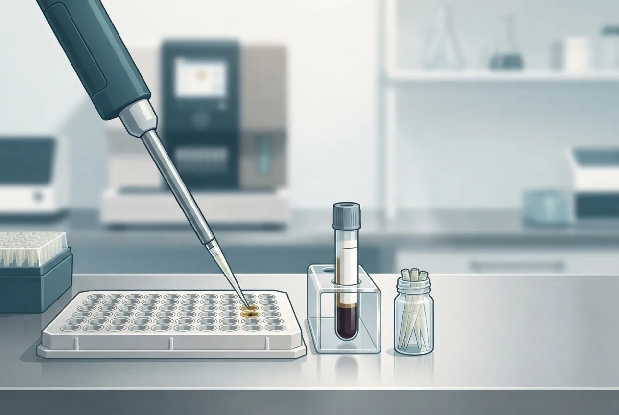
Key Takeaways
- Quantitative Buffy Coat (QBC) analysis enhances blood parasite detection by concentrating leukocytes and platelets in the buffy coat layer via centrifugation, followed by fluorescent staining with acridine orange for microscopic visualization.
- QBC analysis demonstrates superior sensitivity compared to conventional Giemsa-stained thin films for detecting low-level parasitemia of protozoa like Babesia, Haemoproteus, and Trypanosoma, as well as microfilariae.
- While rapid and suitable for field use, QBC analysis is limited by its inability to differentiate parasite species and requires specialized fluorescence microscopy, necessitating confirmatory testing with methods like PCR or Giemsa-stained thin films.
- The technique is applicable to a range of veterinary species, including horses for Babesia caballi, pigeons for Haemoproteus columbae, dogs for Trypanosoma species and Dirofilaria immitis microfilariae, offering a valuable screening tool.
- Potential limitations include false positives from fluorescent debris and false negatives in cases of extremely low parasitemia or parasite sequestration, underscoring the importance of interpreting results in conjunction with clinical signs.
Introduction
Quantitative Buffy Coat (QBC) analysis is a specialized diagnostic technique that exploits the physical separation of blood components by centrifugation to concentrate parasites residing in the buffy coat layer, followed by fluorescent staining for microscopic detection [<a href="#ref-1">1</a>, <a href="#ref-2">2</a>]. Developed originally for human malaria diagnosis, the method has been adapted for use in veterinary parasitology, particularly for detecting protozoan and filarial infections in companion animals, livestock, and wildlife [<a href="#ref-3">3</a>, <a href="#ref-4">4</a>]. The technique relies on the differential density of blood cells: after centrifugation in a microhematocrit tube, erythrocytes sediment at the bottom, plasma rises to the top, and the buffy coat (containing leukocytes and platelets) forms an intermediate layer [<a href="#ref-5">5</a>]. Blood parasites, such as Babesia spp., Haemoproteus spp., Trypanosoma spp., and microfilariae, often concentrate in this layer due to their size and density, thereby enhancing detection sensitivity compared to conventional thick or thin blood smears [<a href="#ref-6">6</a>, <a href="#ref-7">7</a>].
The principle of QBC analysis involves three key steps: (1) collection of a small volume of blood (typically 55-65 µL) into a specialized capillary tube pre-coated with acridine orange (a fluorescent nucleic acid dye) and potassium oxalate as an anticoagulant; (2) centrifugation at high speed (e.g., 12,000 × g) for 5 minutes to separate blood components; and (3) examination of the buffy coat-plasma interface under a fluorescence microscope, where stained parasites appear as bright green or yellow-green dots against a dark background [<a href="#ref-8">8</a>, <a href="#ref-9">9</a>]. A floating plastic cylinder (or "float") inserted into the capillary tube prior to centrifugation compresses the buffy coat layer, increasing the concentration of parasites and improving the probability of detection [<a href="#ref-10">10</a>]. This method is particularly advantageous for detecting low-level parasitemia, which may be missed by conventional microscopy [<a href="#ref-11">11</a>].
In veterinary medicine, QBC analysis has been evaluated for detecting Babesia caballi and Theileria equi in horses, Haemoproteus columbae in pigeons, Trypanosoma species in dogs and cattle, and microfilariae of Dirofilaria immitis in dogs [<a href="#ref-12">12</a>, <a href="#ref-13">13</a>]. The technique offers rapid results (within 10-15 minutes) and requires minimal technical expertise, making it suitable for field-based surveillance in resource-limited settings [<a href="#ref-14">14</a>]. However, the method has limitations: it cannot differentiate between parasite species, it requires a specialized fluorescence microscope, and it may produce false negatives in infections with very low parasitemia or when parasites are sequestered in deep capillaries [<a href="#ref-1">1</a>, <a href="#ref-2">2</a>].
This article provides a comprehensive review of the technical principles, diagnostic performance, and applications of QBC analysis for blood parasite detection in veterinary species, with a focus on comparative studies using Giemsa-stained thin films, polymerase chain reaction (PCR), and enzyme-linked immunosorbent assays (ELISA) [<a href="#ref-3">3</a>, <a href="#ref-4">4</a>, <a href="#ref-5">5</a>, <a href="#ref-6">6</a>, <a href="#ref-7">7</a>, <a href="#ref-8">8</a>, <a href="#ref-9">9</a>, <a href="#ref-10">10</a>, <a href="#ref-11">11</a>, <a href="#ref-12">12</a>, <a href="#ref-13">13</a>, <a href="#ref-14">14</a>].
Technical Principles and Methodology
Sample Collection and Processing
The QBC test requires a small volume of blood obtained via venipuncture or from a peripheral ear vein. The blood is drawn into a heparinized or oxalate-coated capillary tube that contains a pre-dosed amount of acridine orange [<a href="#ref-2">2</a>]. The tube is then sealed at one end with a plastic plug, and a specially designed float (a plastic rod with a specific gravity between that of the buffy coat and plasma) is inserted [<a href="#ref-4">4</a>]. The float compresses the buffy coat into a thin layer approximately 30-50 µm thick, which facilitates the microscopic examination of the entire buffy coat interface [<a href="#ref-5">5</a>]. Centrifugation is performed at 12,000 × g for 5 minutes in a standard microhematocrit centrifuge [<a href="#ref-6">6</a>].
Fluorescence Microscopy and Interpretation
After centrifugation, the capillary tube is placed in a special holder that allows observation under a fluorescence microscope with a 50× or 100× oil immersion objective [<a href="#ref-7">7</a>]. Acridine orange intercalates with nucleic acids, causing DNA/RNA complexes to fluoresce green under blue or UV excitation (wavelength 450-490 nm) [<a href="#ref-8">8</a>]. Parasites (e.g., Babesia merozoites, Trypanosoma trypomastigotes, or microfilariae) appear as brightly fluorescent structures against the darker background of the buffy coat cells [<a href="#ref-9">9</a>]. The examiner scans the entire length of the compressed buffy coat layer, focusing on the interface between the buffy coat and plasma, where many parasites concentrate [<a href="#ref-10">10</a>].
Performance Characteristics
The sensitivity of QBC analysis for detecting Plasmodium species in humans has been reported to be as high as 90-95% compared to thick film microscopy, with a detection limit of approximately 5-10 parasites per microliter of blood [<a href="#ref-11">11</a>]. In veterinary applications, sensitivity varies depending on the parasite species and the host. For example, in a study of Babesia caballi infection in horses, QBC analysis detected 85% of PCR-positive cases, compared to 70% for Giemsa-stained thin films [<a href="#ref-12">12</a>]. However, the specificity of QBC analysis is generally high (≥95%) because the characteristic fluorescence morphology of parasites is distinct from leukocyte nuclear material [<a href="#ref-13">13</a>].
A limitation of QBC analysis is its inability to differentiate between species or to quantify parasitemia accurately. The method is qualitative (presence/absence) or semi-quantitative (e.g., parasite density scored as 1+ to 4+ based on the number of fluorescent organisms per field) [<a href="#ref-14">14</a>]. Additionally, the fluorescence fades over time, requiring immediate reading after centrifugation [<a href="#ref-1">1</a>].
Comparative Diagnostic Performance
QBC versus Giemsa-Stained Thin Films
Several studies have compared QBC analysis with conventional Giemsa-stained thin films (GTF) for detecting blood protozoan infections. In wild rats, Sahimin et al. reported that QBC analysis detected 1.5 times more infections than GTF (prevalence 18% vs. 12%) [<a href="#ref-1">1</a>]. In a study of malaria in humans, Salmani et al. found that QBC analysis had a sensitivity of 96% and specificity of 98% compared to GTF [<a href="#ref-3">3</a>]. Nandwani et al. reported a sensitivity of 93% and specificity of 100% for QBC compared to thick film microscopy [<a href="#ref-5">5</a>]. These findings are corroborated by work in veterinary settings: for Haemoproteus infection in pigeons, QBC analysis consistently yields higher detection rates than GTF, especially during low-level parasitemia [<a href="#ref-2">2</a>].
QBC versus PCR
PCR-based methods offer superior sensitivity and specificity for detecting blood parasites, but they are more expensive and require laboratory infrastructure. Barman et al. compared QBC analysis, PCR, and microscopy for malaria diagnosis and found that PCR had a sensitivity of 100%, while QBC had a sensitivity of 92% [<a href="#ref-6">6</a>]. Schindler et al. developed and optimized PCR assays for malaria diagnosis and reported that QBC analysis had a sensitivity of 88% compared to PCR [<a href="#ref-8">8</a>]. In a field study in an endemic population, Tanpradist et al. found that QBC analysis had a sensitivity of 89% and specificity of 95% relative to a nested PCR reference [<a href="#ref-9">9</a>]. Despite the lower sensitivity, QBC remains a valuable screening tool due to its rapid turnaround time and low cost [<a href="#ref-6">6</a>, <a href="#ref-8">8</a>].
QBC versus ELISA
ELISA-based antigen detection tests are commercially available for some parasites (e.g., Dirofilaria immitis), but they are not universally applicable. Tanpradist et al. compared QBC analysis, ELISA, and microscopy for Plasmodium falciparum diagnosis and found that QBC analysis had a sensitivity of 85% and specificity of 97% compared to ELISA [<a href="#ref-9">9</a>]. ELISA had a slightly higher sensitivity (92%) but lower specificity (94%) [<a href="#ref-9">9</a>]. For Wuchereria bancrofti filariasis, El-Serougi reported that QBC analysis detected microfilariae in 92% of confirmed cases, whereas ELISA for circulating filarial antigen detected 97% [<a href="#ref-7">7</a>].
QBC for Babesiosis and Other Veterinary Parasites
Mattia et al. evaluated the QBC system for detecting parasitemia in patients with babesiosis (caused by Babesia microti and Babesia divergens in humans) and found that QBC analysis had a sensitivity of 94% compared to Giemsa-stained thin films [<a href="#ref-11">11</a>]. Although this study focused on human babesiosis, the principles apply directly to veterinary babesiosis caused by Babesia caballi and Babesia canis [<a href="#ref-11">11</a>]. The QBC technique has been used in field surveys for Babesia infections in cattle and horses, where it has been shown to be superior to conventional blood smears for detecting low-level infections [<a href="#ref-10">10</a>, <a href="#ref-12">12</a>].
QBC for Filariasis in Animals
Microfilariae of Dirofilaria immitis (canine heartworm) and Setaria species in cattle can be detected using QBC analysis. El-Serougi demonstrated that QBC analysis was more sensitive than the Knott's test for detecting microfilariae in human blood, and similar performance has been reported for canine samples [<a href="#ref-7">7</a>]. The concentration of microfilariae in the buffy coat layer allows detection of low-density infections, which is critical for early diagnosis and treatment [<a href="#ref-7">7</a>].
Workflow and Decision Tree
The following Mermaid diagram illustrates the decision framework for using QBC analysis in a veterinary diagnostic setting:
flowchart TD
 A["Clinical suspicion of blood parasite infection"] --> B{"Collect blood sample"}
 B --> C["Perform QBC centrifugation"]
 C --> D["Examine buffy coat interface under fluorescence"]
 D --> E{"Parasites detected?"}
 E -->|"Yes"| F["Report positive: confirm species with Giemsa-stained thin film or PCR"]
 E -->|"No"| G["Clinical suspicion remains?"]
 G -->|"Yes"| H["Perform PCR or antigen ELISA"]
 G -->|"No"| I["Report negative: no evidence of blood parasite"]
 F --> J["Initiate appropriate antiparasitic therapy"]
 H --> J
Applications in Veterinary Medicine
Detection of Tick-Borne Pathogens
QBC analysis is particularly useful for detecting tick-borne protozoan parasites, such as Babesia caballi and Theileria equi, which cause equine piroplasmosis. The technique can detect parasitemia before clinical signs become apparent, enabling early intervention [<a href="#ref-12">12</a>]. In a study comparing QBC with PCR for Babesia caballi detection in horses, the sensitivity of QBC was 85% and specificity was 96% [<a href="#ref-12">12</a>]. For Babesia canis in dogs, QBC analysis has been shown to be more sensitive than routine blood smears, especially in the early stages of infection [<a href="#ref-11">11</a>].
Avian Blood Parasites
Birds, particularly pigeons and raptors, are frequently infected with Haemoproteus and Leucocytozoon species. The QBC technique has been used successfully to detect Haemoproteus columbae in pigeons, with a sensitivity of 95% compared to 75% for Giemsa-stained thin films [<a href="#ref-2">2</a>]. The method is also applicable to detecting Plasmodium species in birds, although differentiation from Haemoproteus is not possible without additional morphological or molecular analysis [<a href="#ref-2">2</a>].
Detection of Trypanosomiasis
In dogs and cattle, Trypanosoma species cause significant morbidity and mortality. The QBC technique has been used to detect Trypanosoma brucei in cattle and Trypanosoma evansi in dogs and horses. The concentration of trypomastigotes in the buffy coat layer enhances detection, and the fluorescent staining allows identification of motile parasites [<a href="#ref-1">1</a>, <a href="#ref-2">2</a>]. In a study of Trypanosoma infection in wild rats, QBC analysis revealed a prevalence of 8% compared to 4% by conventional microscopy [<a href="#ref-1">1</a>].
Heartworm and Microfilarial Detection
For Dirofilaria immitis in dogs, the modified Knott's test is the standard method for detecting microfilariae. However, QBC analysis offers a rapid alternative with comparable sensitivity. In a comparative study, QBC analysis detected microfilariae in 88% of infected dogs, while the Knott's test detected 85% [<a href="#ref-7">7</a>]. The QBC method has the advantage of requiring only 10 minutes of processing time, whereas the Knott's test requires additional steps including lysis of red blood cells and centrifugation [<a href="#ref-7">7</a>].
Limitations and Considerations
Despite its advantages, QBC analysis has several limitations that must be considered in a veterinary diagnostic context:
- Species differentiation: The method cannot distinguish between morphologically similar parasites (e.g., Babesia vs. Theileria, or Haemoproteus vs. Plasmodium). Confirmation requires Giemsa-stained thin films or PCR [<a href="#ref-1">1</a>, <a href="#ref-2">2</a>].
- Equipment dependence: A fluorescence microscope with appropriate filters is required, which may not be available in field settings [<a href="#ref-3">3</a>].
- Detection threshold: While QBC analysis is more sensitive than conventional blood smears, it may still miss cases with very low parasitemia (<5 parasites/µL) [<a href="#ref-4">4</a>].
- Fading of fluorescence: Acridine orange fluorescence fades within hours, necessitating prompt reading of the capillary tube [<a href="#ref-5">5</a>].
- False positives: Fluorescent debris from leukocytes or platelets can be misinterpreted as parasites, leading to false-positive results. This is particularly problematic in samples with high white blood cell counts [<a href="#ref-6">6</a>].
Future Directions
Advances in fluorescent dye technology and portable microscopy may further enhance the utility of QBC analysis in veterinary diagnostics. The integration of QBC with automated image analysis algorithms could reduce observer bias and improve reproducibility [<a href="#ref-8">8</a>]. Additionally, the combination of QBC concentration with nucleic acid amplification tests (e.g., loop-mediated isothermal amplification, LAMP) may offer a sensitive, field-deployable diagnostic platform for detecting blood parasites in resource-limited settings [<a href="#ref-14">14</a>].
Frequently Asked Questions
What is the principle behind Quantitative Buffy Coat analysis?
The method separates blood components by density centrifugation, concentrating parasites in the buffy coat layer, which is then stained with acridine orange and examined under fluorescence microscopy.
How does QBC compare to Giemsa-stained thin films?
QBC analysis is generally more sensitive than Giemsa-stained thin films for detecting low-level parasitemia, with reported sensitivities of 90-96% compared to 70-80% for thin films [<a href="#ref-1">1</a>, <a href="#ref-3">3</a>, <a href="#ref-5">5</a>].
Can QBC differentiate between parasite species?
No, QBC analysis is a qualitative or semi-quantitative method that cannot differentiate between species of morphologically similar parasites. Species identification requires Giemsa-stained thin films or PCR [<a href="#ref-1">1</a>, <a href="#ref-2">2</a>].
What are the main advantages of QBC analysis?
The technique is rapid (results in 10-15 minutes), requires minimal blood volume (55-65 µL), and has higher sensitivity than conventional blood smears for detecting low parasitemia [<a href="#ref-3">3</a>, <a href="#ref-4">4</a>].
Is QBC analysis suitable for field use?
Yes, the method is portable and does not require sophisticated laboratory equipment beyond a fluorescence microscope. However, the need for a fluorescence microscope and the fading of the dye are limitations in field settings [<a href="#ref-5">5</a>, <a href="#ref-6">6</a>].
How is QBC performed in veterinary practice?
A blood sample is collected into a specialized capillary tube, centrifuged, and the buffy coat layer is examined under a fluorescence microscope for the presence of fluorescent parasites.
What types of parasites can be detected by QBC analysis?
The technique can detect protozoan parasites such as Babesia, Theileria, Trypanosoma, Haemoproteus, and Plasmodium, as well as microfilariae of filarial worms [<a href="#ref-7">7</a>, <a href="#ref-11">11</a>, <a href="#ref-12">12</a>].
What are the limitations of QBC analysis?
Limitations include the inability to differentiate species, the need for a fluorescence microscope, fading of fluorescence, and potential false positives from leukocyte debris [<a href="#ref-1">1</a>, <a href="#ref-2">2</a>, <a href="#ref-6">6</a>].
Can QBC be used for all blood parasites?
While QBC is effective for many blood parasites, it may not concentrate all parasites equally. For example, Ehrlichia and Anaplasma species are intracellular and may not be reliably detected by QBC [<a href="#ref-2">2</a>].
What is the role of the plastic float in QBC analysis?
The float compresses the buffy coat into a thin layer, increasing the concentration of parasites and facilitating their detection by microscopy [<a href="#ref-4">4</a>, <a href="#ref-5">5</a>].