UniProt vs. GeneCards vs. Human Protein Atlas
By Dr. Zubair Khalid, DVM, MS, PhD ·

UniProt is a manually curated protein knowledgebase, the Human Protein Atlas (HPA) is an experiment-driven map of where human proteins appear in tissues and cells, and GeneCards is a gene-centric aggregator that pulls summaries of both into a single page. The rule of thumb: start at UniProt when you need curated protein-level facts and batch annotation, go to HPA when tissue context, cell type, pathology, or image-level evidence matters, and use GeneCards for a fast gene-centered overview or to find the right search terms, not as a citable primary source.
These three resources get lumped together because they all seem to answer "what does this protein do and where does it live." In practice they sit at different points in the evidence chain. UniProt summarizes published literature into structured, controlled annotations. HPA generates its own large-scale imaging and transcriptomics data and publishes the raw pictures. GeneCards aggregates and links. The practical question of UniProt vs. Human Protein Atlas subcellular localization, which trips up nearly everyone who annotates a proteomics table, comes down to what kind of evidence you want to stand on. This article covers that comparison in depth, then places GeneCards in the same picture, gives you a UniProt database tutorial you can actually run, and works through the scenarios where each choice wins.
What Each Database Actually Is
UniProt: the curated protein knowledgebase
UniProt is a collaboration between EMBL-EBI, the SIB Swiss Institute of Bioinformatics, and the Protein Information Resource. Its core product is UniProtKB, split into two sections. UniProtKB/Swiss-Prot contains manually reviewed entries curated by expert biologists who read the literature and attach structured annotations, including function, domains, post-translational modifications, and subcellular location. UniProtKB/TrEMBL contains computationally analyzed records awaiting full manual curation, largely derived from the coding sequences of submitted genomes.
Every curated claim in a UniProt entry carries an evidence tag, for example "experimental evidence at the protein level" versus "inferred by electronic annotation," so you can see whether a fact rests on published experiments or on automated transfer from related proteins. UniProt also assigns each entry a protein existence level from 1 (experimental evidence at the protein level) down to 5 (uncertain), which tells you whether the protein itself has ever been physically observed.
For human proteins, UniProt provides a reference proteome of roughly 20,000 protein-coding gene products, consistent with the standard textbook estimate of the size of the human gene set. Each entry also carries Gene Ontology (GO) cellular component terms, a controlled vocabulary that describes where in the cell a protein acts, and cross-references to dozens of other resources, including HPA itself.
Human Protein Atlas: experiment-generated tissue and cell mapping
The Human Protein Atlas is a Swedish research program that profiles human proteins using three main data types. First, antibody-based immunohistochemistry (IHC), which detects a specific protein in thin tissue sections using a labeled antibody and a chromogenic stain, profiles tissue expression across normal organs, cancer specimens, and brain regions. Second, RNA-sequencing and single-cell transcriptomics place each gene in tissue and cell type context. Third, confocal immunofluorescence (IF), which uses fluorescently labeled antibodies to visualize a protein inside fixed cultured cells, underpins the subcellular localization resource.
The subcellular resource annotates protein location inside cells using a defined set of cellular compartments such as the nucleus, nucleoli, cytosol, mitochondria, endoplasmic reticulum, and plasma membrane. Every localization is graded on a reliability scale of Enhanced, Supported, Approved, or Uncertain, and the underlying microscopy images are published so you can judge the staining yourself. Because the images are first-party experimental data, HPA lets you verify a claim rather than trust a summary of it.
GeneCards: the gene-centric meta-search
GeneCards, developed at the Weizmann Institute of Science, is built around the gene rather than the protein. A GeneCards entry ("gene card") aggregates information from dozens of external sources, including UniProt, HPA, NCBI Gene, Ensembl, OMIM, Reactome, and literature mining, and organizes it into sections covering expression, function, pathways, diseases, paralogs, and compounds. Each fact on the card is hyperlinked to the underlying source record, which makes the card a router to primary data rather than a primary record itself. Genes flagged as "Elite" have particularly strong, well-supported evidence across sources.
Recent database-building work treats all three resources as complementary layers. MultifacetedProtDB, a database of multifunctional human proteins, integrates information from UniProt, GeneCards, HPA, the Human Phenotype Ontology, and MONDO to assemble annotations that no single source provides alone [1]. That design reflects the division of labor described in this section. UniProt supplies protein annotations, GeneCards supplies gene-centric linkage, and HPA supplies tissue and cell expression.
Side-by-Side Comparison
| Criterion | UniProt | Human Protein Atlas | GeneCards |
|---|---|---|---|
| Primary unit | Protein entry (per canonical sequence and isoforms) | Gene product per tissue, cell type, and cellular compartment | Gene card (one page per gene) |
| Core evidence | Curated literature plus automatic annotation, with evidence tags | First-party IHC, IF, and RNA-seq experiments with published images | Aggregated summaries from other databases and literature mining |
| Subcellular localization source | Manual curation of published experiments, controlled vocabulary of locations, GO cellular component terms | Confocal IF in cultured cell lines, graded Enhanced to Uncertain, images viewable | Inherited from UniProt, HPA, COMPARTMENTS, and related sources |
| Tissue and cell type context | Brief free-text tissue specificity where available | Central strength, normal tissues, cancer, brain, single-cell resolution | Summary views linking back to HPA and GTEx-style sources |
| Disease and pathology | Disease entries and variant annotations | Dedicated pathology atlas with cancer staging and prognosis views | Broad disease summary sections with links |
| Batch programmatic access | Full REST API, bulk downloads, ID mapping, SPARQL endpoint | Downloadable datasets and search API | Free web access, bulk programmatic use requires a licensed arrangement |
| Best for | Curated facts, sequence analysis, batch annotation of protein lists | Where a protein is in real tissues and cells, pathology, image evidence | Rapid gene overview, finding the right resources and terms |
| Not designed for | Tissue-specific protein abundance, visual verification | Sequence analysis, domain architecture | Primary citation, deep single-question answers |
Read the table this way. UniProt answers "what is known about this protein, and how well is it supported." HPA answers "where is this protein observed, and can I look at the evidence myself." GeneCards answers "what is the overall picture for this gene and where do I go next."
UniProt vs. Human Protein Atlas for Subcellular Localization
This is the comparison that decides most real annotation tasks, so it deserves its own treatment.
Where each localization claim comes from
A UniProt subcellular location annotation is assembled by curators from published experimental literature, transferred by similarity from homologs, or generated through automatic annotation systems, and each claim carries an evidence tag. Locations use a controlled vocabulary maintained by UniProt, so "nucleus" means the same thing in every entry, and GO cellular component terms provide a second structured view. What UniProt does not provide is a photograph. The claim is a summary of literature, not an image you can inspect.
An HPA subcellular localization is a direct experimental observation. The program applies fluorescently labeled antibodies to a fixed panel of cultured cell lines, acquires confocal micrographs, and annotates which compartments show staining. Each localization carries a reliability grade, and the images are published alongside the annotation. What HPA does not claim is comprehensiveness across biology, because IF is performed in a limited panel of cell lines rather than in every human cell type and state.
The difference between UniProt and Human Protein Atlas subcellular localization, in practice
Three structural differences drive almost every discrepancy you will see.
First, the evidence types differ. UniProt aggregates what the literature has shown, which can include locations demonstrated in cell types, organisms, or conditions far removed from your experiment. HPA shows you what one antibody saw in three cultured cell lines under one condition. A protein curated as nuclear based on decade-old biochemistry can look cytoplasmic in an HPA micrograph, and both records can be correct in their own contexts.
Second, localization is conditional. Proteins shuttle between compartments in response to signaling, cell cycle phase, stress, and differentiation state. A multi-label reality sits behind every single-line answer, and the growing literature on multifunctional and moonlighting proteins, which have two or more molecular functions often in different cellular locations, shows that single-label answers systematically underdescribe real biology [1]. UniProt tends to list all literature-supported locations, while HPA lists what the fixed imaging snapshot showed.
Third, the granularity differs. UniProt uses a curated vocabulary that distinguishes, for example, the nuclear envelope from nuclear speckles. HPA's annotation of the same protein may collapse that detail into "nucleus" or capture finer detail if staining quality supports it. Neither is wrong, they just carve the cell at different joints.
A recent computational study illustrates how the two resources complement each other. ProLoc-IHS, a multi-label model for predicting protein subcellular localization, builds its training data by combining IHC images from HPA with protein sequences from UniProt, fusing the two representations to improve prediction [2]. The authors of that work treated HPA as the image evidence layer and UniProt as the sequence and annotation layer, which is exactly how a careful human analyst should use them.
A resolution protocol when the two databases disagree
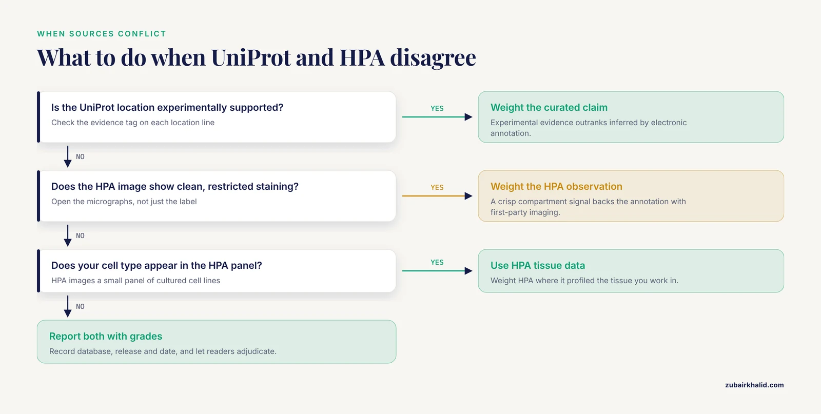
Run this checklist in order when UniProt and HPA conflict on a protein.
- Read the UniProt evidence tags. A location supported by experimental evidence outranks one marked as inferred by electronic annotation.
- Open the HPA subcellular page and look at the images, not just the label. Judge whether staining is specific and compartment-restricted, and check the reliability grade.
- Check the GO cellular component terms in UniProt for a third, ontology-based opinion.
- Consider your experimental context. If you work in a tissue HPA has profiled by IHC, weight HPA tissue data accordingly. If you work in a cell type HPA never imaged, weight curated literature.
- Record which database, release, and date you relied on, because all three resources revise continuously.
How to Use UniProt: A Practical Tutorial
This section is a working UniProt database tutorial. Everything below runs against the live REST API, which requires no authentication.
Reading a UniProt entry
Search uniprot.org for a gene symbol, filter to the reviewed human entry, and open it. Using EGFR (accession P00533) as a worked example, the sections that matter for localization questions are:
- Subcellular location. A structured list of compartments, for EGFR primarily the cell membrane as a single-pass type I membrane protein, with additional locations such as the endoplasmic reticulum listed where literature supports them. Each line ends with an evidence tag you can click to see the source publications or automatic annotation rule.
- GO annotation. Cellular component terms that give an ontology-based view of location.
- Tissue specificity. Short free text, often cross-linked to expression resources, but not a substitute for HPA tissue data.
- Isoforms. Alternative sequences produced by alternative splicing, which matter because isoforms can differ in localization signals.
- Cross-references. Links out to HPA, PDB, STRING, and dozens of other resources.
Website search syntax
The UniProt search box accepts field-limited queries. Useful examples:
gene:EGFR AND organism_id:9606restricts to the human EGFR gene by NCBI taxonomy identifier 9606.reviewed:true AND organism_id:9606returns all reviewed human entries.- To find proteins annotated to a specific compartment, search the subcellular location vocabulary on the UniProt site, note its identifier, and filter with the
slt_idfield in the query box.
Programmatic access with the REST API
Pull a compact TSV table of subcellular locations for a single gene:
curl "https://rest.uniprot.org/uniprotkb/search?query=gene:EGFR+AND+organism_id:9606+AND+reviewed:true&fields=accession,id,protein_name,cc_subcellular_location,go_id,cc_tissue_specificity&format=tsv"Retrieve the full JSON record for one accession:
curl "https://rest.uniprot.org/uniprotkb/P00533.json"Download all reviewed human proteins annotated to the plasma membrane, as a TSV:
curl "https://rest.uniprot.org/uniprotkb/stream?query=reviewed:true+AND+organism_id:9606+AND+slt_id:SL-0039&format=tsv&fields=accession,id,protein_name,cc_subcellular_location"Convert a list of gene symbols into UniProt accessions using ID mapping, which is the standard first step for annotating a proteomics result list:
curl -X POST -H "Accept: application/json" \
  https://rest.uniprot.org/idmapping/run \
  -d "ids=EGFR,TP53,KRAS,MYC" \
  -d "from=Gene_Name" \
  -d "to=UniProtKB"
The POST returns a job identifier. Poll https://rest.uniprot.org/idmapping/status/{job_id} until it completes, then fetch results from the uniprotkb/results/stream endpoint. In R, the Bioconductor package UniProt.ws wraps much of this. UniProt also exposes a SPARQL endpoint at sparql.uniprot.org for users who prefer graph queries.
When to use UniProt
Use UniProt when you need curated function and localization in one record, evidence-tagged claims you can defend in a manuscript, batch annotation of protein or gene lists, canonical sequences and isoforms for downstream tools, or a controlled vocabulary that makes cross-protein comparison consistent. The phrase "when to use UniProt" resolves to this: use it whenever you need a structured, citable, evidence-tagged summary rather than a picture or a link farm.
How to Use the Human Protein Atlas
Navigating a gene page
Enter a gene symbol at proteinatlas.org. Each gene page splits into tissue, brain, single cell type, pathology, and subcellular location views.
- Tissue. IHC images across normal human tissues, scored by pathologists for staining intensity and proportion of positive cells, paired with consensus RNA expression levels.
- Cell type. Single-cell RNA-seq assigning expression to specific cell populations.
- Pathology. IHC across cancer specimens with staining scored per cancer type, plus survival analysis in many cancer cohorts. If your question is "where does this protein show up in colon adenocarcinoma tissue," this tab answers it in a way UniProt cannot.
- Subcellular location. Confocal IF images in cultured cell lines, compartment annotations, and the reliability grade.
Reading the subcellular location tab
The IF resource images proteins in a small panel of fixed human cell lines, including U-2 OS osteosarcoma cells, U-251 MG glioma cells, and A-431 epidermoid carcinoma cells, which is an established design choice of the program. Each annotation is graded Enhanced, Supported, Approved, or Uncertain, reflecting staining specificity and compartment clarity. Treat Uncertain as a hypothesis, and always open the micrographs. A grainy, diffuse image backing an Approved grade is a different fact than a crisp, punctate nuclear signal backing an Enhanced grade, and only looking at images reveals that difference.
For localization in intact tissues, the IHC images in the tissue and pathology tabs show whether staining is nuclear, cytoplasmic, or membranous in each tissue, which is often the practical localization question in a cancer or biomarker project. That imaging modality is now itself a machine learning substrate. ProLoc-IHS demonstrates that IHC images alone carry enough localization signal to train a Vision Transformer-based predictor, and that fusing those images with ProtT5 sequence embeddings from UniProt improves multi-label localization prediction beyond image-only or sequence-only models [2].
Downloads and programmatic access
HPA publishes bulk downloadable datasets for tissue, pathology, and subcellular data from its download page, and provides a search API for scripted queries of tabular data. Image files are available per antibody and per tissue. For reproducible pipelines, download a dated snapshot rather than scraping live pages, and record the HPA release version in your methods.
How to Use GeneCards
GeneCards answers a different question, "give me everything about this gene on one page," and it answers it fast. Search a symbol and you land on a card with function, expression summaries, diseases, pathways, paralogs, and compounds, each hyperlinked to its source. As a practical habit, use GeneCards in three ways:
- As a router. Follow the embedded links to the UniProt entry, the HPA page, or the OMIM record, and work from those primary sources.
- As a term generator. Disease names, pathway names, and aliases on the card make good queries for PubMed and for database searches elsewhere.
- As a triage tool. When you know nothing about a gene, one card read tells you whether it is membrane-bound, enzyme-coding, or transcriptional, which tells you which deep dive to make next.
Do not cite GeneCards itself in a manuscript. Cite the underlying record it links to. And note that bulk programmatic use of GeneCards is a licensed offering, so scripts that need gene-centric aggregation at scale usually rebuild it from the primary APIs of UniProt, HPA, and NCBI instead. The MultifacetedProtDB project shows what such a rebuild looks like in practice, integrating UniProt, GeneCards, HPA, HPO, and MONDO records under one searchable interface with filters for structure, function, and interactors [1].
Worked Scenarios
Scenario 1: Annotate a proteomics results table
You have 500 protein groups from a mass spectrometry run and need a subcellular localization column. Use UniProt. Map gene symbols to accessions with the ID mapping endpoint shown above, then stream a TSV with cc_subcellular_location and go_id fields for the full hit list. The evidence tags travel with the annotation, so you can later filter to experimentally supported claims only. HPA tissue pages are wrong for this task because they are built one gene at a time and emphasize tissue, not compartment.
Scenario 2: Where is the protein in tumor tissue?
You are writing a biomarker proposal and need to know whether EGFR staining in lung squamous carcinoma is membranous and at what frequency. Use the HPA pathology tab, which scores IHC in cancer specimens by type and stage. UniProt will tell you EGFR is a cell membrane protein but says nothing about staining patterns in lung tumor sections.
Scenario 3: Curating a secretome list
You need a defensible list of human secreted proteins, with tissue-of-origin context. UniProt subcellular annotations flag secreted proteins, and HPA adds tissue expression. A purpose-built integration demonstrates the combined approach. SEPDB compiles human, mouse, and rat secreted protein datasets from serum, exosomes, and cell culture media, integrating UniProt and HPA to annotate secreted proteomics data based on subcellular localization and disease markers, and refines signal peptide prediction by excluding proteins with transmembrane domains [3]. When a curated integration exists for your niche, using it beats rebuilding one.
Scenario 4: A protein with two jobs
You found that a metabolic enzyme also regulates transcription and you wonder whether this is an established moonlighting case. Check MultifacetedProtDB, which collects human proteins labeled as pleiotropic, multidomain, promiscuous, or moonlighting by drawing on UniProt, GeneCards, HPA, HPO, and MONDO, precisely because such proteins are hard to retrieve by direct search in any single database [1]. Expect its entries to show multiple GO cellular component terms and multi-compartment localization records.
Scenario 5: Building a localization predictor
You want to train or evaluate a subcellular localization model. ProLoc-IHS shows the current recipe, pairing HPA IHC images with UniProt sequences, encoding images with a Vision Transformer and sequences with the ProtT5 protein language model, fusing them with a cross-attention module, and handling multi-label targets with binary cross entropy and Focal loss [2]. The choice of both databases in that pipeline is not incidental. Images alone carry morphology, sequences alone carry motifs like nuclear localization signals, and the labels are strongest when the two evidence types agree.
Scenario 6: Ten-minute background check on an unfamiliar gene
You are reading a paper that mentions a gene you have never seen. Open GeneCards for the one-page overview, click through to UniProt for curated function and localization, and open HPA if you need to see tissue expression or pathology staining. Total time, under ten minutes, and you end on primary records rather than the aggregator.
Combining the Three in a Single Pipeline
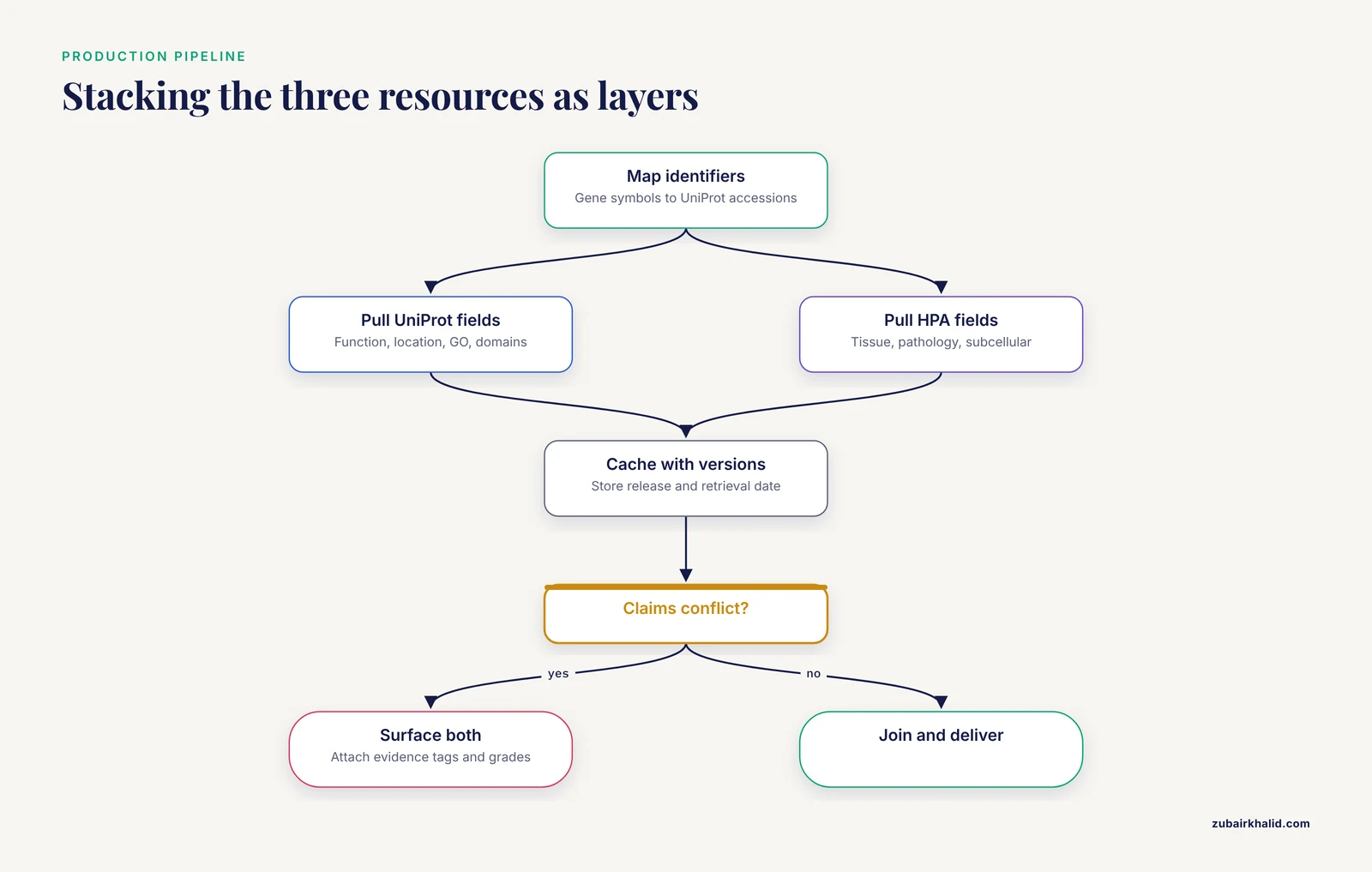
Production pipelines treat these resources as layers. A common architecture for a human protein annotation service looks like this:
- Ingest protein identifiers and map them to UniProt accessions with the UniProt ID mapping service.
- Pull UniProt fields for function, subcellular location, GO terms, domains, and disease annotations via the REST API.
- Pull HPA tissue, single-cell, pathology, and subcellular fields from the HPA downloads or search API, keyed on Ensembl gene IDs.
- Cache both pulls with version numbers and retrieval dates, then join on stable identifiers.
- Where claims conflict, surface both, with evidence tags and HPA reliability grades attached, and let downstream users adjudicate.
The integration literature validates this pattern. SEPDB stands on UniProt plus HPA for secreted protein annotation [3]. MultifacetedProtDB stands on UniProt, GeneCards, HPA, HPO, and MONDO for multifunctional protein annotation [1]. ProLoc-IHS stands on HPA images plus UniProt sequences for machine learning [2]. No published resource in this comparison position tries to replace the others, they all stack them.
Common Mistakes and Limitations
- Treating UniProt localization as tissue-specific. UniProt summarizes what the literature shows anywhere, in any cell type or species context. Pair it with HPA tissue data when cell type matters.
- Reading HPA grades without reading images. An Uncertain grade backed by clean staining can be more informative than an Approved grade backed by weak signal. The images are the data, use them.
- Ignoring evidence tags. A claim marked as inferred by electronic annotation is a prediction, not an observation. Filter or flag these in batch work.
- Citing the aggregator. Cite UniProt, HPA, and primary papers. GeneCards links to sources, it is not a source.
- Isoform blindness. UniProt lists isoforms explicitly, while HPA antibodies may bind epitopes shared by several isoforms. If isoforms differ in localization signals, both records can appear contradictory when they are not.
- Snapshot drift. All three resources revise continuously. Record release versions and retrieval dates in your methods, or your results will not reproduce a year later.
- Expecting single labels from multi-label biology. Proteins occupy multiple compartments, and moonlighting proteins carry multiple functions, so any one-line localization answer is a compression of a richer reality [1].
- Overlooking the cell line limitation. HPA IF localizations come from a small panel of cultured cell lines, so a compartment your cell type uses heavily may be invisible there. Tissue IHC and literature fill that gap.
- Confusing prediction with curation. Both UniProt automatic annotations and computational predictors such as ProLoc-IHS produce predictions [2]. Useful, but a different epistemic class from a manually curated experiment or a published micrograph.
Frequently Asked Questions
What is the difference between UniProt and Human Protein Atlas subcellular localization?
UniProt subcellular locations are curated summaries of published literature, written in a controlled vocabulary and tagged with evidence levels. HPA subcellular locations are direct immunofluorescence observations in a panel of cultured cell lines, graded for reliability and backed by published micrographs. One summarizes literature, the other shows first-party imaging.
Should I use UniProt or Human Protein Atlas for subcellular localization?
Use UniProt for batch annotation, curated and evidence-tagged claims, and sequence-linked analysis. Use HPA when tissue context, cancer pathology, or visual image evidence is the point. For a single protein in a specific tissue or disease, HPA usually wins. For a 500-protein results table, UniProt wins.
When should I use GeneCards instead of UniProt or HPA?
Use GeneCards for a fast, one-page overview of an unfamiliar gene and as a router to primary records. Do not use it as your final evidence source, because its entries aggregate other databases and link to them rather than generating or curating experimental data themselves.
How do I batch-annotate subcellular localization for a list of proteins?
Map your gene symbols to UniProt accessions with the UniProt ID mapping service, then retrieve a TSV with the cc_subcellular_location and GO fields through the REST API. Commands for both steps appear in the UniProt tutorial section above, and neither requires authentication.
Why do UniProt and HPA show different locations for the same protein?
Different evidence, different conditions, and different granularity. UniProt aggregates literature from many systems, HPA shows one antibody in a few cell lines under one condition, and proteins genuinely shuttle between compartments. Run the resolution protocol in this article, weigh evidence tags against HPA reliability grades, and report both when they conflict.
Can I download subcellular localization data programmatically?
Yes. UniProt offers a REST API, bulk stream downloads, ID mapping, and a SPARQL endpoint, all without authentication. HPA offers bulk dataset downloads and a search API. GeneCards web access is free but bulk programmatic use requires a licensed arrangement, so pipelines typically pull gene-centric data from the UniProt and HPA APIs directly.