Subcellular Localization Annotations in UniProt Explained
By Dr. Zubair Khalid, DVM, MS, PhD ·

After working through this guide, you will be able to read any UniProt entry and understand exactly what its subcellular location annotation claims, judge how strong the evidence behind that claim is, retrieve every protein in a given cellular compartment with a single command, parse those results into a usable table, and decide when an external prediction tool is worth running. You will also learn the mistakes that most often corrupt downstream analyses that depend on localization data.
To follow the hands-on parts, you need a web browser, a Unix-style shell (Linux, macOS, or WSL on Windows), curl installed, and Python 3.8 or newer. No other software is required. Every command shown runs as written against the live UniProt REST API.
Subcellular localization (often abbreviated SCL in the prediction literature) describes which compartment or structure inside or outside the cell a protein occupies, such as the cytosol, the mitochondrial inner membrane, or the extracellular space. This single piece of information constrains what a protein can do. A protein cannot phosphorylate a nuclear receptor from the Golgi lumen, and a protease secreted into the extracellular space has a very different candidate substrate set from one sitting in the lysosome. That is why reviews of functional genome annotation have long treated localization prediction as a shortcut to function, and why so many downstream pipelines, from essential-protein discovery to network visualization, consume localization as an input feature [1], [2], [3].
UniProt is the central hub for this information. Knowing how to use UniProt correctly for subcellular localization data is therefore a foundational skill for protein annotation work of any kind.
What a UniProt Subcellular Location Annotation Actually Contains
A UniProt entry carries subcellular information in several distinct places, and confusing them is the root of many analysis errors. Open any well-annotated entry, for example human serum albumin (accession P02768), and you will see a "Subcellular location" section in the entry view. That section is backed by four things in the underlying data: the structured subcellular location comment, the controlled-vocabulary keywords, the Gene Ontology (GO) cellular component annotations, and the sequence-feature lines that describe the targeting machinery.
The structured subcellular location comment
The core annotation lives in the CC -!- SUBCELLULAR LOCATION block of the flat file. It contains three kinds of statements:
- Location names. Controlled terms such as
Secreted,Cytoplasm,Nucleus,Cell membrane, orMitochondrion inner membrane. A protein with more than one location gets one statement per location. - Topology. For membrane proteins, the annotation states how the protein is embedded, for example
Single-pass type I membrane proteinorMulti-pass membrane protein, together with topological domain annotations (extracellular, cytoplasmic, luminal). Human serum albumin carries a signal peptide spanning residues 1 to 18 and a propeptide at residues 19 to 24, which is the classic secretory-route architecture you will see reflected in its feature lines. - Isoform and qualifier notes. If one isoform of a protein goes somewhere the canonical sequence does not, the annotation says so explicitly with isoform identifiers. Qualifiers such as "translocated to the nucleus upon ligand binding" describe regulated relocalization.
Retrieve the flat file for albumin and look at the relevant lines:
curl -s https://rest.uniprot.org/uniprotkb/P02768.txt | grep -A3 "SUBCELLULAR LOCATION"You will see the location line with an evidence tag wrapped in braces, in a form like this (the exact citation behind the braces changes as the entry is updated):
CC -!- SUBCELLULAR LOCATION: Secreted {ECO:0000269|PubMed:...}.The braces matter enormously and are discussed in the next section.
Keywords: the searchable, filterable vocabulary
Every curated subcellular location is also assigned one or more UniProt keywords, short controlled terms in the "Subcellular location" keyword category such as Secreted, Cytoplasm, Nucleus, Mitochondrion, or Cell membrane. Keywords are what power the "Keywords" filter in the UniProt web interface and the keyword: field in the search API. They are coarser than the location comments (the keyword Cell membrane does not tell you which leaflet or whether the protein is single-pass), but they are stable, well-defined, and ideal for bulk queries.
Each keyword has its own page at uniprot.org/keywords/ listing its definition and the number of proteins carrying it, and each keyword is cross-linked to one or more GO cellular component terms.
GO cellular component annotations
UniProt entries carry a DR GO line for Gene Ontology annotations. The cellular component branch of GO (the C: aspect) describes the same biology in a different vocabulary, with finer granularity and its own evidence codes such as IDA (inferred from direct assay) or ISS (inferred from sequence or structural similarity). The GO cellular component terms and the UniProt location keywords are mapped to each other but are not identical. A GO annotation is attached to the entry by an evidence trail to the source database, so the same protein can carry a location keyword with one evidence level and a GO term with another. Treat them as complementary views, and never assume that the presence of a GO term implies the presence of a UniProt keyword or vice versa.
Sequence features that explain the annotation
Feature (FT) lines record the machinery that gets a protein where it goes: SIGNAL for N-terminal signal peptides, TRANSIT for transit peptides that target mitochondria, chloroplasts, or peroxisomes, PROPEP for propeptides removed after sorting, and TRANSMEM for membrane-spanning helices with their residue ranges. When you evaluate a localization annotation, these lines are your first sanity check. A protein annotated Secreted with no signal peptide, or annotated as an integral membrane protein with no TRANSMEM helix, deserves a second look before you trust it.
Who Wrote the Annotation: Reading the Evidence Tags
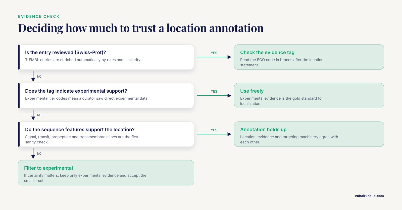
This is the single most consequential thing to understand about UniProt subcellular localization annotations. Every statement carries an evidence tag from the Evidence and Conclusion Ontology (ECO), and the tag tells you whether a human curator saw experimental data or a computational rule fired.
UniProt has two sections. Swiss-Prot (reviewed) entries are annotated by expert curators with literature-level scrutiny. TrEMBL (unreviewed) entries are enriched automatically by rule-based and similarity-based pipelines. A TrEMBL entry can carry a plausible-looking subcellular location that traces back entirely to an automated inference from an ortholog, and treating that the same way as a literature-backed experimental annotation is a serious error. The distinction between reviewed and unreviewed status is the first filter you should apply in almost any analysis.
Within curated entries, the common evidence tiers for localization look like this. Check the entry page for the exact current tag, because UniProt refines evidence mapping over time.
| Evidence tier | Typical ECO code | What it means | How to treat it |
|---|---|---|---|
| Experimental | ECO:0000269 | Direct experimental evidence reported in a cited paper | Gold standard, use freely |
| Proteomics | ECO:0000313 | Large-scale mass spectrometry or similar data | Strong, but check organelle purity |
| Sequence similarity | ECO:0000250 | Inferred from a characterized ortholog | Reasonable, verify taxonomic distance |
| Curator inference | ECO:0000305 | Logical inference from background knowledge | Usually sound, read the rationale |
| Sequence-based prediction | ECO:0000255 | Signal peptide, transit peptide, or transmembrane helix predicted from sequence | Useful, treat as prediction |
| Automatic annotation | ECO:0000256 | Rule-based propagation into unreviewed entries | Treat as a hypothesis |
The experimental tier is what separates annotation from prediction, and it is scarce by nature. Large-scale experimental localization efforts remain expensive and slow, which is precisely why computational prediction has grown into its own field. A recent example of the experimental approach at scale is a study of Giardia intestinalis that fluorescently tagged and imaged 608 proteins, about 12 percent of that organism's proteome, including 240 proteins of unknown function, and turned those imaging results into a structured localization-informed gene annotation [4]. That study illustrates what the gold standard actually costs: careful cloning under native promoters, live-cell imaging, and per-protein inspection. UniProt's experimental tags reflect that kind of work. Most entries in the database do not have it.
For machine-learning researchers, this tiering has a direct practical consequence. Prediction models that want a trustworthy training set restrict themselves to proteins with experimentally annotated localization, as the PSLCNN deep-learning predictor did when it trained and tested only on UniProt data carrying experimental annotation [5]. If you build training sets, replicate that discipline. If you consume predictions, ask what data the model was trained on.
A Step-by-Step Workflow for Working with UniProt Subcellular Localization Annotations
The following five steps take you from reading a single entry to producing a compartment-level summary of a whole proteome and validating it with independent prediction tools.
Step 1: Read a single entry and interrogate it
Go to uniprot.org, search for a protein of interest, and open the entry. Locate the "Subcellular location" section and answer four questions in order:
- Which locations are listed?
- What evidence tag accompanies each one?
- Is the entry reviewed (Swiss-Prot) or unreviewed (TrEMBL)?
- Do the sequence features (signal peptide, transit peptide, transmembrane helices) mechanistically support the stated location?
For a membrane protein such as the human epidermal growth factor receptor (EGFR, P00533), you should find Cell membrane combined with a Single-pass type I membrane protein topology statement and matching TRANSMEM and signal-chain features. If any of those four pieces disagree, dig into the cited literature before using the entry.
Step 2: Retrieve every annotated protein in a compartment with the REST API
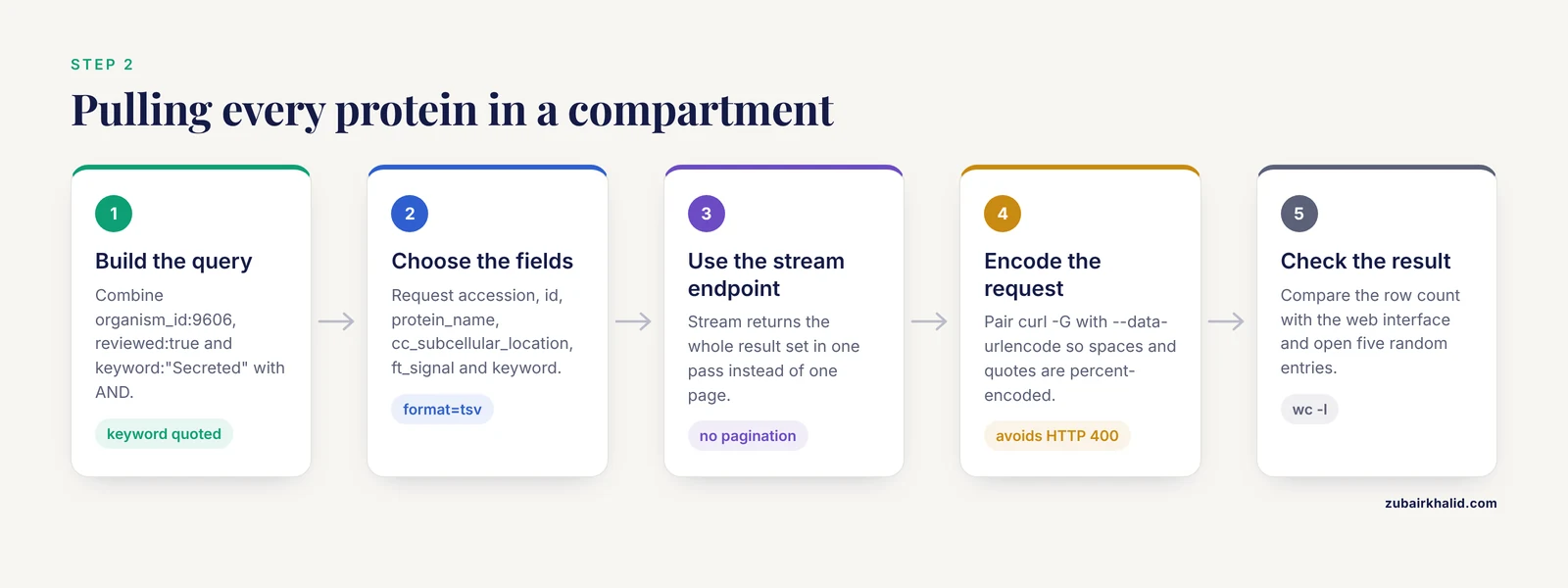
The UniProt REST API lets you pull the complete, current set of proteins matching any localization query. The example below retrieves every reviewed human protein carrying the Secreted keyword, along with the full subcellular location comment and the signal peptide feature ranges:
curl -sG "https://rest.uniprot.org/uniprotkb/stream" \
  --data-urlencode 'query=(organism_id:9606) AND (reviewed:true) AND (keyword:"Secreted")' \
  --data-urlencode 'format=tsv' \
  --data-urlencode 'fields=accession,id,protein_name,cc_subcellular_location,ft_signal,keyword' \
  -o human_secreted.tsv

wc -l human_secreted.tsv
The stream endpoint returns the full result set in one pass, which is the right choice for a filtered set like this. For queries expected to return tens of thousands of rows or more, you can add -data-urlencode 'compress=true' and gunzip the output.
The output is a tab-separated table. It looks like this (row content shown for one entry, exact values change with each UniProt release):
Entry   Entry Name  Protein names   Subcellular location [CC]   Signal peptide  Keywords
P02768  ALBU_HUMAN  Albumin ...     SUBCELLULAR LOCATION: Secreted.     SIGNAL 1..18;   ...
Four parameters in this command do the real work:
querycombines clauses with AND.organism_id:9606restricts to human (the NCBI taxonomy identifier),reviewed:truerestricts to Swiss-Prot, andkeyword:"Secreted"restricts to entries carrying that exact keyword. Keyword names must match the controlled term exactly, in quotes.fieldscontrols which columns you get. Omitting it returns only accessions, which is fast but throws away the information you came for.format=tsvgives you a parseable table.format=fastagives sequences, andformat=jsongives the full structured record.streamversussearch:searchpaginates and returns a default page of results, so a naive call tosearchwithout pagination will silently give you only the first page. Usestreamfor whole-result-set downloads.
Step 3: Parse and summarize with Python
Localization data becomes more useful when summarized per compartment. The following script counts the primary stated location for each retrieved entry:
import csv
from collections import Counter

counts = Counter()
with open("human_secreted.tsv") as handle:
    rows = csv.DictReader(handle, delimiter="\t")
    for row in rows:
        text = row.get("Subcellular location [CC]") or ""
        text = text.replace("SUBCELLULAR LOCATION: ", "")
        primary = text.split(".").strip()
        if primary:
            counts[primary] += 1

for name, n in counts.most_common(10):
    print(n, name)
A run against the live database produces a table headed by the dominant compartment, for example:
Secreted
Secreted, Extracellular space
The exact counts differ between releases, so always record the release date and the date of your download alongside any numbers you publish. Note that many secreted proteins carry qualifiers or additional locations (for example, secretion followed by documented cell-surface association), which is why the counts above report the first stated location rather than the full picture.
How to check the result is right. Three cheap checks catch most retrieval errors. First, cross-check the row count against the number reported by the UniProt web interface for the same query string. Second, sample five random rows and open their entries in a browser to confirm the keyword and location match what you downloaded. Third, run the same query with reviewed:true removed and confirm the count increases, which verifies your boolean logic is doing something.
Step 4: Cross-check unannotated or weakly annotated proteins with prediction tools
A large fraction of any proteome, especially in less-studied organisms, has no subcellular annotation at all. Prediction tools fill this gap, and the field has matured along several tracks that a systematic review of the area classifies as sequence-based versus annotation-based approaches [1].
For a quick sanity check on a protein of interest, CELLO2GO is a well-established web server that combines two independent lines of evidence. Given a query sequence, it runs BLAST against an in-house database derived from the UniProt KnowledgeBase to find GO-annotated homologs, and in parallel runs the CELLO predictor to suggest at least one localization based on the species of the query. Homolog-based GO term counts, including the cellular compartment category, are summarized as pie charts [6]. Running a protein through CELLO2GO and comparing its output against the UniProt entry takes minutes and instantly reveals cases where homology and de novo prediction disagree.
For organism-scale work, species-specialized deep-learning predictors are the current state of the art in their domains. AtSubP-2.0 annotates the Arabidopsis proteome and explicitly handles the difference between singly and dually localized proteins in its first phase, reporting 97.66 percent accuracy in five-fold cross-validation and 98.10 percent on independent data for that step, before classifying singly localized proteins into 12 locations at 98.37 percent cross-validated accuracy [7]. RSLpred-2.0 does the same for the rice proteome, with a four-level architecture that resolves single versus dual localization (97.66 percent cross-validated accuracy), then classifies singly localized proteins into ten classes at 98.33 percent, and dually localized proteins into six classes at 99.20 percent in five-fold testing [8]. Both illustrate a point that general-purpose tools often miss: dual localization is common enough in plants that a predictor which ignores it will systematically mislabel a meaningful slice of the proteome.
The choice between sequence-based and annotation-based prediction matters for organisms with few annotated relatives. Sequence-only models such as PSLCNN, which needs nothing but a FASTA file and applies one-dimensional convolutions to the raw sequence, work anywhere. Annotation-based models gain power by consuming functional information such as GO terms, and transfer-learning approaches now pre-train a network to predict GO annotation in well-annotated species such as human, mouse, and Arabidopsis, then fine-tune it for localization prediction in species where experimental annotations are scarce [9]. Methods built on GO annotation combined with support vector machines follow the same logic. Multi-label frameworks that treat localization as a set of simultaneous destinations, as in the multi-label learning approach based on GO annotation and profile alignment evaluated under Chou's 5-steps rule for bacterial proteins, push further in the same direction [10]. For non-model organisms, the practical recipe is to combine a refined genome annotation with localization prediction, as was done to catalog carbonic anhydrases and their expected compartments in the marine haptophyte Tisochrysis lutea [11].
Bacterial localization deserves its own caution. Gram-negative proteins may reside in the cytoplasm, the inner membrane, the periplasm, the outer membrane, or the extracellular space, and bacterial predictors have their own lineage and pitfalls, reviewed in detail for bacterial methods generally [12] and specifically for mycobacterial proteins, where localization prediction feeds drug-target discovery [13].
Step 5: Put the annotations to work downstream
Localization annotations earn their keep as features in larger analyses, and the following examples show the range.
- Network visualization. The Cerebral plugin for Cytoscape takes an interaction network plus subcellular localization annotations and lays the nodes out in layers by compartment, producing a pathway-style view that scales to networks with thousands of nodes [14]. Localization turns an unreadable hairball into a diagram that matches how biologists think about signaling.
- Essential-protein prediction. Methods that identify essential genes from protein-protein interaction networks get a measurable boost when they integrate localization information. TEGS combines network topology, gene expression profiles, GO annotation, and subcellular localization essentiality into a data-fusion score evaluated in Saccharomyces cerevisiae [3]. The RSG method similarly weights an interaction network using RNA-Seq correlation and GO similarity, then incorporates localization information when ranking essentiality [15].
- Expression and functional annotation pipelines. Localization can be layered onto transcriptomic data so that differential expression can be interpreted compartment by compartment, an approach used in functional annotation pipelines built around microarray analysis [16].
- Beyond proteins. Localization is not a protein-only concept. RNALocate v2.0 catalogs RNA subcellular localization with expanded species coverage, integrated RNA-seq datasets containing localization information, and bundled prediction tools such as DM3Loc, iLoc-lncRNA, and iLoc-mRNA, which matters when your pathway of interest involves RNA trafficking as well as protein localization [17].
Key Query Fields and What They Return
| Task | Query or field | Notes |
|---|---|---|
| Restrict to curated entries | reviewed:true in the query | The single most important quality filter |
| Restrict to an organism | organism_id:9606 | NCBI taxonomy identifier, 9606 is human |
| Select by location keyword | keyword:"Secreted" | Exact controlled term, quoted |
| Full location comment | fields=cc_subcellular_location | Includes topology, isoform notes, evidence |
| Membrane-spanning ranges | fields=ft_transmem | Residue ranges of helices |
| Signal peptide | fields=ft_signal | Residue range of the signal peptide |
| Transit peptide | fields=ft_transit | Mitochondrial, chloroplastic, or peroxisomal targeting |
| Keyword column | fields=keyword | All keyword names as a pipe-delimited string |
| GO terms | fields=go | GO identifiers and term names |
| Whole-result download | uniprotkb/stream endpoint | Single pass, no pagination, supports compression |
Common Mistakes and Limitations
These are the errors that most frequently corrupt analyses built on UniProt localization data, with concrete fixes.
- Reading prediction as experiment. The most damaging mistake. An evidence tag of
ECO:0000255(sequence-based prediction) orECO:0000256(automatic annotation) is a computational inference, not an observation. Always record the evidence tier, not just the location, in any derived dataset. If your downstream claim depends on certainty, filter toECO:0000269experimental evidence and accept the much smaller protein set that survives.
- Treating TrEMBL like Swiss-Prot. Unreviewed entries carry automatic annotations that can be perfectly fine or badly propagated from a distant relative. For localization specifically, the safest habit is to query with
reviewed:trueunless you have a specific reason not to, and to state that filter in your methods.
- Assuming one protein, one compartment. Dual and multi-localization are real biology, not annotation noise. Plant predictors explicitly model dual targeting because it is frequent in chloroplasts and mitochondria [7], [8], human predictors have been built specifically to handle multi-localization in the annotation feature space [18], and multi-label bacterial frameworks exist for the same reason [10]. If you collapse every protein to a single primary location in your pipeline, document that decision, because it deletes biologically meaningful proteins from specific compartments.
- Conflating GO cellular component terms with UniProt location keywords. They are mapped, not identical. A keyword query and a GO query for nominally the same compartment can return different protein sets. Pick one vocabulary per analysis, state it, and check the mapping if you switch.
- Ignoring membrane topology.
Cell membranealone tells you almost nothing about whether the N-terminus faces outside or inside, how many helices span the membrane, or whether there is a soluble domain. Always pullft_transmemalongside the location comment when you care about domains.
- Interpreting missing annotation as cytoplasmic localization. Absence of a subcellular location statement means the annotation does not exist, not that the protein is cytosolic. In poorly characterized proteomes this matters a lot. Roughly 40 percent of the Giardia proteome remained functionally uncharacterized before a recent large-scale imaging effort, which gives a sense of how much of a proteome can sit in annotation darkness even in a well-studied pathogen [4]. The honest label for an unannotated protein is "unannotated," and imputing cytoplasm is a choice you must make and report explicitly, never silently.
- Circular validation of prediction tools. Many predictors train on UniProt experimental annotations, and CELLO2GO literally searches UniProt-derived homologs as part of its pipeline [6],. If you validate a new predictor against the same UniProt annotations it was trained on, your benchmark is inflated. Reserve held-out entries with recent experimental evidence, or test on data the training set could not have included.
- Hard-coding against one release. UniProt updates continuously. Counts, entries, and even keyword assignments change between releases. Freeze a release date, download once, archive the file, and cite the release in your methods so someone can reproduce your numbers.
- API errors from unencoded queries. A curl command with raw spaces and quotes in the URL returns an HTTP 400 with a JSON error body from the REST API. The fix is
curl -Gcombined with-data-urlencodeas shown in Step 2, which percent-encodes every clause correctly. A related silent failure is a typo in afieldsvalue, which returns a TSV missing that column, and then a PythonKeyErrorwhen you access it. Printrow.keys()before debugging anything downstream.
A broader limitation applies to everything above. Computational localization, however good, is a statement about likelihood, and any specific protein's behavior in a specific experiment can differ from both the annotation and the prediction. Treat UniProt annotations as strong priors that direct your experiments, never as substitutes for them.
Frequently Asked Questions
What exactly is a subcellular localization annotation in UniProt?
It is a structured statement in a UniProt entry declaring which cellular compartment or structure a protein occupies, backed by an evidence tag, a controlled keyword, and often GO cellular component terms and sequence features such as signal peptides.
How do I tell whether a UniProt localization annotation is experimental or predicted?
Look at the evidence tag in braces after the location statement. Codes beginning with the experimental tier indicate direct experimental support, while sequence-based prediction and automatic annotation codes indicate computational inference. The entry page also summarizes the evidence tier visually.
Do Swiss-Prot and TrEMBL annotations differ in quality for localization?
Yes. Swiss-Prot entries are reviewed by curators with literature scrutiny, while TrEMBL annotations are assigned automatically by rules and similarity. For localization analyses, restrict to reviewed entries unless you have a specific reason to accept lower-confidence data.
Can a UniProt protein have more than one subcellular location?
Yes, and this is normal biology rather than an error. Multi-localized proteins carry one location statement per compartment, and specialized predictors exist precisely because dual targeting is common enough to break single-label methods.
How do I download all UniProt proteins annotated with a specific location?
Use the REST API with a query such as keyword:"Secreted" AND reviewed:true AND organism_id:9606 and the stream endpoint, requesting format=tsv with the cc_subcellular_location and keyword fields. The command in Step 2 above works as written.
Which prediction tools should I use alongside UniProt annotations?
Use CELLO2GO for a quick per-protein check combining homology and de novo prediction, and organism-specific deep-learning tools such as AtSubP-2.0 for Arabidopsis or RSLpred-2.0 for rice at proteome scale. Always compare predictions against existing UniProt annotations rather than replacing them.
Why does my protein have no subcellular location annotation in UniProt?
Most likely it has no experimental localization data and no sufficiently close, well-annotated relative for automated inference. Unannotated does not mean cytoplasmic. Run an independent predictor and treat its output as a hypothesis for experimental follow-up.
Are GO cellular component annotations the same thing as UniProt location keywords?
No. They are separate vocabularies covering overlapping biology, with UniProt keywords being coarser and directly integrated into entry filtering, and GO terms being finer-grained with their own evidence codes. UniProt maps between them, but a query on one can legitimately return a different set than a query on the other.
Sources
- Prediction Methods of the Protein Subcellular Localization: A Systematic Reviews
- The prediction of protein subcellular localization from sequence: a shortcut to functional genome annotation.
- Predicting Essential Proteins by Integrating Network Topology, Subcellular Localization Information, Gene Expression Profile and GO Annotation Data
- Breaking the annotation barrier: An initial subcellular localization atlas of Giardia’s hypothetical and conserved hypothetical proteins provides a resource for functional discovery
- PSLCNN: Protein Subcellular Localization Prediction for Eukaryotes and Prokaryotes Using Deep Learning
- CELLO2GO: A Web Server for Protein subCELlular LOcalization Prediction with Functional Gene Ontology Annotation
- AtSubP-2.0: An integrated web server for the annotation of Arabidopsis proteome subcellular localization using deep learning
- RSLpred2: An Integrated Web Server for the Annotation of Rice Proteome Subcellular Localization Using Deep Learning
- Deep Protein Subcellular Localization Predictor Enhanced with Transfer Learning of GO Annotation
- Use of Chou’s 5-steps rule to predict the subcellular localization of gram-negative and gram-positive bacterial proteins by multi-label learning based on gene ontology annotation and profile alignment
- Comprehensive identification and subcellular localization prediction of carbonic anhydrases in the marine haptophyte Tisochrysis lutea based on a refined genome annotation
- Methods for predicting bacterial protein subcellular localization
- Recent Advance in Predicting Subcellular Localization of Mycobacterial Protein with Machine Learning Methods.
- Cerebral: a Cytoscape plugin for layout of and interaction with biological networks using subcellular localization annotation
- Predicting essential proteins based on RNA-Seq, subcellular localization and GO annotation datasets
- Functional annotation pipeline for the analisys of microarray data with regards to subcellular protein localization
- RNALocate v2.0: an updated resource for RNA subcellular localization with increased coverage and annotation
- A New Subcellular Localization Predictor for Human Proteins Considering the Correlation of Annotation Features and Protein Multi-localization