How to Find Protein Domains and Motifs in UniProt
By Dr. Zubair Khalid, DVM, MS, PhD ·

Protein domains are conserved, independently folding units of protein structure that usually carry a discrete molecular function, while motifs are much shorter conserved sequence patterns, often three to twenty residues, that flag active sites, binding surfaces, or targeting signals. UniProt records both kinds of features on every entry in a position-annotated feature table, so one page shows what a protein is built from, where each part sits, and how strong the evidence is.
This matters because domains are the working parts of biology. Proteins carry out signaling, metabolism, and transport through specific interactions that occur within their structural domains, and domains act as conserved folding units that combine and recombine over evolution [1]. Because domains are conserved across species, they can be detected in uncharacterized proteins using profile hidden Markov models (HMMs), which are statistical models of a sequence family built from multiple alignments [2]. Domain and motif annotations also change practical outcomes. Pathogenicity predictors for human protein kinases rely partly on Pfam domain annotations [3], and clinical genetics studies routinely sort patient variants by the protein domains defined in UniProt [4]. Learning the UniProt feature system once therefore pays off in every downstream project, from annotation of a new genome to interpretation of a variant list.
UniProt Protein Domains and Motifs Explained
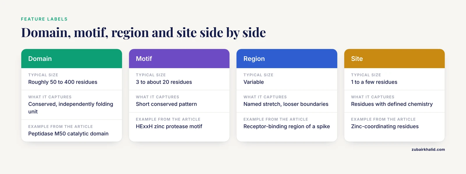
A domain is a compact, conserved part of a protein that folds largely on its own and often retains function when separated from the rest of the chain. Proteins are built by combining domains in different orders and copy numbers, and this combinatorics is a major driver of evolutionary innovation [1]. A motif is a short, recurring sequence pattern. Some motifs are catalytic, such as the HExxH motif (histidine, glutamic acid, any two residues, histidine) that coordinates the catalytic zinc of metalloproteases [5]. Others are interaction signals, such as the LXXLL and PXXP motifs that mediate receptor and protein-protein interactions in nuclear receptor signaling proteins [6].
UniProt also annotates several neighboring concepts that students often blur together. A family is a whole-protein grouping descended from a common ancestor. A repeat is a short segment present in tandem copies. A region is a named stretch of the protein with a shared property whose boundaries are drawn less strictly than a domain. A site is one or a few residues with defined chemistry, such as an active site residue or a binding site residue. All of these appear as labeled, positioned features on the entry page.
Where does the annotation come from? UniProt combines expert curation in the reviewed section (Swiss-Prot) with automatic annotation in the unreviewed section (TrEMBL), and it imports domain and family matches from member databases. The most widely used are Pfam, PROSITE, SMART, SUPERFAMILY, CDD, PANTHER, TIGRFAMs, and the structure-based Gene3D. Pfam and SUPERFAMILY maintain libraries of profile HMMs for known domains, which is what allows a conserved domain to be recognized in a novel sequence [2]. PROSITE additionally curates short patterns written in a regular-expression-like syntax, which is how short motifs such as the HExxH zinc metalloprotease signature are represented. In the underlying flat file, these features carry standardized keys such as DOMAIN, MOTIF, REGION, REPEAT, SITE, ACT_SITE, BINDING, COILED, COMPBIAS, SIGNAL, TRANSMEM, CHAIN, and PROPEP.
| Term | UniProt feature label | What it captures | Typical size | Example |
|---|---|---|---|---|
| Domain | Domain | Conserved, independently folding structural and functional unit | Roughly 50 to 400 residues | Peptidase M50 catalytic domain [5] |
| Motif | Motif | Short conserved pattern, often catalytic or an interaction signal | 3 to about 20 residues | HExxH zinc protease motif [5], LXXLL receptor binding motif [6] |
| Family | Cross-reference, not a positioned feature | Whole-protein evolutionary group | Entire proteins | UPF0235 uncharacterized protein family [7] |
| Repeat | Repeat | Tandem copies of a recurring segment | 20 to 40 residues per copy | Tandem CBS domain pairs [5] |
| Region | Region | Named stretch with a shared property, looser boundaries | Variable | Receptor-binding region of a coronavirus spike [8] |
| Site | Site, Active site, Binding site | Individual residues with defined chemistry | 1 to a few residues | Zinc-coordinating residues of a metalloprotease [5] |
Reading a UniProt Entry Like an Annotator
Anyone learning how to use UniProt for annotation work should start by dissecting one entry end to end. Use the SARS-CoV-2 spike glycoprotein, accession P0DTC2, as the practice case because it is heavily studied and richly annotated.
Open uniprot.org, paste P0DTC2 into the search bar, and open the entry. The header shows the suggested entry name, the reviewed status badge, the organism, and the sequence length. The page is organized into collapsible sections: Function, Names and Taxonomy, Subcellular Location, PTM and Processing, Expression, Interaction, Structure, Family and Domains, Sequence, and External references. The graphical feature viewer sits near the sequence and draws every annotation as a track along the protein. Switch on the Domain, Motif, Region, and Site tracks and the anatomy of the spike becomes visible in one screen: an N-terminal signal peptide, the S1 subunit containing the receptor-binding region that engages ACE2, the S2 subunit with the fusion machinery, and a transmembrane anchor near the C-terminus [8].
Click any feature. A panel opens with the exact residue range, a description, and, critically, the evidence. UniProt marks features as experimental, inferred by similarity from homologs, predicted by rule or by signature match, or imported from an external resource. Two features can look identical in the graphics and differ completely in reliability. The Family and Domains section below the viewer lists cross-references grouped by member database, so you can jump from the spike entry to its Pfam family page, its PROSITE profile hits, and its PDB and AlphaFold structure links.
Two habits prevent most coordinate errors. First, all positions refer to the canonical sequence displayed on the entry, and many entries carry multiple isoforms with their own numbering, so always confirm which isoform a paper or a variant refers to. Second, mature chain numbering starts after signal peptide or propeptide removal, so a residue numbered 50 in the mature protein may sit at position 63 in the canonical sequence. Check the Chain and Propeptide features before quoting any coordinate.
Unreviewed entries deserve a separate habit. Accessions beginning with A0A belong to TrEMBL and carry automatically transferred annotation. A mussel sperm isoform of the protein kinase A catalytic subunit, accession A0A8B6GIP6, illustrates the pattern: its catalytic core is strongly conserved with mammalian counterparts, but its N-terminal region is a distinctive acidic extension with two phosphorylation sites found by mass spectrometry [9]. Automatic annotation captured the conserved core, while the isoform-specific N-terminus is exactly the kind of feature you must verify yourself.
How to Find Protein Domains and Motifs in UniProt: Six Workflows
Workflow 1. Look up a single accession
- Paste the accession into the search bar at uniprot.org and open the entry.
- Confirm the reviewed badge and the organism before reading further.
- In the feature viewer, enable the Domain, Motif, Region, Repeat, and Site tracks.
- Click each feature and record its residue range and evidence tag.
- Open the Family and Domains section and follow the Pfam link for family and clan context and the PROSITE links for pattern-level signatures.
- Cross-check the Sequence section for isoforms and processing events before quoting any position.
This workflow answers most day-to-day questions and doubles as the fastest uniprot database tutorial for new students, because every concept on the page is grounded in one concrete protein.
Workflow 2. Search outward from a domain or motif
To move from one protein to all proteins sharing its parts, work through the family databases linked from the entry. Pfam and InterPro let you browse a signature and retrieve the list of matched proteins, which you can then filter by taxonomy and reviewed status. Within UniProt itself, the query builder supports fielded searches, for example restricting to reviewed entries and a taxonomy identifier, and the cross-reference filters let you pull entries annotated to a given member database family.
Short motifs need different tools because they are too degenerate for a plain text search. The UniProt Peptide Search service finds exact matches to a submitted peptide across UniProtKB, which is useful when you have a short conserved stretch from a mass spectrometry peptide or a binding assay. ScanProsite scans sequences against PROSITE patterns and profiles and reports every hit with its match positions.
This workflow scales to evolutionary questions. In one published analysis, researchers mined UniProt together with NCBI and EukProt to collect every detectable PELP1 homolog, recovering 646 proteins found exclusively in eukaryotes within the Opisthokonta clade of fungi, filastereans, and animals, with no bacterial, viral, plant, or oomycete homologs [6]. A domain-centered search turned one human protein into a testable statement about the origin of a whole signaling architecture.
Workflow 3. Search by sequence with BLAST
When you have a raw sequence rather than an accession, run UniProt BLAST from the website against UniProtKB, preferably the reviewed Swiss-Prot subset first. Inspect the domain architecture of the top hits rather than only the percent identity. A hit that shares your full domain arrangement supports functional transfer, while a hit that matches one domain in isolation does not. Transfer annotation conservatively, and treat hits that agree on architecture but differ in the details of the feature table as a prompt to check the primary literature.
Workflow 4. Run your own HMMER scans and map results back to UniProt
UniProt feature tables are precomputed, so there are times you must scan yourself: when your profile is newer than the database release, when you are working with fragments from metagenomes or surveillance samples, or when you want genome-scale control over thresholds. The HMMER suite is the standard toolset, and Pfam and SUPERFAMILY supply the HMM libraries that power domain detection in uncharacterized sequences [2].
A published protocol demonstrates the full loop with the SARS-CoV-2 receptor-binding domain. The author built an HMM profile for the domain, used HMMER to search for homologs within the UniProt database, built a multiple sequence alignment (MSA) of the hits, and used Jalview to visualize and edit the alignment for downstream phylogenetic analysis, revealing conserved features across species [2]. The core commands look like this:
hmmbuild rbd.hmm rbd_msa.sto
hmmsearch rbd.hmm uniprotkb_reviewed.fasta > rbd_results.txt
Here rbd_msa.sto is a Stockholm-format alignment of your seed domain, hmmbuild converts it into a profile, and hmmsearch scans a downloaded UniProt FASTA. To download the reviewed set for scanning:
curl "https://rest.uniprot.org/uniprotkb/stream?query=reviewed:true&format=fasta" > uniprotkb_reviewed.fastaYou can also pull a ready-made HMM straight from a Pfam release with hmmfetch. For example, the Rhodococcus site-2 protease-like protein 2 study identified its metalloprotease machinery through two peptidase M50 domains (Pfam PF02163) alongside conserved HExxH and LDG motifs and two CBS domains [5], and a Pfam HMM for that family is what an InterProScan or HMMER run would apply to your own sequences. For batch annotation of many proteins, InterProScan runs the member database libraries in one pass:
interproscan.sh -i proteins.fasta -f TSVAfter scanning, map your hits back to UniProt accessions so that every custom result stays connected to the curated feature table, the cross-references, and the variant data that live there.
Workflow 5. Add structural and ligand context
Sequence-based domain calls gain enormous power when checked against structure. The AlphaFold Protein Structure Database now aligns its entries with UniProt releases, and its redesigned entry pages integrate annotations directly with an interactive 3D viewer and add dedicated domains and summary tabs, with coverage extended to isoforms and the underlying multiple sequence alignments available through the website, FTP, cloud, and APIs [10]. Open the AlphaFold page for your accession from the UniProt entry and ask whether the predicted structure supports the domain boundaries drawn on the sequence.
For ligand-level questions, specialized resources built on UniProt accessions do the heavy lifting. DrugDomain 2.0 links evolutionary domain classifications from ECOD, the Evolutionary Classification Of protein Domains, to ligand binding events across the Protein Data Bank, and it extends domain-ligand annotations to AlphaFold models for human drug targets that lack experimental structures [1]. Its scale gives a sense of what is now connectable: interactions with more than 37,000 PDB ligands and 7,560 DrugBank molecules, over 6,000 small-molecule-associated post-translational modifications, 14,000-plus PTM-modified human protein models with docked ligands, 43,023 unique UniProt accessions, and 174,545 PDB structures [1]. The spike protein again provides a concrete case: a curated compilation holds 1,797 entries for P0DTC2 structures spanning full-length molecules and individual domains bound to ligands including ACE2 and antibodies, with antibody binding motifs on the receptor-binding domain classified into four epitope classes [8].
Workflow 6. Retrieve features in bulk through the REST API
For a protein set rather than a single entry, the UniProt REST API returns feature annotations in machine-readable form. Two commands cover most needs. Fetch one entry in flat file or FASTA format:
curl "https://rest.uniprot.org/uniprotkb/P0DTC2.txt"
curl "https://rest.uniprot.org/uniprotkb/P0DTC2.fasta" > spike.fasta
Search and download a table of features for many entries at once:
curl "https://rest.uniprot.org/uniprotkb/search?query=reviewed:true+AND+organism_id:9606&format=tsv&fields=accession,id,protein_name,length,ft_domain,ft_motif"The fields parameter selects the columns, and feature fields such as ft_domain and ft_motif return the annotated ranges directly in the table. If your input identifiers come from Ensembl or RefSeq, run them through the UniProt ID mapping tool first so that every row lands on a stable accession. For reproducibility, record the UniProt release version alongside your downloads, since the database is updated on a dated release cycle.
Interpreting Domain and Motif Annotations in UniProt
Finding features is the easy half. UniProt protein domains and motifs interpretation rests on four habits.
Read the evidence tag first
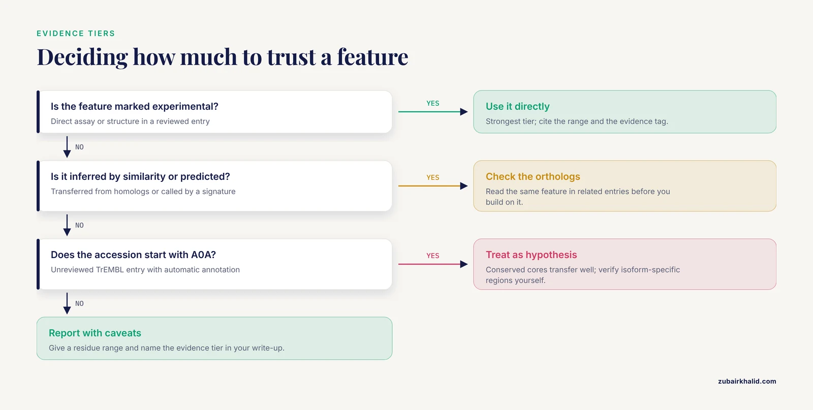
Every feature carries provenance. Experimental evidence outranks inference by similarity, which outranks prediction. Rule-based automatic annotation, common in TrEMBL, transfers annotations from related entries and inherits their uncertainty. Before you build an argument on a domain boundary or a motif position, check which tier supports it. A domain marked experimental in one entry may appear only as a signature match in its unreviewed orthologs.
Expect boundary disagreement between databases
Pfam, SMART, PROSITE, CDD, and Gene3D use different algorithms, alignment seeds, and family definitions, so they frequently draw different edges around the same domain. This is not an error, it is a reflection of method differences. Use the InterPro integration, which consolidates member database matches, as a consensus view, and when you report coordinates in a manuscript, give a range rather than a single residue set. Structure provides a tiebreaker, and the AlphaFold domains tab makes that check fast [10].
Weigh motifs in context
A short motif hit is weak evidence on its own because the patterns are degenerate. The PELP1 analysis found LXXLL and PXXP motifs across its protein set even though the leucine and proline residues were not universally conserved within the motifs [6], which is exactly the fuzziness you should expect. Motif calls gain weight when they sit inside the right domain architecture and cellular context. The site-2 protease-like protein 2 study is a model: the HExxH and LDG motifs were interpreted as meaningful because they occurred within two peptidase M50 domains and two CBS domains in a hydrophobic transmembrane protein, and that combined picture was then tested experimentally [5]. A HExxH match in a soluble protein with no protease context deserves skepticism, not a metalloprotease claim.
Domain architecture also explains biology that single domains miss. Among protein kinase A catalytic subunits, the catalytic core is conserved across species while the N-terminal region varies, and the sperm-specific mussel isoform carries a longer acidic N-terminal extension with its own phosphorylation sites [9]. The functionally interesting differences live outside the recognizable domain, which is why the full feature table, not just the domain track, deserves a read.
Map variants and active sites onto features
Variant interpretation is where domain annotation earns its keep. One arrhythmogenic right ventricular cardiomyopathy study collected 1,358 reported PKP2 variants, extracted topology information from the UniProt database, categorized the 497 nonsynonymous variants by the structural domains they hit, and analyzed how pathogenic classifications and hotspot variants distributed across those domains relative to clinical phenotypes [4]. Computational predictors work the same way. KinMut, a support vector machine classifier for pathogenic kinase mutations, combined gene-level membership, domain-level Pfam annotations, and residue-level physicochemical and functional features, and it was trained on 3,492 human kinase mutations with experimentally validated pathogenic or neutral status in UniProt [3]. Purpose-built viewers extend this further: the Human Protein Mutation Viewer accepts mutations as UniProt accessions with HGVS nomenclature, genome coordinates, or FASTA sequences, and renders them against globular domains, targeting signals, and post-translational modification sites with linked alignments and 3D mapping [11].
Active sites follow the same logic. UniProt curates ACT_SITE and BINDING features with evidence tags, and machine learning systems now push residue-level active-site classification further. M3Site integrates sequence embeddings, structural graphs, and functional text to classify active sites at residue level, using a curated dataset of 25,883 proteins sourced from UniProt and AlphaFold2 [12]. Treat such predictions as candidates to check against the curated feature table, not replacements for it.
Finally, treat missing annotations as unknowns rather than negatives. A survey of hydroxyproline modifications in HeLa cells identified 24 unique hydroxyproline sites in 21 proteins, of which only Pro62 of the ribosomal protein RPS23 had previously been reported in UniProt [13]. Rare post-translational modifications lag in curated databases, so an empty PTM track is a statement about annotation coverage, not about biology.
When Sequence Patterns Are Not Enough
Some proteins resist every pattern-based tool, and UniProt itself tells you so honestly. The UPF0235 family collects conserved proteins across archaea, bacteria, and eukaryotes whose functions remain uncharacterized. The Pyrococcus furiosus member PF1765, accession Q8U052, had its crystal structure solved at 1.3 angstrom, revealing a compact α+β fold with no significant similarity to known functional proteins, and biochemical assays showed that it binds double-stranded DNA, single-stranded DNA, and RNA despite lacking any canonical nucleic acid binding domain [7]. The family label in UniProt was accurate and useful, but only structure and experiment could assign function.
Two developments broaden what you can detect beyond conserved sequence. Zero-shot Protein Segmentation uses embeddings from the protein language model ProtT5 to predict the boundaries of functional segments without training on labeled examples, and its boundary predictions for the human proteome reproduce UniProt reviewed annotations better than established bioinformatics tools while categorizing over 200 of the most common UniProt annotation types, including folded domains, sub-domains, and intrinsically disordered regions [14]. At the other end of the scale, a proteome-wide structural analysis of ocean archaea using AlphaFold2-predicted structures identified 255 protein domain clusters not defined by any domain database [15], a reminder that the domain universes in Pfam and its siblings are still incomplete. When a protein of interest returns no domain hits, triangulate: run an HMMER scan with relaxed gathering thresholds, inspect the AlphaFold model for repeat-like or globular substructures, and check whether language-model segmentation flags coherent segments [14].
Visualizing Domain Architecture for Figures and Reports
Publication figures need clean topology schematics, and manual drawing does not scale. ProToDeviseR is an R package with a Shiny graphical interface that generates protein topology schemes automatically from database accession numbers, interrogating UniProt or NCBI GenPept and combining features from multiple prediction resources into one figure [16]. Feed it a UniProt accession and it renders the domains, motifs, and regions in a consistent style you can export.
For alignment-based figures, Jalview remains the standard companion to HMMER workflows, letting you visualize, edit, and annotate the MSAs that underpin domain definitions [2]. The AlphaFold viewer adds a structural panel with annotations overlaid on the 3D model [10]. Whichever tools you choose, label each figure with the feature source and the database release, because a domain diagram without a version stamp cannot be reproduced.
Common Mistakes and Limitations
- Treating a motif hit as a domain call. A short pattern match is a hint. The HExxH motif only implies metalloprotease function when embedded in a peptidase domain and appropriate context [5].
- Copying residue numbers across isoforms. Feature positions refer to the canonical sequence of that entry. Isoforms, signal peptide removal, and propeptide cleavage all shift numbering, so verify the sequence context before quoting a coordinate.
- Ignoring evidence tags. Two identically drawn features can carry experimental versus purely predicted evidence. Read the tag before building a claim on it.
- Trusting TrEMBL annotation as curated fact. Accessions beginning with A0A are unreviewed and automatically annotated. Conserved cores transfer reliably, but isoform-specific regions such as the divergent N-terminus of the mussel PKA isoform [9] demand independent verification.
- Expecting one true boundary. Member databases legitimately disagree on domain edges. Report ranges and use structural evidence to arbitrate.
- Concluding no function from no domain. Q8U052 binds three classes of nucleic acid with no canonical binding domain [7], and hundreds of structural domain clusters remain absent from every domain database [15]. Absence of annotation is absence of knowledge, not absence of function.
- Mistaking low-complexity and coiled-coil tracks for domains. These compositional features are annotated separately and do not represent folded globular units.
- Skipping the release version. UniProt updates continuously, and feature tables change between releases. Record the release date with every download or figure.
- Over-reading automated variant-to-domain mappings. Clinical mapping studies such as the PKP2 analysis [4] show that domain-based grouping is informative, but automated annotation does not replace experimental characterization of an individual protein or variant.
Quick Review
- Domains are conserved, independently folding functional units, motifs are short conserved patterns, and UniProt stores both as positioned features with evidence tags.
- Reviewed Swiss-Prot entries carry expert curation, while A0A-prefix TrEMBL entries carry automatic annotation.
- Member databases such as Pfam, PROSITE, and SMART disagree on boundaries, so report ranges and consult structure.
- Motif hits need domain architecture and cellular context before you infer function.
- HMMER with Pfam or custom profiles lets you scan UniProt sequences yourself and map results back to accessions.
- The REST API exports feature tables in bulk, and recording the UniProt release keeps analyses reproducible.
- Absence of a known domain is not absence of function, as uncharacterized families and newly discovered domain clusters keep demonstrating.
Frequently Asked Questions
What do protein domains and motifs mean in a UniProt entry?
They are the labeled, position-annotated parts of a protein listed in the entry's feature table. A domain is a conserved, independently folding unit that typically carries a discrete function, while a motif is a short conserved pattern, often a few residues, marking an active site or interaction signal. Each feature shows its residue range and an evidence tag telling you whether it is experimental, inferred by similarity, or predicted.
How do I find the domains and motifs of a single protein in UniProt?
Search the accession at uniprot.org, open the entry, and enable the Domain, Motif, Region, and Site tracks in the feature viewer. Click each feature to read its exact residue range and evidence, then open the Family and Domains section for cross-references to Pfam, PROSITE, and other member databases. Confirm the isoform and any signal peptide processing before quoting coordinates.
Can I search UniProt for every protein that carries a given domain?
Yes, by working through the family databases linked from any UniProt entry. Browse the domain in Pfam or InterPro, retrieve the list of matched proteins, and filter by organism or reviewed status, or use UniProt's query builder with cross-reference filters. For short motifs, use UniProt Peptide Search for exact peptide matches or ScanProsite for pattern-based scans.
Why do Pfam, SMART, and PROSITE disagree about my protein?
Because they use different algorithms, alignment seeds, and family definitions, so their domain boundaries legitimately differ. Treat the InterPro consolidation as a consensus view and report a coordinate range rather than a single boundary. When agreement matters, check the AlphaFold model or an experimental structure to arbitrate.
Do I still need to run HMMER if UniProt already shows Pfam domains?
Yes, when your question involves a custom profile, newly published families, fragmented sequences, or thresholds you control. UniProt feature tables are precomputed against a fixed release, so HMMER lets you scan the latest sequences with your own models and then map hits back to UniProt accessions for curated context.
How reliable are UniProt domain and motif annotations?
Reliability varies with the evidence tier, which is printed on every feature. Experimental annotations in reviewed entries are the strongest, similarity-based and predicted annotations are progressively weaker, and automatic TrEMBL annotation should be treated as a starting hypothesis. Missing annotations are also common for rare features, so absence of a feature is not evidence of absence.
Sources
- DrugDomain 2.0: Comprehensive database of protein domains-ligands/drugs interactions across the whole Protein Data Bank.
- Exploring Virus Protein Domains: Detection and Evolutionary Insights.
- Prioritization of pathogenic mutations in the protein kinase superfamily.
- [[Distribution characteristics of PKP2 non-synonymous variations in protein domain and genotype-phenotype relationship in patients with arrhythmogenic right ventricular cardiomyopathy].](https://pubmed.ncbi.nlm.nih.gov/41529851/)
- Site-2 protease-like protein 2 mediates phenol tolerance in Rhodococcus ruber through transcriptomic and functional analyses.
- Proline-, Glutamic Acid-, Leucine-Rich Protein 1 (PELP1): Diversity, Structural Conservation, and Evolutionary Origins Across the Species.
- Crystal structure and nucleic acid binding activity of the conserved UPF0235 family protein PF1765 from Pyrococcus furiosus.
- In Silico Ligand-Based Screening of PDB Database for Searching Unique Motifs Against SARS-CoV-2.
- Mussel Spermatozoa Express a Specific Isoform of the Catalytic Subunit of cAMP-Dependent Protein Kinase.
- AlphaFold Protein Structure Database 2025: a redesigned interface and updated structural coverage.
- HPMV: human protein mutation viewer - relating sequence mutations to protein sequence architecture and function changes.
- M3Site: multiclass multimodal learning for protein active site identification and classification.
- The Hydroxyproline Proteome of HeLa Cells with Emphasis on the Active Sites of Protein Disulfide Isomerases.
- Zero-shot segmentation using embeddings from a protein language model identifies functional regions in the human proteome.
- A comprehensive proteome structural analysis suggests undiscovered functional domains in ocean archaea.
- ProToDeviseR: an automated protein topology scheme generator.