Post-Translational Modifications in UniProt
By Dr. Zubair Khalid, DVM, MS, PhD ·

Post-translational modifications in UniProt are curated sequence annotations that record chemically modified residues and processing events, such as phosphorylation, glycosylation, disulfide bonding, and lipidation, at specific positions on a protein sequence. Every annotation carries the modified position, a controlled vocabulary term for the modification type, and an evidence tag that tells you whether the claim is experimental, inferred by similarity, or computationally predicted.
This matters because a single gene can yield many proteoforms, meaning distinct molecular forms of a protein produced through modifications, splicing, and mutations, and those proteoforms frequently carry the biology that sequence alone cannot explain. PTMs regulate cell signaling, protein degradation, enzyme activity, and localization, and the UniProt knowledge base has become a standard reference layer that mass spectrometry analysts, computational biologists, and experimentalists use to name, localize, and verify modifications. When a mass spectrometry pipeline reports an unexplained mass shift on a peptide, a UniProt annotation is often the fastest way to decide whether that shift corresponds to a known biological modification or an artifact [1][2]. This article explains how UniProt post-translational modifications annotation is structured, how to read it, how to query it programmatically, and how to use it in real proteomics and proteogenomics workflows.
What UniProt Post-Translational Modifications Annotation Means
UniProtKB (the UniProt Knowledgebase) has two sections. Swiss-Prot contains manually curated, reviewed entries. TrEMBL contains unreviewed entries annotated largely by automated methods. PTM annotations appear in both sections, but the depth and reliability differ substantially between them, so the first step in interpreting any UniProt PTM record is checking whether the entry is reviewed.
PTM information appears in three main places on a UniProt entry:
- The feature table. Features such as MOD_RES (modified residue), CARBOHYD (glycosylation site), LIPID (lipidation site), DISULFID (disulfide bond), and CROSSLNK (cross-link) record the modified positions directly on the sequence. MOD_RES is the workhorse feature for phosphorylation, acetylation, methylation, hydroxylation, and most other residue-level modifications.
- The PTM notes and keywords. Keyword lines place each protein into a controlled category such as Phosphorylation, Acetylation, Glycoprotein, or Ubl conjugation, which lets you retrieve whole families of modified proteins with a single query.
- Cross-references. Each entry links out to specialized PTM resources so you can dig deeper into site-level evidence, orthologous sites, and disease associations.
The foundation for all of this is a controlled vocabulary, meaning a fixed, standardized list of terms for each modification type rather than free text. Swiss-Prot curators standardized PTM feature annotation this way precisely so that both expert biologists and computer programs could retrieve and interpret the data consistently, a design goal the UniProt curation team described in their foundational paper on PTM annotation in the Swiss-Prot knowledge base [3]. That paper also sketched a companion resource devoted to the biology of modifications, including the modifying enzyme, the taxonomic range, and the mass change, an idea that shaped how later PTM knowledge bases were built [3].
A useful mental model for UniProt PTM annotation has three layers:
| Layer | What it records | Example | Typical evidence |
|---|---|---|---|
| Feature position | A specific modified residue and modification type | MOD_RES Phosphoserine at position 768 | Experiment, similarity, prediction |
| Modification event | A broad process level annotation in notes | Autophosphorylation of the kinase | Literature curation |
| Protein-level keyword | The protein carries this class of modification | Keyword Phosphorylation | Any of the above |
Students most often confuse UniProt PTM features with variant features. A VARIANT feature records a residue that differs between protein isoforms or individuals because of genetic change. A MOD_RES feature records a chemical change to a residue that is encoded normally. Phosphorylation of a serine does not change the DNA or the translated sequence. It changes the chemistry of the finished protein. Keeping that distinction straight prevents many downstream interpretation errors.
How UniProt Builds and Curates PTM Annotations
UniProt assembles PTM annotations through four routes, and each route leaves a different footprint on the entry.
Route 1: Literature curation. UniProt curators read primary papers and add features with position, modification type, and supporting citations. When a paper demonstrates that a specific residue is phosphorylated in vivo, the curator adds a MOD_RES feature with the experimental evidence tag. This is the gold standard route and applies mainly to reviewed Swiss-Prot entries.
Route 2: Similarity and rule-based annotation. If a residue is experimentally shown to be modified in one species, curators or automated pipelines may project that annotation to orthologous residues in related species, tagged as "inferred by similarity." Curated rules, such as those derived from PROSITE patterns and expert-written UniRule entries, can also propagate annotations automatically.
Route 3: Computational prediction. Some annotations are predictions. The classic example is potential N-glycosylation sites. The sequon motif, meaning the Asn-X-Ser/Thr sequence context required for N-linked glycosylation, is easy to detect in sequence, so UniProt marks matching positions as "potential" glycosylation sites. A potential site is a prediction about a motif, not evidence that glycosylation happens there. This distinction is the single most common source of misinterpretation for new users.
Route 4: Cross-reference import. UniProt links entries to external PTM databases that specialize in site-level data. The scale of this external ecosystem is large. PTMD 2.0, for example, catalogs 342,624 PTM-disease associations covering 15,105 proteins, 93 types of PTMs, and 2,083 diseases, with each association classified into categories such as upregulation, downregulation, absence, presence, site creation, or site disruption [4]. Prediction resources also feed the ecosystem. MIND-S, a deep-learning platform built on multi-head attention and graph neural networks, predicts 26 types of PTMs across datasets of roughly 50,000 proteins and includes an interpretation module that scores the relevance of each amino acid to the prediction [5]. None of these resources replace UniProt annotations, but following the cross-references on a UniProt entry is how you move from "this residue is annotated as phosphorylated" to "here is the experiment, the disease association, and the predicted kinase."
A good example of cross-resource integration comes from kinase biology. Investigators built federated queries linking the Protein Kinase Ontology, iPTMnet, Protein Ontology, neXtProt, and Mouse Genome Informatics to find gaps in functional coverage of the human kinome. Those queries identified 32 functional domains enriched in both cancer variants and PTMs, and generated mechanistic hypotheses at the residue, protein, pathway, and species level [6]. That study is a template for how to treat UniProt PTM annotation: not as an endpoint, but as one node in a network of resources you query together.
Reading a UniProt PTM Entry, Step by Step
Open a reviewed human protein with rich PTM annotation, such as the epidermal growth factor receptor (EGFR, accession P00533), and work from the top of the entry down. This walkthrough doubles as a short uniprot database tutorial for the PTM-specific parts of the interface.
Step 1. Check the entry status and evidence level. Confirm the entry is reviewed. The protein evidence level (PE line) tells you whether protein existence is supported by evidence at the protein level, transcript level, homology, or prediction. PTM annotations on a protein whose existence itself is only predicted deserve extra skepticism.
Step 2. Scan the keywords. EGFR carries keyword annotations including Phosphorylation and Glycoprotein. Keywords are retrieval handles, so note them for later queries.
Step 3. Read the feature table by category. Features are grouped under headings. Under the PTM category you will find DISULFID features for extracellular domain disulfide bonds, CARBOHYD features for N-linked glycosylation sites, and a long block of MOD_RES features for phosphorylation sites. Each MOD_RES feature shows the position, the modification name (for example, Phosphoserine or Phosphotyrosine), and usually a short description with the functional context.
Step 4. Read the PTM notes. UniProt places larger narrative annotations in note blocks labeled PTM. For a receptor kinase, these notes summarize autophosphorylation behavior, which residues serve as docking sites for downstream adaptors, and in some cases the functional consequence of specific site mutations. The federated kinase study used exactly this kind of site-level information to test hypotheses. It found that serine 768 phosphorylation in the C-helix of EGFR is inhibitory, because oncogenic variants that alter S768 phosphorylation increase basal EGFR activity, whereas the reverse pattern holds for the conserved hydrophobic motif site of PKCβII, where variants at S660 reduce kinase activity and enhance membrane translocation [6]. When you read a UniProt PTM note on a kinase, you are reading the distilled version of evidence like this.
Step 5. Inspect the evidence tags. Every feature carries evidence tags. An experimental tag means the modification was demonstrated, typically by mass spectrometry, mutagenesis, or biochemical assay. A similarity tag means the annotation was projected from another species. A potential tag means it is predicted. Mixed evidence on one protein is normal, and reading the tags per feature rather than per entry is the correct habit.
Step 6. Follow the cross-references. The DR (database cross-reference) lines point to specialized resources, including PTM-focused databases. If you need site-level spectral evidence, disease links, or kinase-substrate relationships, these cross-references are the bridges.
Step 7. Watch the numbering. UniProt annotates features on the precursor sequence, which includes the signal peptide, propeptide, and transit peptides. EGFR carries a signal peptide at the start of the sequence, so residues numbered in mature protein literature can differ from UniProt precursor numbering by a fixed offset. This offset problem is covered in detail in the mistakes section below because it causes real errors in practice.
How to Query and Export PTM Annotations
Once you understand how to read a single entry, the next skill is retrieving PTM annotations at scale. UniProt provides a website query builder and a REST API, and the API is what you will use in practice.
Retrieving all reviewed human proteins with a specific modified residue
The REST API accepts feature fields as queryable terms prefixed with ft_. To pull every reviewed human entry that carries an annotated phosphoserine feature:
curl "https://rest.uniprot.org/uniprotkb/search?query=(organism_id:9606) AND (reviewed:true) AND (ft_mod_res:Phosphoserine)&fields=accession,id,protein_name,ft_mod_res&format=tsv&size=500"Swap Phosphoserine for Phosphotyrosine, N-acetylalanine, or any other term from the UniProt PTM controlled vocabulary. Change organism_id to target another species. The fields parameter controls which columns the TSV output contains, so you can request ft_carbohyd, ft_disulfid, ft_lipid, or ft_crosslnk in the same way.
Retrieving proteins by PTM keyword
Keywords are the coarse filter. The query keyword:Phosphorylation AND reviewed:true AND organism_id:9606 returns all reviewed human entries annotated with the Phosphorylation keyword. This returns more proteins than the site-level query because the keyword can be supported by any annotated site or curated evidence.
Downloading full PTM feature sets
For bulk work, stream results with the UniProt ID mapping or stream endpoints and parse the TSV in Python or R. A typical pipeline downloads accession and ft_mod_res columns for a full proteome, splits the position ranges, and builds a residue-level table. That table then serves as the reference set for validation analyses described below.
Verifying what the vocabulary means
The PTM controlled vocabulary is published as a downloadable list, and every MOD_RES description string is drawn from it. When you encounter an unfamiliar term, such as Cysteine S-nitrosylation or Lysine succinylation, check the vocabulary rather than guessing at the chemistry, because the term encodes the exact modified atom and mass shift. This vocabulary-driven approach is the direct descendant of the Swiss-Prot standardization effort that introduced a controlled term for every described PTM so that retrieval would stay consistent for both humans and programs [3].
Using UniProt Annotations to Validate Mass Spectrometry PTM Calls
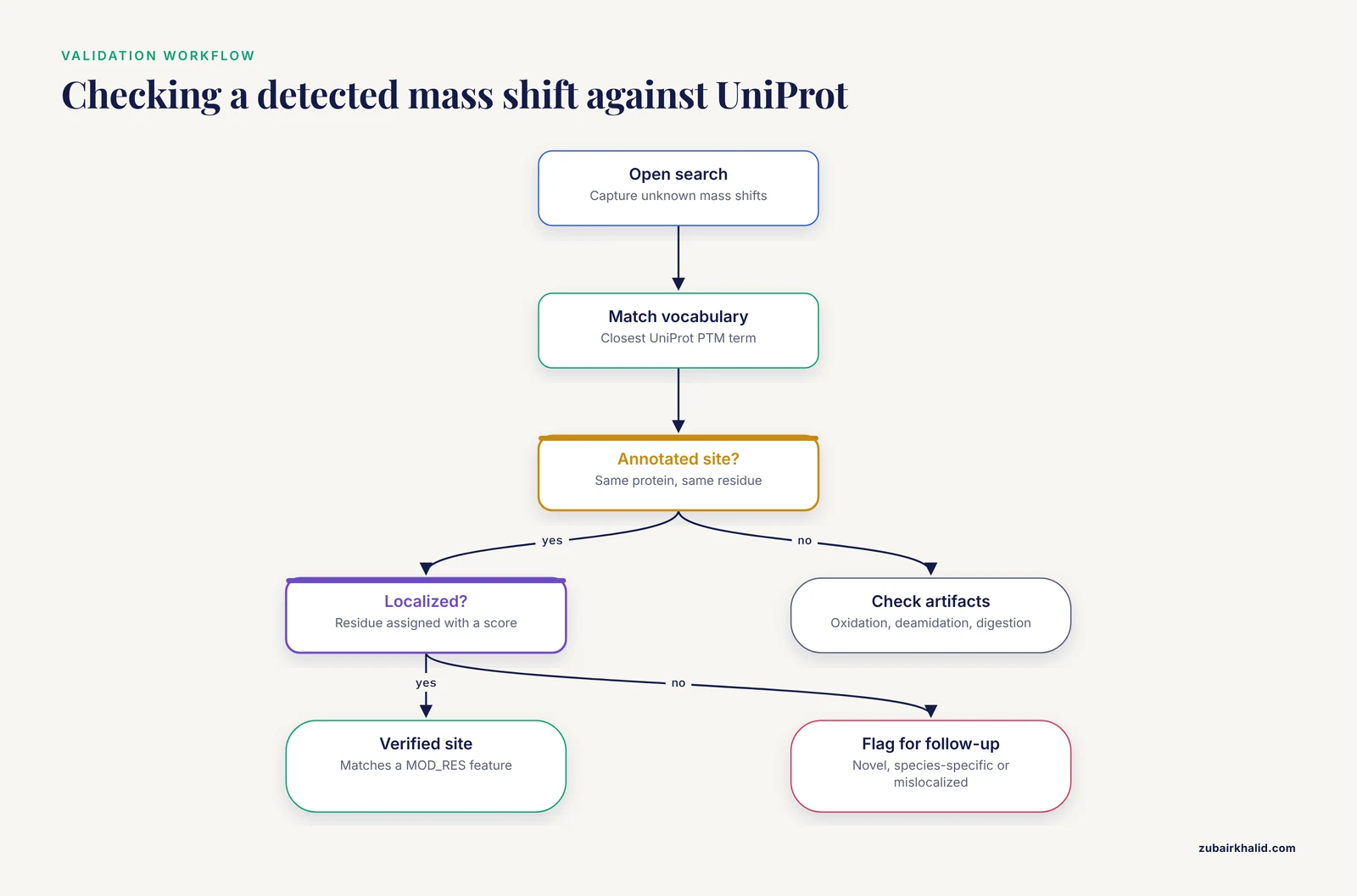
The most practical application of UniProt PTM annotation for many readers is validating modifications detected in their own mass spectrometry data. Understanding why requires a quick look at how MS-based PTM analysis works.
In bottom-up mass spectrometry, proteins are digested into peptides, and the mass of each peptide is measured. A modified peptide appears with a mass shift, for example approximately +79.97 Da for phosphorylation, +42.01 Da for acetylation, or +15.99 Da for oxidation. The problem is that many different modifications produce similar or identical mass shifts, and open modification searches report shifts without naming the chemistry. This is where annotation layers earn their keep.
The PTM-TBA pipeline demonstrates the approach directly. The authors combined top-down mass spectrometry, which measures intact proteoforms, with bottom-up mass spectrometry and UniProt annotations to identify, localize, and verify PTMs in SW480 cell proteoforms [1]. The numbers illustrate the scale of the matching problem. In the top-down dataset, the search identified 1,662 mass shifts, of which 545, about 33 percent, matched to 12 common PTMs, and 351 could be localized to specific residues. PTM-TBA then validated 346 of the 1,662 shifts using UniProt annotations or the bottom-up dataset [1]. In the journal version of the study, built on technical triplicates, database search of top-down data identified on average 2,000 mass shifts per run, 814.5 of them, or 40.7 percent, matched to 11 common PTMs, and 423 were localized, with 435 shifts verified using bottom-up data and UniProt annotations [2]. Two lessons follow. First, a large fraction of raw mass shifts do not correspond to canonical PTMs, so annotation matching is a genuine filter rather than a formality. Second, UniProt annotations alone verified a meaningful share of detected shifts, which means every proteomics analysis should include this step.
A practical validation workflow looks like this:
- Run your database search with open or ultratolerant modification searching to capture unknown shifts. Ultratolerant searching loosens the mass tolerance so unexpected modifications can be detected without prior knowledge, an approach that now enables hypothesis-free PTM analysis [7].
- Convert detected mass shifts to the closest matching terms in the UniProt PTM vocabulary and mark every shift that matches an annotated site on the same protein.
- For localized sites, compare your residue position against UniProt MOD_RES features, adjusting for precursor numbering as described above.
- Flag discrepancies. If your localized phosphorylation site has no UniProt annotation, it is either novel, species-specific, or a localization error, and each possibility needs different follow-up.
- Account for artifacts. Recent multi-layer workflows capture confounders such as variation in trypsin digestion efficiency and hypermodified regions that can masquerade as biological PTM signals, and they reanalyzed public datasets to confirm oxidative modifications on Met and Cys residues that earlier pipelines missed [7].
The PTM-TBA study reinforces a broader point about complementary evidence. Top-down mass spectrometry excels at detecting modifications on intact proteoforms and at localizing them across the whole protein, while bottom-up mass spectrometry provides deeper sequence coverage and confident site localization for individual peptides. Combining both with a knowledgebase layer increases the sensitivity and accuracy of PTM identification compared with either approach alone [1][2]. UniProt annotation is the common reference frame that makes the combination work.
PTM Annotation in Proteogenomics and Non-Model Organisms
PTM annotation in UniProt depends on underlying protein annotation, and protein annotation itself depends on genome annotation. Proteogenomics closes this loop by using mass spectrometry evidence to refine predicted gene models, and PTM profiling rides along with the same data.
GAPP, a proteogenomic software package for prokaryotes, shows the workflow in a single command. Applied to proteomic data from Helicobacter pylori, GAPP confirmed 84.9 percent of the predicted proteins, identified 20 novel protein-coding genes, and corrected four existing gene models with respect to translation initiation sites, while also revealing a large repertoire of PTM events from the same data [8]. The pipeline is applicable to any sequenced prokaryotic organism, which matters because PTM annotation in UniProt is much sparser for non-model bacteria than for humans, and proteogenomics is how that gap gets filled [8].
Plant proteomics illustrates the same principle at proteome scale. The Arabidopsis thaliana PeptideAtlas build matched 70 million MS/MS spectra to the Araport11 genome annotation, identifying about 0.6 million unique peptides and 18,267 proteins at the highest confidence level, which represents 78.6 percent of the predicted proteome. The release mapped sites for 5,198 phosphorylated proteins, 668 ubiquitinated proteins, 3,050 N-terminally acetylated proteins, and 864 lysine-acetylated proteins. The remaining 21.4 percent of the predicted proteome, 5,896 proteins, lacked MS support, a fraction called the dark proteome, and it is strongly enriched for E3 ligases and transcription factors, classes of proteins that are short-lived and hard to detect [9]. For anyone using UniProt PTM annotations in Arabidopsis, the practical takeaway is that absence of PTM annotation on a protein may reflect absence of MS evidence for the protein at all, not absence of modification.
Linking PTMs to Function and Disease
UniProt PTM annotations become most valuable when connected to functional and disease context, and several approved resources show what those connections look like in practice.
Functional hypothesis generation. The federated kinase study mentioned above did not stop at annotation retrieval. It used cross-resource queries to prioritize understudied kinases, variants, and PTMs, then experimentally tested the resulting hypotheses for EGFR S768 and PKCβII S660 [6]. This is the correct posture toward UniProt annotations. They are hypotheses with assigned evidence levels, suitable for driving experiments, not endpoints in themselves.
Disease association. PTMD 2.0 provides structured PTM-disease associations at scale, with 342,624 associations annotated across 13 additional data layers per disease-associated protein, including mutation, interaction, pathway, structure, and expression information [4]. When a UniProt MOD_RES site on your protein of interest links to a disease association in a resource like this, the annotation stops being a sequence detail and becomes a mechanistic lead.
Residue-level functional inference. The CysDuF project attacked the problem of domains of unknown function (DUF), meaning protein families whose activities are uncharacterized, by annotating cysteine residues based on PTMs, biochemical pathways, taxonomy, diseases, and computed protein microenvironments around cysteines from 70,342 protein structures. Their DeepCys prediction server, validated against 18,626 experimental cysteine PTMs, achieved an accuracy of 0.79 [10]. This example shows the direction the field is moving. Sequence context plus structure plus PTM knowledge from resources like UniProt can generate functional hypotheses for proteins that have no experimental characterization at all.
Multi-omics PTM crosstalk. Large-scale PTM profiling increasingly treats modifications as an interacting system rather than independent events. In head and neck squamous cell carcinoma cells exposed to X-ray or carbon-ion irradiation, integrated proteomics, phosphoproteomics, and acetylomics profiling at 30 minutes post-irradiation found that carbon ions induced 21 percent more phosphosites (2,781 versus 2,301) and 61 percent more acetyl sites (3,324 versus 2,070) than X-rays, along with histone modifications that potentially alter chromatin accessibility and kinase activity shifts including ATM hyperactivation [11]. Reviews of macrophage polarization make the same systems point, describing interconnected PTM circuits between signaling, epigenetic, and metabolic pathways where phosphorylation, ubiquitination, methylation, acetylation, and lactylation jointly shape cell state [12]. UniProt PTM annotation feeds these analyses as the prior knowledge layer that anchors newly detected sites to known biology.
Visualization and Interpretation Tools
A PTM list is hard to reason about. Visualization turns it into insight, and several tools connect directly to UniProt-style annotations.
PTMVision. This web server provides interactive exploration of PTMs identified in mass spectrometry experiments, with views including 3D structures, contact maps, UniMod classification summaries, and site-specific overviews. It was built specifically because open modification searches now reveal hundreds to thousands of distinct modifications per sample, which outstripped available visualization capacity [13].
Protein Visualizer 2.0. This web-based tool visualizes topology together with co- and post-translational features for all human proteins, with particular attention to relationships between N-glycosylation, disulfide bonds, free cysteine sites, and membrane topology. It exists because tools such as UniProt PTM viewing do not depict conflicts between predicted topology and known modifications, for example a glycosylation site annotated inside a transmembrane segment [14].
STRAP PTM. For labs processing liquid chromatography-tandem mass spectrometry output in protXML format from the Trans-Proteomic Pipeline, STRAP PTM collates, scores, and ranks PTMs across multiple samples using spectral counting, supporting differential comparison at protein, peptide, and PTM levels [15].
PTM-Psi on the cloud. For structural questions, PTM-Psi simulates the impact of modifications on protein structures and interactions. Refactored as a cloud-compatible workflow, it enabled high-throughput simulation across the combinatorial explosion of thiol protein PTMs on a protein megacomplex in the cyanobacterial Calvin-Benson cycle, demonstrating how PTM-aware structural modeling scales when coupled to cloud resources [16].
Single-cell detection. PTM detection is also moving toward single cells. Optimized MS data interpretation applied to single-cell and single-nucleus proteomics revealed cell-state heterogeneity in PTMs, protein-protein interactions, and variants, including epigenetic drug-induced changes detectable in single nuclei, information inaccessible through RNA profiling [17].
Common Mistakes and Limitations
The following errors appear repeatedly in real projects. Each one is avoidable with a specific habit.
Confusing potential sites with verified sites. A "potential N-glycosylation site" in UniProt is a sequon prediction. It does not mean the residue is glycosylated. Always read the evidence tag on the feature before citing the site.
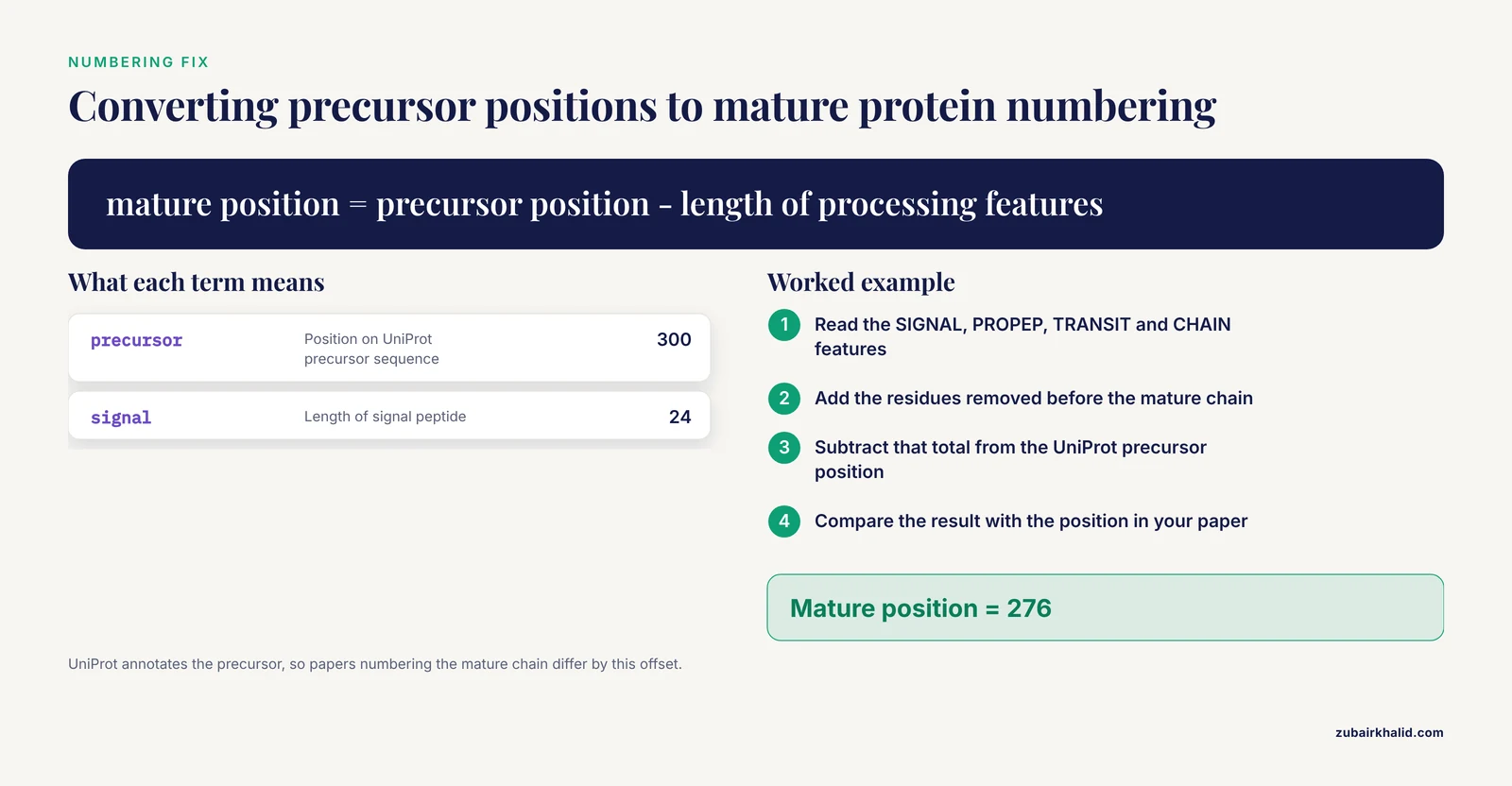
Ignoring precursor numbering. UniProt positions include signal peptides, propeptides, and transit peptides. A phosphorylation site at precursor position 300 on a protein with a 24-residue signal peptide corresponds to position 276 in mature protein numbering. Mass spectrometry papers and UniProt entries frequently disagree by exactly this kind of offset, and analysts who do not check the CHAIN and SIGNAL features will report false novel sites.
Treating reviewed and unreviewed entries the same. TrEMBL annotations are largely automatic. A PTM feature on an unreviewed entry may come from a prediction rule applied to a similar sequence. For high-stakes conclusions, restrict to reviewed entries or trace the evidence tag on the specific feature.
Reading keywords as site evidence. The keyword Phosphorylation on an entry means the protein is known or predicted to be phosphorylated. It does not tell you which residue, whether the site is active in your tissue, or whether the evidence is direct. Keywords are for retrieval, features are for positions, notes are for function.
Assuming absence of annotation means absence of modification. PTM annotation tracks what has been measured and curated. The Arabidopsis PeptideAtlas example makes this concrete. More than one fifth of the predicted proteome had no MS support at all, and the dark proteome is enriched for exactly the protein classes, E3 ligases and transcription factors, whose PTM regulation matters most [9]. An unannotated site on a poorly covered protein may simply be unobserved.
Overtrusting localization from a single experiment. Peptide-level mass spectrometry can identify a modified peptide without pinpointing which of several residues carries the modification. Localization scores exist for a reason, and annotation matching helps, but the PTM-TBA results show that even with combined top-down and bottom-up evidence only a minority of raw mass shifts get confidently localized to a residue [1][2].
Ignoring modification chemistry and artifacts. Oxidation of methionine during sample preparation, deamidation during storage, and digestion-related variation can all masquerade as biological PTMs. Multi-layer workflows that control for trypsin efficiency changes and hypermodified regions exist precisely because these artifacts are common enough to distort conclusions [7]. A detected shift that matches the correct mass is a starting point, not proof of biology.
Forgetting cross-talk and context. A site annotated as phosphorylated may function only in combination with a nearby acetylation or ubiquitination event. Studies of macrophage polarization and radiation response both show PTMs acting as interacting networks rather than isolated switches [11][12]. Interpreting one MOD_RES feature in isolation can miss the actual regulatory logic.
Skipping the sequence conflict. Some UniProt entries list sequence conflicts where literature sequences disagree with the reference. If your peptide maps to a conflicted region, the mass shift you attribute to a PTM may actually reflect a sequence difference.
Quick Review
- UniProt PTM annotation lives mainly in features (MOD_RES, CARBOHYD, LIPID, DISULFID, CROSSLNK), PTM notes, keywords, and cross-references, all built on a controlled vocabulary for every modification type [3].
- Every feature carries an evidence tag. Experimental, similarity-based, and potential (predicted) annotations look similar in the table but mean very different things.
- Feature positions refer to the precursor sequence, including signal peptides and propeptides. Check CHAIN and SIGNAL features before comparing positions with mature protein numbering.
- Reviewed Swiss-Prot entries and unreviewed TrEMBL entries differ in curation depth. Check review status first.
- REST queries using ft_ fields, for example ft_mod_res:Phosphoserine, retrieve PTM annotations at scale, and keyword queries retrieve protein-level PTM categories.
- Mass spectrometry pipelines use UniProt annotations to match raw mass shifts to known PTMs. In one top-down dataset of SW480 cells, only about a third of 1,662 mass shifts matched 12 common PTMs, and UniProt annotations helped verify 346 shifts [1].
- Absence of PTM annotation often reflects absence of measurement. The dark proteome concept from Arabidopsis PeptideAtlas, with 5,896 of roughly 27,000 predicted proteins lacking MS support, applies across species [9].
Frequently Asked Questions
What does UniProt post-translational modifications annotation mean?
It means curated records of chemically modified residues and processing events, such as phosphorylation, acetylation, glycosylation, and disulfide bonding, attached to specific positions on UniProtKB protein sequences, each with a controlled vocabulary term, a position, and an evidence tag indicating whether the claim is experimental, inferred, or predicted.
How do I tell whether a PTM in UniProt is experimentally proven or predicted?
Read the evidence tag on the specific feature, not the entry as a whole. Experimental tags indicate demonstrated modifications, similarity tags indicate projection from another species, and "potential" annotations, such as potential N-glycosylation sites, are motif-based predictions with no direct measurement behind them.
How do I search UniProt for all phosphorylated human proteins?
Use the REST API with a query such as (organism_id:9606) AND (reviewed:true) AND (ft_mod_res:Phosphoserine) for site-level results, or keyword:Phosphorylation for protein-level retrieval. The ft_ form returns proteins with annotated phosphoserine features and lets you export positions directly in TSV format.
Why does the PTM position in UniProt differ from the numbering in my paper?
UniProt annotates the precursor sequence, which includes signal peptides, propeptides, and transit peptides, while many papers number the mature chain. Subtract the length of the processing features, which you can read from the SIGNAL, PROPEP, TRANSIT, and CHAIN features, to convert between the two numbering schemes.
Can UniProt annotations validate the PTMs I detected in my mass spectrometry data?
Yes, and this is a standard practice. Pipelines such as PTM-TBA match detected mass shifts to common PTMs and verify localized sites against UniProt annotations, and in their SW480 analysis UniProt annotations or bottom-up data verified 346 of 1,662 top-down mass shifts [1]. Use annotation matching as one evidence layer alongside localization scores and artifact controls [7].
What is the difference between the MOD_RES feature and the Phosphorylation keyword?
MOD_RES marks a specific modified residue at a defined position with a defined chemistry. The Phosphorylation keyword says the protein is phosphorylated somewhere without committing to a site. Use keywords to find proteins and MOD_RES features to find residues, and never treat a keyword as evidence for a particular site.
Sources
- Characterization of proteoform post-translational modifications by top-down and bottom-up mass spectrometry in conjunction with UniProt annotations.
- Characterization of Proteoform Post-Translational Modifications by Top-Down and Bottom-Up Mass Spectrometry in Conjunction with Annotations.
- Annotation of post-translational modifications in the Swiss-Prot knowledge base
- PTMD 2.0: an updated database of disease-associated post-translational modifications
- MIND-S is a deep-learning prediction model for elucidating protein post-translational modifications in human diseases.
- Integrative annotation and knowledge discovery of kinase post-translational modifications and cancer-associated mutations through federated protein ontologies and resources
- Integrative multi-layer workflow for quantitative analysis of post-translational modifications
- GAPP: A Proteogenomic Software for Genome Annotation and Global Profiling of Post-translational Modifications in Prokaryotes*
- Detection of the Arabidopsis Proteome and Its Post-translational Modifications and the Nature of the Unobserved (Dark) Proteome in PeptideAtlas.
- CysDuF database: annotation and characterization of cysteine residues in domain of unknown function proteins based on cysteine post-translational modifications, their protein microenvironments, biochemical pathways, taxonomy, and diseases
- Integrated multi-omics analysis of post-translational modifications in head and neck squamous cell carcinoma: elucidating x-ray and carbon ion irradiation effects
- The multifaceted role of post-translational modifications in macrophage polarization: from mechanisms to therapeutic targets
- PTMVision: An Interactive Visualization Webserver for Post-translational Modifications of Proteins
- Protein Visualizer 2.0: Intuitive and Interactive Visualization of Protein Topology and Co/Post-Translational Modifications.
- STRAP PTM: Software Tool for Rapid Annotation and Differential Comparison of Protein Post-Translational Modifications
- PTM-Psi on the Cloud: A Cloud-Compatible Workflow for Scalable, High-Throughput Simulation of Post-Translational Modifications in Protein Complexes
- Diversity of post-translational modifications and cell signaling revealed by single cell and single organelle mass spectrometry