Viewing Peptide Interactions in the UniProt Feature Viewer
By Dr. Zubair Khalid, DVM, MS, PhD ·

By the end of this guide you will be able to open any UniProt entry, locate every peptide-related feature on its graphical feature viewer, read the interaction annotations attached to those features, export the same annotations as text or JSON, and check the evidence behind each claim. You will also know which common mistakes produce wrong residue numbers or false interaction calls.
You need very little to follow along. A modern web browser is enough for the interactive workflow. For the scripted parts you need one of the following on your computer: curl plus jq (any Linux or macOS shell), or Python 3 with only its standard library. No installs beyond that. Everything runs against the public UniProt REST API at rest.uniprot.org, so the workflow is reproducible on any machine with internet access.
How to Use UniProt Before You Touch the Viewer
Many people open UniProt, get lost in the tabs, and give up before reaching the viewer. A short orientation fixes that. UniProt is the collaborative protein sequence and function database maintained by the UniProt Consortium, and its manually reviewed section is called UniProtKB/Swiss-Prot while the automatically annotated section is UniProtKB/TrEMBL. Each protein entry carries a stable accession, for example P01308 for human insulin. That accession is the key to everything in this guide.
The entry page is organized into sections you scroll or tab through. The ones that matter for peptide work are:
- Entry information, which gives the accession, entry name, and the reviewed or unreviewed status.
- Function, which holds free-text functional annotations, including interaction statements written as curated comments.
- Features, the graphical feature viewer plus the full feature table.
- Interaction, a structured list of binary protein-protein interactions with partner accessions and curated publication counts.
- Cross-references, which link out to structural databases such as PDBe, interaction databases such as IntAct, and proteomics resources.
To find an entry, type the accession if you know it. If you only have a gene name, type the gene symbol plus the organism, for example INS human. UniProt ranks reviewed human entries first in most cases, but you should always confirm the organism and the reviewed badge before you trust a page. The distinction matters because TrEMBL entries inherit much of their annotation from automated pipelines and from the reviewed ortholog, so feature positions and interaction records are thinner and less reliable.
One habit worth building early: note the accession and the release you used in your notebook or script. UniProt updates continuously, and a feature set you exported last month can change. Reproducibility in UniProt work means recording the accession, the sequence version if you care about exact residues, and the date of retrieval.
What the Feature Viewer Is and Where Peptide Data Lives
The feature viewer is the horizontal graphic on a UniProt entry page that maps annotation categories onto the amino acid sequence. Each category is a track, each feature is a colored bar or a marker on that track, and clicking a feature opens a panel with its label, positions, notes, and evidence. The viewer is interactive. You can zoom into a region by dragging on the ruler, click a feature for details, and toggle whole tracks on and off.
For peptide interactions, the tracks that carry the relevant information fall into four groups.
Molecule processing features. These describe how a precursor becomes a mature peptide. A signal peptide directs secretion and is cleaved. A propeptide is an inhibitory or spacer segment cleaved during maturation. A chain is a distinct mature polypeptide released from the precursor, and a peptide feature marks a biologically active mature peptide shorter than a conventional chain. Insulin is the classic case. Its precursor is cut three times to yield the signal peptide, the B chain, the connecting C-peptide, and the A chain, and the A and B chains remain linked by disulfide bonds.
Sites and bonds. The binding site track marks residues involved in binding another molecule, and UniProt frequently annotates those residues with notes naming the partner, for example a peptide hormone binding its receptor. Disulfide bond features connect cysteine pairs and, for peptides assembled from multiple chains, carry the note "Interchain." Cross-link features record covalent links such as isopeptide bonds, which matter for peptide-derived cross-linked structures. Intramolecular bond features cover other covalent internal links.
Proteomics-derived peptide evidence. UniProt ingests peptide identifications from mass spectrometry through the PEFF format and displays them in the Proteomics section of the viewer. These features tell you that a specific stretch of the protein was observed as a peptide in an experiment. They are observations of the sequence, not statements about interactions, and confusing the two is one of the most common reader errors described later in this guide.
Variant and mutagenesis features. Natural variants and engineered mutagenesis results sit on their own tracks. When a variant or a mutagenesis change falls inside a binding site or a processing site, the interplay between tracks is often exactly what you need to evaluate the functional consequence.
A note on terminology. When UniProt curators annotate a binding site they use the controlled vocabulary of the ECO evidence and conclusion ontology to attach evidence codes, so every feature on the viewer can be traced to an experiment type and, where present, a publication. Reading those evidence tags is part of reading the interaction correctly, and a later section shows you how.
Finding the Right Entry Before You Open the Viewer
Search discipline saves time. UniProt lets you build structured queries, and the same query syntax works on the website and in the REST API. The following query returns only reviewed human insulin entries.
curl -s "https://rest.uniprot.org/uniprotkb/search?query=gene:INS+AND+organism_id:9606+AND+reviewed:true&fields=accession,id,protein_name,organism_name,length&format=tsv"The expected output is a small tab-separated table with a header row.
Entry	Entry Name	Protein Names	Organism	Length
P01308	INS_HUMAN	Insulin ...	Homo sapiens (Human)	110
Three query elements do the heavy lifting. organism_id:9606 restricts the taxonomy to human, using the NCBI taxonomy identifier. reviewed:true restricts the search to Swiss-Prot. gene: restricts the gene symbol. You can swap in any organism identifier or gene symbol for your target.
Two decisions to make at this stage, because they change everything downstream. First, reviewed versus unreviewed. For interaction work, start with reviewed entries whenever one exists for your organism. Second, canonical sequence versus isoforms. The feature viewer displays features positioned on the canonical sequence shown at the top of the entry page. If a given isoform differs by an exon splice event, features from other isoforms can appear shifted or absent. The viewer lets you select isoforms, but the default view is canonical, and most interaction annotation refers to the canonical numbering.
Uniprot Feature Viewer Peptide Interaction Step by Step
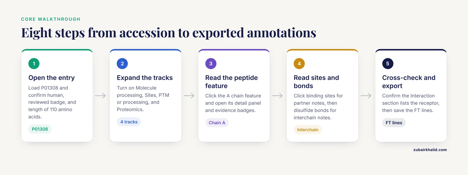
This is the core walkthrough. It uses human insulin, accession P01308, because it is the cleanest single example of a peptide whose interactions depend on processing features. The same eight steps work for any entry.
Step 1. Open the entry
Go to www.uniprot.org and enter the accession P01308 in the search box, or go directly to www.uniprot.org/uniprotkb/P01308. Confirm three things on the loaded page: the organism is human, the reviewed badge is present, and the length shown near the sequence is 110 amino acids for the canonical precursor.
Step 2. Locate the feature viewer
Scroll to the Features section. The viewer renders the sequence as a horizontal bar with stacked tracks grouped by category, including Molecule processing, Sites, PTM or processing, Variants, Structure, and Proteomics. Tracks are collapsed or expanded depending on the current interface state, so click a category header to expand it if the track you want is hidden.
Step 3. Turn on the peptide-relevant tracks
For peptide interaction work, enable these tracks and ignore the rest for the moment.
- Molecule processing, which shows the signal peptide, the chains, and the peptide features.
- Sites, which shows binding sites with partner notes.
- PTM or processing, which shows disulfide bonds and cross-links.
- Proteomics, which shows experimentally observed peptides if the entry has them.
Each track header includes a count of features in that track. A track with zero features displays as empty, which is itself a finding. If your protein of interest shows no binding site track content, the curated interaction may live only in the Interaction section or in a cross-referenced database, not as a positional feature.
Step 4. Click a peptide feature and read the detail panel
Click the peptide feature covering the A chain region. The panel that opens shows the feature type, the start and end positions, a label such as "Chain A," and evidence badges. Evidence badges are the colored tags that link the annotation to its evidence type, for example experimental evidence with a publication, evidence by similarity from an ortholog, or predictive evidence from a sequence model. Clicking an evidence badge expands the source reference. This is where you separate a measured fact from a propagated one.
For insulin, the processing layout you should see, reading left to right on the sequence, is a signal peptide at the N-terminus, then the B chain, then the C-peptide, then the A chain. The mature active hormone is the two chains held together by disulfide bonds, and the C-peptide is the removed connector. That single visual sentence, read off the viewer, is the entire maturation story.
Step 5. Read the interaction annotations on the site features
Click each binding site marker. For entries where a peptide binds a protein partner, the note on the binding site names the partner and often the region involved. The note text tells you what interacts. The position tells you where. The evidence badge tells you how we know. If the note says the residue is involved in binding a specific molecule, that statement, not the mere existence of the marker, is the interaction claim you can carry into your own analysis.
Step 6. Check the disulfide bonds that hold the peptide together
Open the PTM or processing track and click the disulfide bond features. For a multi-chain peptide like insulin, the interchain bonds are the ones that explain why two separate chains behave as one molecule. The note on each bond tells you whether it is interchain and names the connected chains. This is a recurring pattern in peptide biology, from insulin to many other processed peptide hormones, and the feature viewer is the fastest way to see it.
Step 7. Cross-check with the Interaction section
Scroll to the Interaction section of the entry page. This section is a table, not a graphic. Each row lists a partner protein with its accession, the interaction property if any, and the number of curated publications supporting the interaction. UniProt imports these binary interactions from partner resources such as IntAct and curates its own records as well. For insulin, the partner you expect to find is the insulin receptor, and the receptor entry carries the reciprocal record. Cross-checking the reciprocal direction is a quick reliability test. If an interaction appears on one side and not the other, treat it with more caution.
Step 8. Export the same annotations so your visual reading is reproducible
The feature viewer is for reading. The flat file and JSON exports are for keeping. Run this command.
curl -s https://rest.uniprot.org/uniprotkb/P01308.txt -o P01308.txt
grep -E "^FT   (SIGNAL|PROPEP|PEPTIDE|CHAIN|DISULFID|CROSSLNK|BINDING|SITE)" P01308.txt
The FT lines are the feature table, the machine-readable equivalent of the viewer. A representative excerpt, with the caveat that you should always read live coordinates from the current entry because annotations are updated between releases, looks like this.
FT   SIGNAL          1..24
FT                   /evidence="ECO:0000250|PROSITE-ProRule:PRU..."
FT   CHAIN           25..54
FT                   /note="Chain B"
FT   PEPTIDE         55..89
FT                   /note="C-peptide"
FT   PEPTIDE         90..110
FT                   /note="Chain A"
FT   DISULFID        31..96
FT                   /note="Interchain (between B7 and A7)"
If the grep returns nothing, the most likely cause is a missing entry, a network error, or a filter that does not match this entry's feature types. Re-run the curl alone and check that the file starts with ID P01308;.
Feature Types That Matter for Peptide Interactions
The table below summarizes the feature types you will encounter, what each one asserts, and why it matters when the question is peptide interaction.
| Feature type | What it asserts | Why it matters for peptide interactions |
|---|---|---|
| Signal peptide | N-terminal targeting segment, cleaved during secretion | Locates where the mature peptide starts, sets the coordinate frame |
| Propeptide | Cleaved segment, often keeping the peptide inactive until removed | Explains activation state and processing dependencies |
| Peptide | Mature active peptide shorter than a standard chain | Identifies the bioactive molecule itself |
| Chain | Mature polypeptide chain from a precursor | Multi-chain peptides require several chain features plus bonds |
| Disulfide bond | Covalent cysteine-cysteine link, interchain or intrachain | Holds multi-chain peptides together, frequently essential for binding |
| Cross-link | Covalent link other than disulfide, such as isopeptide | Marks covalent linkage in peptide-derived structures |
| Binding site | Residues involved in binding another molecule | The positional interaction annotation, with partner named in the note |
| Site | Any site of interest with a descriptive note | Catch-all for interaction-relevant residues without a dedicated type |
| Region | Extent of a region of interest | Context for where a binding region sits within a domain |
| Variant | Natural sequence variation | Tests whether variation hits an interaction interface |
| Mutagenesis | Experimentally altered residue and its measured effect | Direct evidence for residue roles in binding |
| Proteomics peptide (PEFF) | Experimentally observed peptide from mass spectrometry | Evidence the region is expressed and detected, not an interaction claim |
The single most useful distinction in that table is the last row. Peptide evidence features answer "was this sequence seen," while binding site features answer "what does this residue touch." Keep the two separate in your notes and your scripts.
Reading Interaction Annotations Correctly
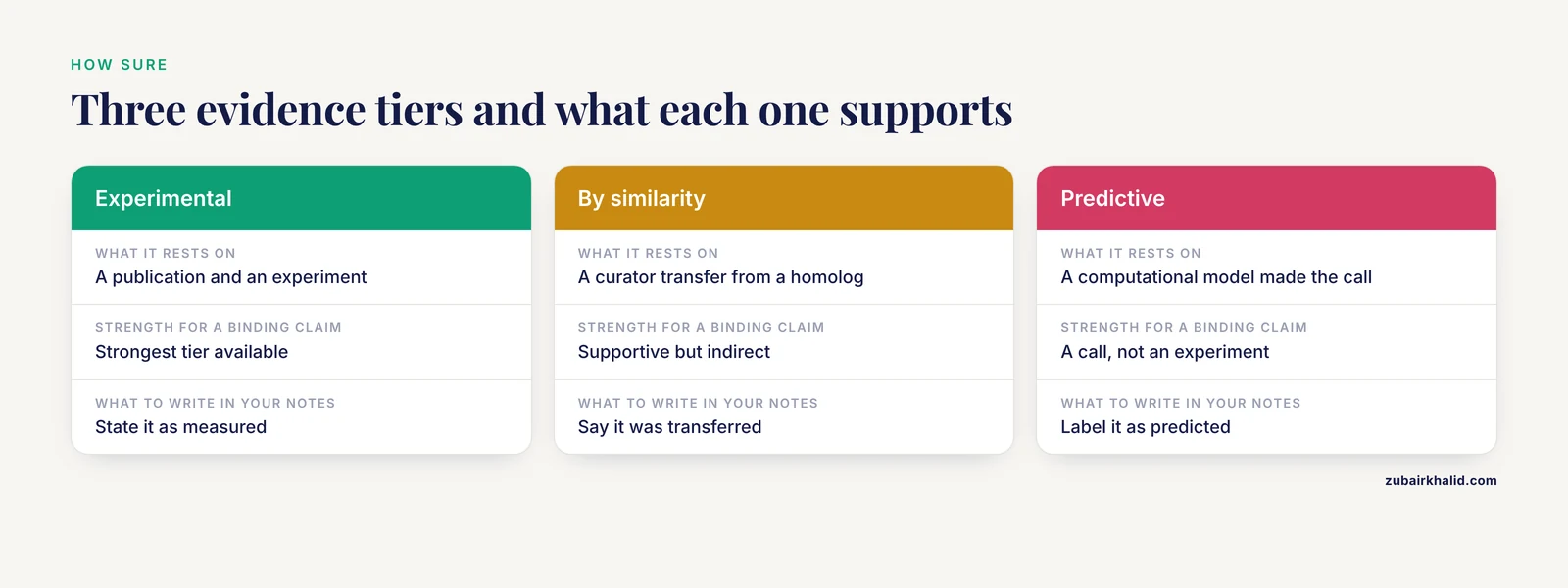
Three layers of information combine to make an interaction annotation trustworthy, and the feature viewer gives you all three if you look.
The positional claim. The start and end coordinates on the canonical sequence. Always verify which sequence version the coordinates refer to, because the entry header lists a sequence version number that increments when the sequence changes. A coordinate from an old export can point at the wrong residue after a sequence update.
The partner claim. The text in the note or the row in the Interaction section. UniProt writes binding site notes that name the partner molecule and, where known, the role of the residue. A note is curated language, so treat it as an assertion with an evidence trail rather than a guess.
The evidence claim. The ECO evidence code and its source. Experimental evidence with a publication is the strongest tier. Evidence by similarity means a curator transferred the annotation from a homolog. Predictive evidence means a computational model made the call. None of these tiers is automatically wrong, but each supports a different strength of conclusion, and any downstream claim you write should match the tier you actually have.
The Interaction section deserves one more paragraph because people underuse it. It is a structured, reciprocal, publication-counted record of binary interactions. For peptide work the practical pattern is to read the peptide entry for its processing features, then read the receptor entry for its binding site features, then confirm that the Interaction sections of both entries list each other. When all three agree, you have a solid interaction annotation. When they disagree, the disagreement is usually a curation scope difference, such as an interaction recorded at the family level on one side.
Command-Line and Scripted Access
For anything beyond a single lookup, script the API. The REST endpoint pattern is simple. Replace the accession and choose a format suffix of .txt for the flat file or .json for structured data. Both are documented by UniProt and stable for scripting.
A compact Python example extracts every feature relevant to peptide interactions and prints one line per feature.
import json
import urllib.request

acc = "P01308"
url = f"https://rest.uniprot.org/uniprotkb/{acc}.json"
data = json.load(urllib.request.urlopen(url))

interesting = {"Peptide", "Chain", "Signal", "Disulfide bond", "Cross-link", "Binding site", "Site"}
for feat in data["features"]:
    if feat["type"] in interesting:
        start = feat["location"]["start"]["value"]
        end = feat["location"]["end"]["value"]
        desc = feat.get("description", "")
        print(f"{feat['type']}\t{start}..{end}\t{desc}")
Expected output is a tab-separated block, one line per feature, for example lines beginning with Peptide, Chain, Disulfide bond, and Binding site followed by their coordinates and descriptions. The JSON feature objects also carry an evidences list with ECO codes and source identifiers, which is the programmatic equivalent of the badges you saw in the viewer.
To filter in a shell pipeline instead, jq does the same job.
curl -s https://rest.uniprot.org/uniprotkb/P01308.json \
  | jq -r '.features[] | select(.type=="Peptide" or .type=="Disulfide bond") | [.type, .location.start.value, .location.end.value, .description] | @tsv'
To pull features for many accessions at once, loop over a file of accessions and be polite to the service by pacing requests.
while read acc
do
  curl -s "https://rest.uniprot.org/uniprotkb/${acc}.json" -o "${acc}.json"
  sleep 1
done < accessions.txt
The one-second sleep keeps a batch of a few hundred accessions within polite use. For thousands, use the streaming endpoint documented by UniProt, which supports retrieving many entries in a single request with a query and a fields selector, rather than hammering the per-entry endpoint in a loop.
Worked Example, Insulin as a Peptide Interaction Case
Walking the full example end to end shows how the pieces fit. Human insulin, P01308, is a 110-residue precursor. The feature viewer shows four processing features. The signal peptide occupies the N-terminus and is removed during secretion. The B chain and the A chain are the mature hormone components. The C-peptide between them is excised during proinsulin processing in the secretory pathway. Three disulfide bond features connect the cysteines, two of them interchain, holding the A and B chains into one functional molecule.
The interaction question, "what does insulin bind," is answered in two places. The Interaction section lists the insulin receptor as a curated binary interaction partner. The receptor's own entry annotates binding and functional consequences on the receptor side. A rigorous peptide interaction reading always takes both sides, because the peptide entry tells you what the ligand is and the receptor entry tells you where and how it binds.
The mutagenesis and variant tracks close the loop. If a residue change sits inside a binding site note or disrupts a disulfide-bonded cysteine, the viewer lets you see that overlap in seconds without writing a line of code. That visual overlap check is the everyday use case for the viewer in peptide work.
Practical Tips for the Feature Viewer
These tips come from repeated use of the viewer in annotation-quality work.
- Read the sequence ruler before quoting coordinates. The viewer numbers residues from the first residue of the displayed sequence. If you switched to an isoform view earlier in the session, switch back and confirm, because quoting coordinates from the wrong frame is the fastest way to invalidate a downstream design, for example a peptide synthesis order.
- Use evidence badges as a sorting key, not decoration. When a track shows many features, expand the two or three with experimental evidence first. This gives you the measured core of the annotation before the predicted additions.
- Check the reciprocal entry for every interaction. Interaction records are curated on both sides. Two minutes on the partner entry catches most one-sided or scope-mismatched records.
- Download features before you need them. The download options on the entry page and the REST API give you a snapshot. If a reviewer or colleague questions a residue number months later, your snapshot resolves it.
- Treat the proteomics peptide track as a coverage layer. When you are deciding whether an annotated peptide interaction region has experimental support at the protein level, observed peptides overlapping the region are supportive context. They are not proof of the interaction, and they do not substitute for the binding site annotation.
- When a feature has no note, follow the cross-references. The entry page cross-references to structure and interaction databases often hold the depth the feature viewer compresses. A binding site with a sparse note usually has a richer record one click away.
- For mutation-focused questions, pair the viewer with a mutation-mapping tool. The Human Protein Mutation Viewer accepts mutations as UniProt accessions with HGVS names, as genome coordinates, or as FASTA-derived input, and it renders an interactive cartoon that relates mutations to sequence-architectural features including domains, targeting signals, and post-translational modification sites, with a tree view of additional information and a simple 3D mapping to known PDB structures where available [1]. Used after the feature viewer establishes which features matter, it answers the question the viewer does not, namely what a specific sequence change does to those features.
Common Mistakes and Limitations
These are the errors that actually happen in day-to-day work, each with its symptom and fix.
Confusing peptide evidence with peptide interactions. The Proteomics track shows peptides observed by mass spectrometry. It does not mean those peptides interact with anything. Symptom, a script that reports "interacting peptides" because it selected the wrong feature type. Fix, filter on Binding site and Site for positional interaction claims and read their notes.
Quoting coordinates from an isoform view. The viewer supports isoform selection, and coordinates change between isoforms. Symptom, a residue number that lands in the wrong domain when you check it against the canonical sequence. Fix, record which sequence the coordinates refer to and convert if needed, using the isoform sequence alignment on the entry page.
Reading a stale export. UniProt entries change between releases. Symptom, a feature list from an old file that no longer matches the live viewer. Fix, re-fetch before every final analysis and record the retrieval date and sequence version in your results file.
Trusting an interaction from one side only. Symptom, a partner listed in one entry's Interaction section with no reciprocal record. Fix, check the partner entry, check the publication counts, and check IntAct through the cross-references before relying on it.
Ignoring evidence tiers. Symptom, a claim written as experimentally proven when the underlying badge is evidence by similarity. Fix, expand every evidence badge you rely on, or read the evidences field in the JSON, and match your written conclusion to the tier.
Assuming absence of a feature means absence of interaction. Symptom, concluding a peptide does not bind its receptor because the binding site track is empty. Fix, search the Function text, the Interaction section, and the cross-referenced interaction resources. UniProt features are one representation of curated knowledge, not the whole of it.
Overwriting files in batch downloads. Symptom, a loop that saves every accession to the same filename, leaving one file behind. Fix, name each output with the accession, as shown in the loop above.
Miscounting mature peptide boundaries. Symptom, designing a construct from precursor coordinates that include the signal peptide or C-peptide. Fix, always subtract the processing features before any design step, and verify the final numbering against the chain and peptide feature labels.
A final limitation belongs here rather than in a separate caveat box. The feature viewer and the underlying annotation describe proteins at the level of curated evidence. Individual experimental questions, especially those that will drive wet-lab design or translational claims, need domain-expert review of the primary literature behind each annotation, and no viewer output substitutes for that review.
Putting the Annotations in Context With Mutation-Centered Tools
The feature viewer answers where interactions are annotated. A complementary class of tools answers what a sequence change does to the annotated architecture. The Human Protein Mutation Viewer is built for that question. It accepts protein mutations as UniProt accessions with mutations expressed in HGVS nomenclature, as genome coordinates, or from FASTA sequences, and it produces an interactive cartoon that places the mutations relative to sequence-architectural features selected for their relevance to mutation interpretation, including globular domains, targeting signals, and post-translational modification sites [1]. Clicking a sequence feature in its cartoon expands a tree view with additional detail, including multiple sequence alignments of conserved domains and a simple 3D viewer that maps the mutation onto known PDB structures when structures exist [1]. The cartoon is also correlated with a multiple sequence alignment of similar sequences from other organisms, which helps distinguish a residue conserved for a functional reason from one free to vary [1].
The division of labor is straightforward. Use the UniProt feature viewer to establish the peptide's processing, its positional interaction annotations, and its evidence. Use a mutation-oriented viewer when variants or engineered changes enter the question, because those tools are designed to show feature-level consequences of residue changes rather than the features alone. For entries where a mutation is likely to have a straightforward interpretation, such as a point change disrupting a well-understood targeting signal, the cartoon-based readout is quick and direct [1].
Frequently Asked Questions
What is the difference between a peptide feature and a peptide interaction in UniProt?
A peptide feature marks a stretch of sequence, such as a mature peptide, a proteomics-observed peptide, or a cleaved propeptide. A peptide interaction is an annotation about binding or contact, such as a binding site note naming a partner or a row in the Interaction section. The feature viewer displays both kinds of information on the same graphic, but they are different claims and you should never report one as the other.
Where do UniProt's peptide interaction annotations come from?
They come from curated literature, structural data, and imported records from partner resources such as IntAct, each attached to features with ECO evidence codes. Every binding site note and interaction row traces back to an evidence type and, where present, a specific publication you can open from the entry page.
Can I view peptide interactions without installing any software?
Yes. The feature viewer, the Interaction section, and the evidence panels all run in a web browser on the UniProt entry page. You only need command-line tools such as curl, jq, or Python when you want to export annotations or process many entries in a batch.
Why do feature coordinates sometimes differ between isoforms and the canonical sequence?
Because the viewer positions features on whichever sequence is displayed, and isoforms differ by exon-level sequence changes. The default view uses the canonical sequence, and most positional annotation refers to it. If you work with a specific isoform, check the isoform sequence and convert coordinates rather than assuming they transfer.
How current is the data in the feature viewer?
UniProt updates continuously and the entry page reflects the current release at the time you load it. Because of that, always note the accession, sequence version, and retrieval date for anything you record, and re-fetch features before a final analysis instead of reusing old exports.
How do I download feature annotations for many UniProt entries at once?
Loop over your accession list calling the REST API per entry, or use UniProt's streaming search endpoint to retrieve many entries in one request with a query and a fields selector. Save each accession to its own file and pace your requests so you stay within polite use of the public service.
What do the evidence badges on features mean?
They are ECO evidence codes that classify how the annotation is supported, with the main tiers being experimental evidence, evidence by similarity from a homolog, and predictive evidence from computational models. Expanding a badge shows the source, including publication identifiers when the annotation is literature-based.
When should I use a different tool instead of the feature viewer?
Use the feature viewer for reading positional features and interaction annotations. Use structure databases when you need 3D context, interaction databases when you need full interaction networks, and mutation-mapping tools such as HPMV when the question involves sequence changes and their effect on domains, signals, and modification sites [1].