How to Choose Between UniProt Expression and Localization Data
By Dr. Zubair Khalid, DVM, MS, PhD ·

UniProt expression annotations tell you where in the body, and under which conditions, a protein is found. UniProt localization annotations tell you which subcellular compartment the protein occupies once it is made. The two answer different questions, and choosing the wrong one for your design decision is one of the most common avoidable errors in protein-focused experimental planning.
The rule of thumb is simple. If your decision is about selection, meaning which proteins, tissues, samples, or candidate genes you will work with, start with UniProt expression data. If your decision is about access and manipulation, meaning where the protein physically sits and how you will reach it, tag it, immunocapture it, or compartment-enrich it, start with localization data. Pipelines that need both work best when expression narrows the candidate list first and localization then fixes the assay format, because that order keeps the expensive filtering step (localization with evidence checking) applied to the smallest possible set.
This guide is a practical UniProt database tutorial for experimental design. It explains what each annotation type actually contains, compares them side by side across the criteria that decide real choices, walks through worked scenarios drawn from published workflows, and lists the mistakes that burn the most time.
The two annotation types, explained from first principles
A UniProtKB entry is organized into labeled sections, and "Expression" and "Subcellular location" are two separate sections of the same entry. Confusing them at the entry level is rare. Confusing them at the design level is common, because both describe "where a protein is found." The difference is scale and mechanism.
What the Expression section contains
The Expression section of a UniProt entry carries qualitative statements about protein abundance in biological contexts. Its main fields are:
- Tissue specificity. Statements such as which tissues or cell types show detectable expression, which show enhanced expression, and which show absent or low expression. For human proteins, a substantial share of this content is imported from large profiling resources rather than from single papers.
- Developmental stage. Statements about expression during development, aging, or life-cycle stages, common in model organism entries.
- Induction. Statements about expression changes in response to stimuli, such as drugs, cytokines, stress, or growth conditions. This field matters when you plan a treatment experiment, because a protein annotated as inducible may be near zero in your untreated baseline sample.
Read every expression line as a curated qualitative claim, not a measurement. UniProt does not store normalized abundance values. If a line says a protein is "expressed at high level in liver," that is a synthesis of cited evidence, not a fold-change you can put into a power calculation. When you need numbers, use the cross-references in the entry (the Human Protein Atlas and similar resources are linked from the DR lines) to reach the underlying quantitative datasets.
Also note what the Expression section does not cover. It says nothing about how easy the protein will be to express recombinantly, how soluble it will be, or whether your construct will express well in E. coli or CHO cells. That confusion is a real and frequent mistake, and it is covered in the limitations section below.
What the Subcellular location section contains
The Subcellular location section carries compartment assignments. Its main content is:
- Location terms. Standardized compartment names such as Nucleus, Cytoplasm, Mitochondrion, Endoplasmic reticulum, Cell membrane, and Secreted. A single protein can carry multiple terms, and entries frequently note that a protein "shuttles" between compartments or translocates in response to a stimulus.
- Topology. For membrane proteins, the entry specifies whether the protein is single-pass or multi-pass, whether it is type I, type II, or type III, and which terminus faces the extracellular side or the organelle lumen. This is the single most design-relevant piece of information for anyone planning epitope tags, surface staining, or extracellular-domain assays.
- Sequence features that explain the localization. The feature table carries SIGNAL (signal peptide), TRANSMEM (transmembrane helices), TOPO_DOM (topological domains), DOMAIN, and related features with residue ranges. A localization claim you can trace back to a signal peptide at residues 1 to 24 is far more actionable than a bare term.
UniProt assigns subcellular locations at three evidence levels, and the entry itself tells you which level applies. Some locations are supported by direct experiments reported in the literature. Some are imported from curated external resources. Some are predicted, most often by similarity to annotated homologs. The annotation text marks the source, and the evidence tags on each line tell you how strong the support is. Treating a predicted location as if it were experimentally confirmed is one of the highest-risk habits in this entire workflow, because downstream decisions like tag placement or compartment enrichment can be wrong from the first step.
Evidence strength, review status, and why they matter before you design anything
UniProtKB contains two sections. Swiss-Prot entries are reviewed, manually annotated, and carry rich, sourced expression and localization content. TrEMBL entries are unreviewed and carry largely computationally derived annotation. The scale difference explains why both exist. A charge-distribution analysis of translation covered 551,705 reviewed proteins totaling roughly 180 million 30-residue protein segments drawn from the Swiss-Prot section of the UniProt database, which gives a sense of the curated corpus available across organisms [1].
For experimental design, the working rule is to make decisions on reviewed entries whenever they exist for your organism, and to treat unreviewed entries as hypothesis-generating only. Within a reviewed entry, check the evidence tags on each expression and localization line. Experimental evidence outranks imported curation, which outranks prediction. If the localization line you need is predicted only, and the decision it drives is expensive (an animal study, a large cohort, a synthetic construct), spend the time to verify it against the primary literature or an orthogonal resource before committing.
How to pull expression and localization data out of UniProt
The website covers exploration. For design at scale, use the REST API and the ID-mapping tool. Three patterns cover most needs.
Targeted search for TSV output. To pull every reviewed human protein annotated as secreted, together with its localization and tissue specificity text:
curl "https://rest.uniprot.org/uniprotkb/search?query=(organism_id:9606) AND (reviewed:true) AND (cc_subcellular_location:Secreted)&fields=accession,id,protein_name,cc_subcellular_location,cc_tissue_specificity&format=tsv&size=500" -o secreted_human.tsvBuild queries incrementally in the web interface first, where the autocomplete shows the valid field values, then port the query into curl or a Python requests call. Field names in the query mirror field names in the output, so a query built around cc_subcellular_location and cc_tissue_specificity returns columns you can filter directly in a spreadsheet or pandas.
Full-proteome download for search databases. Mass spectrometry pipelines need a complete, versioned FASTA. The stream endpoint returns the whole set:
curl "https://rest.uniprot.org/uniprotkb/stream?query=(organism_id:9606) AND (reviewed:true)&format=fasta" -o human_swissprot.fastaDecide before downloading whether you want reviewed-only or reviewed-plus-isoforms, write the release version and date into your methods, and never mix releases between discovery and validation runs.
ID mapping. When your candidate list arrives as gene symbols from a microarray or RNA-seq analysis, map identifiers through the "Retrieve/ID mapping" tool at uniprot.org/id-mapping or the corresponding REST endpoint before joining expression or localization annotations. Gene-symbol joins are the leading source of silent one-to-many errors in this workflow, because several symbols map to multiple entries with different localizations.
Side-by-side comparison table
This table is the core decision aid. Match your design question to the criterion row, not to whichever column sounds more familiar.
| Criterion | UniProt Expression annotations | UniProt Localization annotations |
|---|---|---|
| Core question answered | Where in the body, when in development, and under which conditions is this protein present | Which compartment does this protein occupy, and how does it get there |
| Typical content | Tissue specificity, developmental stage, induction by stimuli | Compartment terms, membrane topology, signal peptides, transmembrane helices, domains |
| Form of the data | Qualitative statements with evidence tags | Structured terms plus residue-level features with evidence tags |
| Main data sources | Manual curation plus imports from large profiling resources | Manual curation, curated imports, homology-based prediction |
| Strength of typical evidence | Mixed, mostly imported and literature-based, always qualitative | Mixed, spans direct experiment to prediction, residue features are the most checkable part |
| Best suited to decide | Which tissues or samples to collect, which candidate genes survive a filter, whether a baseline will be nonzero | Tag terminus choice, surface-accessibility assays, compartment enrichment, secreted-protein screening, topology-dependent assay design |
| Poor fit for | Quantitative abundance values, cell-to-cell variation within a tissue, disease-state changes not already curated | Predicting expression levels, deciding sample type, any abundance-based prioritization |
| Format inside the entry | Free-text annotation lines in the Expression section | Structured terms plus numbered sequence features |
| Natural downstream partners | Cross-referenced quantitative resources, GEO-style datasets, spectral libraries | Structure databases, conservation mappers, topology viewers |
| Key risk when misused | Treating "expressed in X" as a fold-change or a threshold | Treating a predicted or isoform-specific location as universal and confirmed |
Design options and their pros and cons
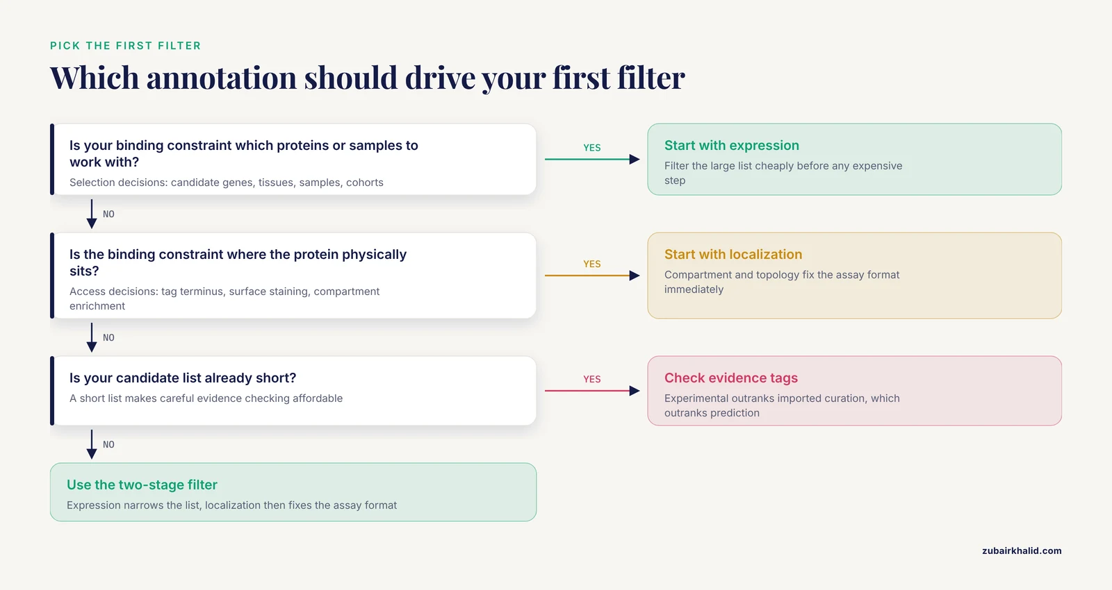
There is no single best UniProt expression and localization experimental design. The right option depends on which decision is binding first. These five options cover the choices that recur in real projects, each with its pros and cons stated plainly.
Option 1: Expression-first candidate selection
You start from a broad gene list, apply expression annotation as the first filter, and proceed with whatever survives.
- Pros. Cheap, fast, and applied early, so it shrinks the list before any expensive step. Expression annotation is usually present for well-studied organisms, and tissue-specificity statements are intuitive to apply even for newcomers learning how to use UniProt.
- Cons. Qualitative thresholds invite inconsistency across a team, because one analyst's "enhanced in" is another's "detected in." Expression at the tissue level can hide cell-type specificity, and annotations lag behind the conditions you may care about.
Use this option when the candidate list is large (hundreds or more), the downstream steps are costly, and your key requirement is only that the protein is plausibly present in your sample.
Option 2: Localization-first candidate selection
You start from a compartment. For example, you pull all reviewed proteins annotated as Secreted, or all single-pass type I membrane proteins with an extracellular N-terminus, and only then ask whether your samples express them.
- Pros. The filter is more decisive than expression filtering. A secreted protein is assayable in plasma regardless of what tissue produced it, so localization fixes the assay class immediately. Topology annotations directly determine whether an antibody or tag will reach its target.
- Cons. Compartment membership says nothing about abundance, so you can assemble a beautiful list of secreted proteins that are vanishingly rare in the fluid you plan to assay. Multi-compartment proteins complicate interpretation, and predicted locations require verification.
Use this option when the physical context of the protein is the binding constraint on your experiment, which is the case for secretome studies, surfaceome studies, and organelle proteomics.
Option 3: The two-stage filter, expression then localization
This is the workhorse design and the best UniProt expression and localization experimental design for most biomarker-style projects. Stage one applies expression criteria to a differential or tissue-specific list. Stage two applies localization criteria to the survivors.
- Pros. Each filter is applied to the smallest possible set, which is where careful evidence-checking is affordable. The order matches the logic of the experiment, because expression decides whether the protein is present where you sample, and localization decides what you can do about it.
- Cons. Order matters, and reversing it wastes effort. Localization-first on a huge list buries you in compartment assignments for proteins that fail expression anyway. The two-stage design also depends on both annotation types being present for every surviving candidate, so budget for manual curation of the final shortlist.
Option 4: Localization plus structure-aware engineering
When the experiment involves physically modifying the protein, localization information extends into sequence features and structure. Tag placement is the classic case.
- Pros. Residue-level features let you reason about the termini, which are the only practical attachment points for short affinity tags. Tools built on this logic accept a UniProt identifier directly, retrieve conservation and structural context, and produce a concrete recommendation.
- Cons. It requires structure or reliable models, which not every protein has, and it adds analysis steps before you order a single primer. It is only worth the effort when tag artifacts are a credible risk for your protein.
Option 5: UniProt as the reference and interpretation layer for mass spectrometry
Here UniProt is not a filter at all. It is the search database against which spectra are identified and the annotation layer through which identified proteins are interpreted.
- Pros. A versioned, organism-correct UniProt database makes identifications reproducible and auditable, and the same entries then supply expression and localization context for the proteins you detect.
- Cons. Database scope is a real experimental parameter. Reviewed-only databases improve false-discovery control but miss proteins absent from curation, and the choice must be recorded and frozen before acquisition.
When expression data should drive your design
Two situations put expression annotations in the driver's seat, and both are common in translational proteomics.
Building a biomarker candidate list from expression
Expression-first filtering is the natural design when your starting point is a differential expression analysis and your endpoint is an assay in an accessible fluid. A published prostate cancer biomarker workflow shows the pattern in full. The team began with a gene expression dataset of 13 prostate tumor and 8 normal tissue samples from GEO, identified differentially expressed genes with thresholds of absolute log2 fold change above 1 and adjusted p below 0.05, then filtered the upregulated candidates for secretory proteins using annotations from the Human Protein Atlas and UniProt databases. Prioritization by receiver operating characteristic analysis selected candidates with area under the curve above 0.85, and the workflow culminated in peptide synthesis and ELISA validation of SPON2 and MSMB against patient and control sera [2].
Notice which annotation did which job. The gene-level differential analysis supplied the expression contrast. The UniProt and Human Protein Atlas annotations for secretion, a localization property, determined that the endpoint could be a blood-based immunoassay at all. The expression data drove selection, the localization data drove the assay format, and the pipeline failed without both. If you are choosing among uniprot expression localization experimental design options for a biomarker project, this two-stage pattern is the most defensible default, because every filter is applied to the smallest surviving set.
Choosing and documenting the search space for proteomics
In quantitative proteomics, the UniProt database is part of the measurement system, not just a lookup table. A lung cancer proteomics study illustrates the full chain. The researchers profiled tumor tissue from 351 patients with stage I non-small cell lung cancer by liquid chromatography-tandem mass spectrometry in data-independent acquisition mode with a 150-minute gradient, analyzed the data against the human UniProt database using DIA-NN version 1.8.1, and identified 4,260 differentially expressed proteins that entered a recurrence-prediction model alongside clinical features, validated in an external cohort of 103 participants [3].
Two design lessons follow. First, the search database choice and its version belong in your methods with the same rigor as the gradient length, because a different database changes identifications and downstream statistics. Second, expression and localization annotations from the same resource become the interpretation layer for the differential proteins, telling you whether a hit is plausibly tumor-derived, secreted, or membrane-associated and therefore whether it belongs in a follow-up assay.
When localization data should drive your design
Localization annotations dominate whenever the experiment touches the protein physically. Three cases cover most of them.
Affinity tag placement on recombinant proteins
A short 6xHis tag placed at the wrong terminus can damage expression, solubility, or function, and the risk is not symmetric between the N-terminus and C-terminus of a given protein. The Terminator software package was built to resolve exactly this choice, and its input design tells you how UniProt fits in. Terminator takes a UniProt identifier or a sequence, identifies related sequences, maps conservation from ConSurf onto structures from the PDB and SWISS-MODEL, and analyzes proximity to cavities and functional sites before recommending the N- or C-terminus for a 6xHis tag shorter than 15 residues. Against published experimental outcomes for short tags, its predictions of relative risk between the two termini were 86 to 100 percent accurate, and its predictions of absolute risk for modifying a given terminus were 92 to 93 percent accurate [4].
The design insight generalizes. Proximity of the terminus to surface cavities and functional sites predicted tag damage better than whether the wild-type terminus was buried in the structure. That means your localization reasoning should include the entry's domain and feature table, not only the compartment terms. This option is among the strongest of the uniprot expression localization experimental design options when protein engineering is part of the plan.
Sequence context at the termini and the translation consequences
Terminus choice has a second, earlier consequence that is easy to miss. As a nascent polypeptide passes through the ribosomal exit tunnel, roughly 30 residues of it are inside the tunnel at any moment, and positively charged stretches in that window can interact with the ribosome walls and slow translation. In an analysis of the reviewed UniProt corpus across organisms, negative stretches outnumbered positive ones, supercharged positive sequences with net charge of 14 or more were underrepresented in proteomes, and proteins were more positively charged at both termini, with ribosome profiling from yeast showing a nonlinear relationship between N-terminal charge and ribosome occupancy once charge became strongly positive [1].
For construct design, the practical reading is that the terminus you choose for a tag is also the terminus the ribosome sees first or last, and positive charge density near the termini is a real variable in translation. A His tag adds consecutive histidines, and histidine is positively charged at physiological pH, so the tag itself shifts the charge profile of the terminus you attach it to. None of this overrides the structural advice from tag-placement tools, but it belongs in the same decision, especially for proteins with already unusual termini.
Verifying binding sites and domain claims against an independent annotation
Any workflow that predicts interactions or functional sites benefits from an independent, curated check, and UniProt entries serve that role. In a hybrid quantum-classical modeling study of surfactant protein A binding to the SARS-CoV-2 spike protein, a graph-pruning algorithm on a quantum device eliminated chemically irrelevant atoms to isolate candidate binding residues, and the team then confirmed that the remaining residues contained the potential binding sites using the UniProt database before comparing dynamics simulations across force fields [5]. The pattern is transferable. When your in silico pipeline nominates residues, domains, or interfaces, the feature table of the reviewed UniProt entry is a fast, free sanity check that the nominated region corresponds to annotated biology rather than a computational artifact.
Three end-to-end worked scenarios
These scenarios assemble the pieces above into complete designs, stated as step sequences you can adapt.
Scenario A: Secreted biomarker discovery from a gene expression dataset
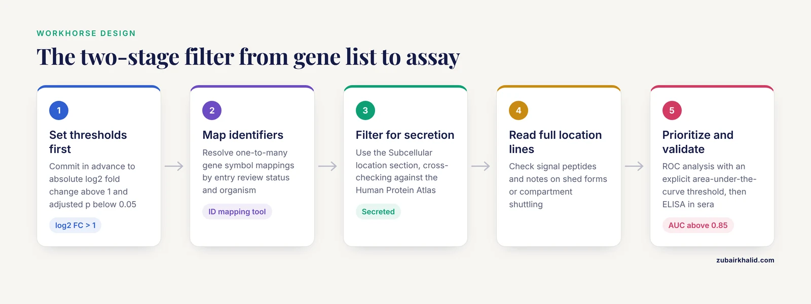
You have RNA-seq or microarray data comparing tumor and normal tissue, and your goal is a blood-based diagnostic.
- Run your differential analysis and set thresholds you commit to in advance, as the prostate workflow did with absolute log2 fold change above 1 and adjusted p below 0.05 [2].
- Keep upregulated candidates only if your assay concept requires elevated levels in the disease state.
- Map gene symbols to UniProt accessions through the ID-mapping tool, resolving one-to-many mappings by entry review status and organism.
- Filter survivors for secretory proteins using the Subcellular location section, cross-checking UniProt annotations against the Human Protein Atlas as that workflow did [2].
- For each remaining candidate, read the full localization lines, not just the term, checking for signal peptides and for notes about shed forms or compartment shuttling.
- Prioritize by diagnostic performance on your expression data, using ROC analysis and an explicit area-under-the-curve threshold.
- Validate the top candidates at the protein level in clinical samples, which in the published example meant peptide synthesis and indirect ELISA against patient and control sera [2].
The design succeeded because the expression filter ran first and the localization filter ran on a short list, and because the final assay format was chosen by localization rather than assumed.
Scenario B: Tumor proteomics from cohort to predictive model
You have tissue samples from a patient cohort and want protein markers of outcome.
- Freeze your search database decision before acquisition. Specify organism, review status, isoform inclusion, and release version, as the lung cancer study did in analyzing DIA data against the human UniProt database with DIA-NN version 1.8.1 [3].
- Match the chromatographic method to the depth you need, recognizing that the published study used a 150-minute gradient in data-independent acquisition mode [3].
- Run differential abundance analysis with your predeclared statistical model, integrating clinical covariates where relevant.
- Annotate every differential protein with UniProt expression and localization content before interpreting it, so that a membrane protein and a nuclear protein are not interpreted identically as tumor markers.
- Carry the annotated shortlist into validation in an independent cohort, as the published model did with 103 external participants [3].
Scenario C: A recombinant protein you can purify without breaking it
You need milligram quantities of an active enzyme or binding protein.
- Start at the reviewed UniProt entry. Read the Subcellular location section and the full feature table, flagging signal peptides, transmembrane regions, and domains near either terminus.
- Run a structure-aware tag-placement analysis such as Terminator, which accepts the UniProt identifier, maps conservation onto available structures, and recommends a terminus for a short 6xHis tag with high reported accuracy against experimental outcomes [4].
- Check the charge profile around the chosen terminus before finalizing the construct, remembering that strongly positive N-terminal stretches associate with slower elongation and that supercharged positive sequences are rare in natural proteomes [1].
- Order the construct with the recommended terminus, express under a standard system, and treat any solubility or activity deficit as a reason to revisit tag placement rather than the folding conditions alone.
Common Mistakes and Limitations
These are the errors that appear repeatedly in real project reviews, each with the fix.
- Reading qualitative annotation as quantitative. "Expression is enhanced in lung" is not a fold-change. For numbers, follow the entry's cross-references to the underlying profiling resources and use their quantified datasets.
- Ignoring evidence tags. An expression or localization line backed by direct experiment and one backed by prediction can look identical at a glance. Filter on the evidence level whenever the downstream step is expensive.
- Designing from unreviewed entries. TrEMBL annotation is largely automatic. If a reviewed entry exists for your protein, it wins. If only an unreviewed entry exists, verify before you commit resources.
- Assuming one localization covers all isoforms. UniProt entries annotate isoform-specific locations where they differ, and an N-terminally extended isoform can carry a signal peptide its canonical form lacks. Check the isoform lines before any topology-dependent decision.
- Treating compartments as exclusive. Many proteins are annotated in two or more locations, and some translocate on stimulus. A dual-annotated protein can appear in your nuclear fraction and your membrane prep, and that is not a contamination artifact by default.
- Confusing UniProt "Expression" with recombinant expression behavior. The Expression section describes endogenous biology. It will not tell you whether your His-tagged construct will express solubly in your host. Tag placement and construct design need the structure-aware approach described above [4].
- Letting the database drift during a project. UniProt updates continuously. Record the release version and date with your search database and annotation extracts, and do not swap releases between discovery and validation, or your identifications will not be comparable.
- Overriding localization with one conflicting observation. If your western blot of the cytosolic fraction finds a protein annotated as membrane-bound, suspect fractionation carryover or an isoform difference before rewriting the annotation.
- Forgetting that annotation lags biology. Condition-specific expression changes, especially in disease, are only in UniProt after they are curated from published work. Your own samples can and will differ from the annotation, and every individual experimental finding needs independent validation before it becomes the basis for further decisions.
Frequently Asked Questions
Should I filter candidates by expression first or localization first?
Filter by expression first in almost all biomarker-style designs, then apply localization to the survivors. Expression filters work on large lists cheaply, while localization filtering rewards careful evidence-checking that is only affordable on a short list. Reverse the order only when compartment membership is your hard constraint from the start, such as a secretome-wide survey.
Are UniProt subcellular location annotations experimentally confirmed?
Some are and some are not, and the entry tells you which. UniProt assigns locations with direct experimental support, by import from curated sources, or by prediction from homology, and each line carries evidence information. Check the evidence level before using a location for a design decision, because a predicted location and an experimentally supported location differ enormously in reliability.
Can I use UniProt expression data as quantification?
No. UniProt expression lines are curated qualitative statements about where and when a protein is found, not normalized abundance values. Use the entry's cross-references to reach quantitative datasets, and treat the UniProt line itself as a plausibility check and an interpretation layer rather than a measurement.
Should my mass spectrometry search database be Swiss-Prot or the full UniProtKB?
Reviewed-only Swiss-Prot entries reduce the search space and improve false-discovery control for well-annotated organisms, while adding unreviewed entries catches proteins absent from curation at the cost of a larger target space. Pick one, record the release version, and keep it identical across discovery and validation. Published quantitative proteomics workflows commonly analyze against the human UniProt database and report the tool and version alongside it.
How do I cite UniProt so my design is reproducible?
Cite the resource, the entry accessions you used, and the release version with its date. For REST or bulk downloads, record the exact query strings and field lists in your methods or repository, because annotations change between releases and your filters should be rerunnable byte for byte.
Can localization annotations change across conditions, and how do I plan for that?
Yes, and UniProt entries frequently say so directly, with notes about translocation on stimulation, cell-cycle-dependent redistribution, or stimulus-dependent secretion. Plan controls around it. If your protein is annotated as conditionally localized, a single-compartment readout in a single condition will underrepresent its biology, so design sampling or fractionation to capture the states your annotation warns you about.
Sources
- Protein charge distribution in proteomes and its impact on translation.
- Immunodiagnostic profiling of SPON2 and MSMB as biomarkers in prostate cancer for nanomaterial- based detection strategies.
- Proteomic profiling of early-stage non-small cell lung cancer identifies a high-performance protein signature associated with postoperative recurrence.
- Terminator: A Software Package for Fast and Local Optimization of His-Tag Placement for Protein Affinity Purification.
- SP-A binding to the SARS-CoV-2 spike protein using hybrid quantum and classical in silico modeling and molecular pruning by Quantum Approximate Optimization Algorithm (QAOA) Based MaxCut with ZDOCK.