Using UniProt Conserved Motifs to Guide Protein Engineering
By Dr. Zubair Khalid, DVM, MS, PhD ·

Conserved motifs are short, recurring sequence patterns, usually 3 to 30 residues long, that mark catalytic residues, ligand contacts, metal coordination sites, or structural hinges that evolution has kept intact across a protein family. UniProt records these patterns as explicit, queryable features on each protein entry, which makes them one of the most practical starting points for deciding which residues to mutate in a protein engineering project. They differ from domain annotations in a specific way. A domain tells you which region of the protein you are working in. A motif points at individual residues you might actually edit.
The rule of thumb is simple. If UniProt or a linked database already annotates motifs for your protein family, start with those annotations, confirm them with a multiple sequence alignment of reviewed homologs, and design mutations only after that confirmation. If the family is uncharacterized, do not trust any residue list until you have built your own evidence with profile hidden Markov models, structural predictions, and cross-species conservation analysis.
This article is a working UniProt database tutorial aimed at protein engineers. It walks through how to use UniProt to find conserved motifs, compares the main analysis options side by side, works through real scenarios drawn from the literature, and catalogs the mistakes that most often derail motif-guided engineering.
What UniProt Conserved Motifs Actually Are
UniProtKB, the core UniProt knowledgebase, contains two sections. Swiss-Prot holds entries reviewed by human curators with rich, standardized annotations. TrEMBL holds unreviewed entries annotated largely by automated pipelines. When you look at a reviewed entry, conserved motifs appear in the feature table under several feature keys:
- MOTIF: a short conserved sequence pattern of biological interest, often imported from PROSITE patterns or profiles.
- DOMAIN: the location of a named domain family, usually cross-referenced to Pfam, PROSITE profiles, or SUPERFAMILY.
- ACT_SITE: catalytic residues, annotated individually.
- BINDING and METAL: residues in direct contact with ligands or metal cofactors.
- SITE: any other interesting single site, such as a phosphorylation switch or a cleavage point.
These feature types are not interchangeable for engineering purposes. A MOTIF line tells you a pattern is conserved across a family. An ACT_SITE or METAL line tells you a specific residue has direct functional evidence. Mutating a catalytic residue usually kills activity, which is sometimes what you want, but more often you want to modify specificity, stability, or expression while keeping catalysis intact. Knowing which feature key you are reading is the first step in using UniProt conserved motifs for protein engineering rather than against your own goals.
Family-level resources linked from UniProt expand this picture. The EVEREST project, for example, built 20,029 domain families automatically from sequence alone using hidden Markov models, scanned over 3,000,000 UniProt and PDB sequences with them, and annotated 64 percent of all sequences and 59 percent of all residues [1]. The practical takeaway from that scale is that most proteins carry some computable family structure, but a meaningful fraction of residues fall outside any annotated family or motif, so absence of an annotation is never proof that a residue is unimportant.
A second takeaway concerns uncharacterized families. UniProt explicitly maintains families labeled UPF (unknown protein family), and these can still be conserved and functionally coherent. The UPF0235 family protein PF1765 from Pyrococcus furiosus, for instance, has a 1.3 angstrom crystal structure, remains monomeric and stable up to 95 degrees Celsius, and binds dsDNA, ssDNA, and RNA despite lacking any canonical nucleic acid binding domain [2]. Motif annotation alone would not have predicted that binding behavior, which is exactly why the comparison table later in this article puts motif features and structure-based evidence in separate rows.
How to Use UniProt to Find Conserved Motifs
This section is the hands-on core of the tutorial. It covers four skills: searching, reading entries, retrieving homologs, and working programmatically.
Searching UniProtKB for Your Protein
Start at the UniProt website and search by protein name, gene name, or sequence. If you have a sequence, paste it into the BLAST tab on the website. BLAST against Swiss-Prot first. A reviewed hit with curated motif features is worth more than a hundred unreviewed TrEMBL hits with predicted-only annotations. Use the Reviewed filter to restrict results to Swiss-Prot, and use the organism filters to narrow to taxa relevant to your engineering goal.
For batch work, the search interface builds a query string you can reuse. A query such as (protein_name:glutaminase) AND (organism_id:562) returns Escherichia coli glutaminase entries, and appending &format=tsv&fields=accession,id,protein_name,length returns a spreadsheet-ready table. Similar fielded searches work for any protein family you plan to engineer.
Reading the Feature Table
Open a reviewed entry and switch the feature viewer to the MOTIF, DOMAIN, ACT_SITE, and BINDING tracks. Read the feature notes, not just the positions. A MOTIF line with a PROSITE cross-reference usually links to a curated pattern page that lists the biological role of each conserved position and how often the pattern is documented to occur in true positives versus false positives. That per-position commentary is exactly the evidence you need to rank candidate mutation sites.
If you prefer plain text, download the entry in text format and inspect the feature lines directly:
curl "https://rest.uniprot.org/uniprotkb/Q8U052.txt"The UPF0235 protein PF1765 from Pyrococcus furiosus is a useful test case because its UniProt entry describes a family that is conserved across archaea, bacteria, and eukaryotes yet functionally uncharacterized [2]. Reading such an entry teaches you to distinguish what is curated from what is merely predicted, which matters enormously when you plan mutations.
Retrieving and Aligning Homologs
From any entry, use the BLAST tab to pull close and remote homologs, then download selected sequences in FASTA format. Align them with your aligner of choice, such as MAFFT or MUSCLE, and overlay the UniProt motif annotations on the alignment. UniProt's own Align tool does a quick pairwise version of this directly on the website.
For deeper homology searches, install HMMER locally and run iterative searching against a local Swiss-Prot or UniRef database:
jackhmmer --incE 1e-5 -N 5 query.fasta uniprot_sprot.fastaIterative search finds remote homologs that a single BLAST round misses, and those remote homologs are often the ones that reveal which motif positions are truly invariant versus merely typical. Invariant positions are your danger zone for mutation. Variable positions flanking invariant ones are usually your best engineering targets.
Working Programmatically with the REST API
Every UniProt search is also a REST call. The same query you build on the website works from the command line, which makes the whole motif-discovery workflow scriptable and reproducible:
curl "https://rest.uniprot.org/uniprotkb/search?query=(protein_name:glutaminase) AND (reviewed:true)&format=tsv&fields=accession,protein_name,organism_name,length"Reproducibility matters here for a practical reason. Motif annotations change as curation progresses, and a residue list you generated six months ago may not match today's entry. Saving the exact query, the date, and the output files in your project repository costs nothing and saves arguments later.
UniProt Conserved Motifs Protein Engineering Options Compared
There are five practical options for turning UniProt motif information into engineering decisions. They differ in evidence quality, effort, and the kinds of proteins for which they work. The table below is the core of this article, and each option gets its own section afterward.
| Approach | Main input | What you get | Strengths | Watch out for |
|---|---|---|---|---|
| Curated motif features | Reviewed UniProt entry, PROSITE links | Per-residue motif positions with functional notes | Fast, curator-verified, citable | Unreviewed entries have predicted-only annotations |
| Multiple sequence alignment | BLAST or jackhmmer homologs | Which motif positions are invariant vs variable across taxa | Reveals plasticity, guides safe mutation choices | Alignment quality depends on homolog selection |
| Profile HMMs from motifs | Curated seed alignment | A reusable family classifier with a numeric threshold | Scales to thousands of sequences, quantifies confidence | Threshold choice changes results dramatically |
| Structure and conservation overlay | AlphaFold model, PrankWeb, PDB | Mapping of motifs onto 3D context and predicted pockets | Distinguishes active-site motifs from structural ones | Predictions, not measurements, need validation |
| Language-model segmentation | Protein sequence only | Segment boundaries and functional-region labels | Works without any prior annotation | Newer evidence base, complements rather than replaces motifs |
A sixth, often underrated option is visualization. When your project involves a whole protein set rather than a single protein, tools that display feature architectures for many proteins at once change what you can see. ProFeatMap takes a user-supplied list of UniProt accession codes, extracts annotated features such as domains, repeats, disorder, and post-translational modification sites from UniProt or InterPro, and renders them as a customizable two-dimensional map that sorts proteins by homologous feature content [3]. That sort of map is how you notice, before any wet-lab work, that the feature you planned to engineer is duplicated in some family members and absent in others.
Option 1: Curated Motif Features in UniProt Entries
This is the fastest option and the right first stop. Open the reviewed entry, read the MOTIF, ACT_SITE, and BINDING annotations, follow the PROSITE and Pfam cross-references, and write down every residue the curators flagged. The entry for a reviewed enzyme typically gives you a short list of individually annotated functional residues plus one or more family motifs, which is a working hypothesis list on day one.
The limitation is coverage and specificity. Curated motifs exist mainly for well-studied families. For a family like UPF0235, conserved across all three domains of life yet uncharacterized, curated functional annotations are minimal and the motif list will be short [2]. In that situation, options 2 through 5 carry the weight.
There is also a subtlety worth internalizing early. Motif conservation at the pattern level does not guarantee conservation of every residue within the pattern. An analysis of 646 PELP1 proteins across eukaryotic genomes found the expected LXXLL and PXXP signaling motifs in most sequences, but the leucine and proline residues inside those motifs were not universally conserved [4]. If you treat a motif string as inviolable, you will under-engineer. If you check per-position conservation across homologs first, you will know which positions tolerate change and which do not.
Option 2: Multiple Sequence Alignments of Homologs
The alignment is where motif annotations meet reality. Pull 20 to 100 homologs spanning the taxonomic range relevant to your goal, align them, and color the UniProt motif positions. Three patterns matter:
- Positions identical in every homolog are load-bearing. Mutating them is a bet against a billion years of selection, and you should have a structural reason.
- Positions conserved within a subgroup but divergent between subgroups mark specificity determinants. These are prime targets when you want to alter substrate or ligand preference.
- Positions variable everywhere are candidate tolerance slots for stability or solubility substitutions.
An instructive example comes from work on parvalbumin-like proteins. Two sequences from the pathogenic fungus Trichophytum violaceum, UniProt IDs A0A178F775 and A0A178F7E4, were found to contain EF-hand domains with calcium-binding sites and conserved amino acid motifs characteristic of parvalbumins, a protein class otherwise associated with vertebrate muscle [5]. Multiple sequence alignment and phylogenetic analysis placed the fungal proteins among known parvalbumin-like sequences, and antigenicity prediction located antigenic sites on both sides of the calcium-binding site of the first EF-hand. For anyone engineering a parvalbumin-like scaffold, that result illustrates two alignment-derived lessons at once: EF-hand motifs can be recognized far outside their usual taxonomic home, and residues immediately flanking a functional motif can matter for properties, such as antigenicity, that the motif string itself says nothing about.
Option 3: Profile HMMs Built from Conserved Motifs
When your engineering project needs to classify many sequences, for example to pick which homologs to order as genes or to confirm that a designed variant still belongs to the target family, a profile hidden Markov model built around conserved motifs is the tool of choice. The workflow is: curate a seed alignment around the motifs of interest, build the HMM with hmmbuild, and search with hmmsearch at a threshold you can defend.
A published example shows how much the threshold matters. Researchers studying protein-tyrosine sulfotransferases built a hidden Markov model, TPST-HMM, specifically from the conserved motifs that bind the substrate PAPS and the ligand tyrosine in metazoan enzymes [6]. At an e-value threshold of 1e-5, the model identified more than 91.8 percent of metazoan TPSTs in UniProt. Tightening the threshold to 1e-20 dropped the metazoan identification rate to 83.9 percent and reduced identification of TPSTs in non-animal species to approximately zero, which turned the motif-based HMM into a phylogenetic specificity filter as well as a family classifier [6]. For engineering, the lesson generalizes. The same motif-derived model can serve two opposite goals depending on threshold: broad recall when you want candidate homologs from across the tree, and tight specificity when you want to guarantee that a variant you built still reads as a member of your target subfamily.
Option 4: Conservation and Binding-Site Predictions on Structures
Motifs gain meaning in three dimensions. Two residues can be adjacent in sequence but distant in structure, and a motif that looks like a binding site in a linear readout may turn out to form a structural core. Overlaying motif positions on an experimental structure, or on an AlphaFold model when no structure exists, resolves most of these ambiguities.
PrankWeb is a good practical anchor here. It predicts ligand-binding sites from structure, accepts a UniProt ID directly and uses the corresponding AlphaFold model when no experimental structure is available, and estimates evolutionary conservation through a pipeline built on the UniRef50 sequence database and the HMMER3 package [7]. It exposes a public REST API and ships as a Docker container, so the prediction step slots into the same scripted workflow as your UniProt queries. The combined output, motif positions plus predicted pockets plus per-residue conservation scores, is the evidence package that lets you argue a residue is a genuine functional target rather than a conserved-but-structural one.
The UniProt entry side has a matching tool. HPMV, the Human Protein Mutation Viewer, accepts mutations in HGVS nomenclature, genome coordinates, or FASTA sequences, and displays them in relation to sequence architecture features including globular domains, targeting signals, and post-translational modification sites, with an expandable tree that includes multiple sequence alignments of conserved domains and a simple 3D viewer mapping each mutation onto known PDB structures [8]. If your engineering project is interpretation-heavy, for example you are enumerating which point mutations plausibly disrupt a targeting signal versus which merely alter a surface residue, this mutation-to-feature mapping is the fastest structured view available.
Option 5: Language-Model Segmentation as a Complement
Protein language models now offer an annotation-free route to functional regions. Zero-shot Protein Segmentation uses embeddings from ProtT5 to predict boundaries of protein segments without training or fine-tuning any parameters, and its boundary predictions for the human proteome reproduce reviewed UniProt annotations better than established bioinformatics tools while categorizing over 200 of the most common UniProt annotation types, including folded domains, sub-domains, and intrinsically disordered regions [9]. Because the method does not depend on conserved patterns in primary sequence, it can propose functional regions where sequence-based motif search returns nothing.
Treat this as a complement, not a replacement. Sequence motifs carry direct, curator-vetted links to function that embeddings do not. But when a region of your protein shows strong structural conservation yet no recognizable motif, language-model segmentation is a legitimate way to flag that region for closer study before you commit mutagenesis resources.
Choosing the Best Option for Your Project
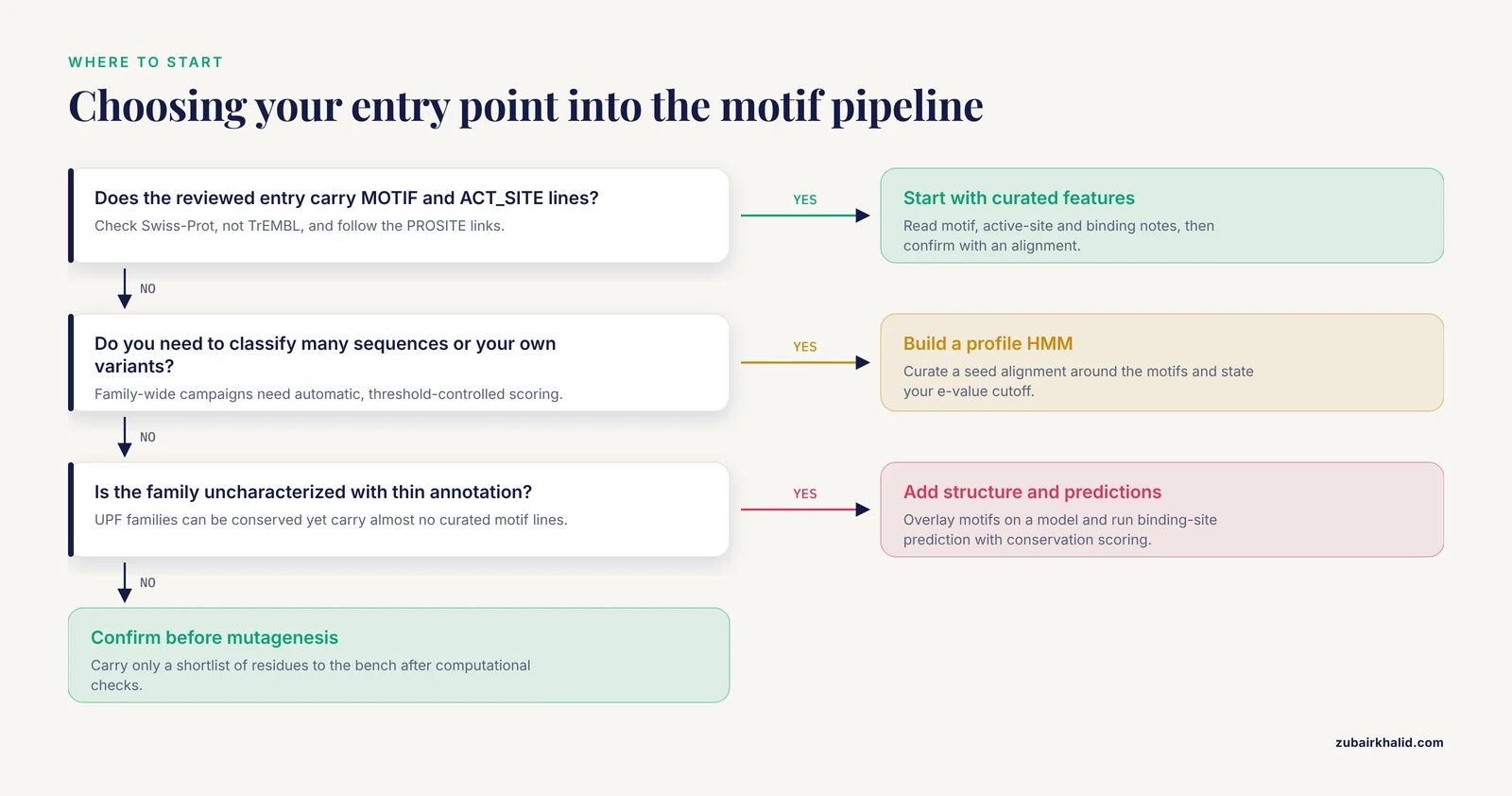
The five options are not competitors. They form a pipeline, and the best starting point depends on how much is already known about your family. Use these criteria to decide where you enter the pipeline.
- Annotation depth. If the reviewed UniProt entry has MOTIF and ACT_SITE lines with literature-backed notes, option 1 plus a confirming alignment handles most single-protein projects. If the entry is unreviewed or family annotations are thin, budget time for options 2 through 4.
- Scale. For one protein, an alignment and a structure overlay are enough. For a family-wide engineering campaign, build a profile HMM so that classification of new sequences, including your own variants, is automatic and threshold-controlled [6].
- Risk tolerance. Options 1 and 2 rest on curated evidence and your own alignment. Options 4 and 5 rest on predictions. Anything you learn from a prediction should be validated before it drives an expensive experiment, and predicted binding pockets from tools like PrankWeb are a hypothesis generator, not a assay result [7].
- Taxonomic scope. If you plan cross-kingdom engineering, expect motif-level behavior to vary. PELP1 motifs kept their architecture across 646 eukaryotic proteins even while individual motif residues drifted [4], and parvalbumin-like motifs surfaced in a fungal genome far outside the vertebrate context [5]. Both cases argue for per-position conservation checks before cross-species transfer of any motif assumption.
One more criterion deserves its own sentence: reversibility. Motif and alignment work is cheap, computational, and repeatable. Mutagenesis is not. Spend your uncertainty budget on the computational side, where iterating costs an afternoon, and carry only a shortlist of residues to the bench.
Worked Scenarios
Scenario 1: Engineering an Enzyme Whose Motifs Are Annotated
Suppose you want to engineer bacterial glutaminases for therapeutic applications. A published in silico analysis pulled 40 glutaminase sequences from UniProt across Escherichia and Bacillus species, ran homology searches, phylogenetic tree construction, motif and superfamily searches, and multiple sequence alignment [10]. The sequences fell into the serine-dependent beta-lactamase and penicillin-binding protein superfamily, phylogenetic analysis resolved two main clusters, and the common beta-lactamase motifs were distributed across the enzymes along with several non-common motifs [10]. The authors concluded that a conserved motif carrying the signature sequence of beta-lactamases could be considered for the genetic engineering of glutaminases, particularly given interest in their application in cancer therapy, while noting that stability and immunogenicity still need improvement [10].
The workflow lessons here transfer directly. First, superfamily identification reframes what motifs you should look for. Once you know your enzyme sits in the beta-lactamase superfamily, every curated beta-lactamase motif in UniProt becomes prior evidence for your protein. Second, the gap between common and non-common motifs across the two phylogenetic clusters is a map of subgroup-specific positions, which is exactly where substrate-specificity engineering usually pays off. Third, motif-guided engineering solves the catalytic framework problem, not the stability and immunogenicity problems, and the published analysis says so plainly [10].
Scenario 2: Building a Motif-Derived Classifier for a Subfamily
Suppose you are working on type II sulfotransferases and need to reliably tell protein-tyrosine sulfotransferases apart from other family members before engineering substrate preference. The TPST-HMM study shows the construction path: identify the conserved motifs that contact PAPS and the tyrosine substrate, curate an alignment around them, build the HMM, and tune the e-value threshold [6]. The resulting tool identified over 91.8 percent of metazoan TPSTs in UniProt at 1e-5, and at 1e-20 it became effectively metazoan-specific, identifying 83.9 percent of metazoan enzymes and almost nothing from other species [6].
For an engineer, that specificity profile is a feature, not a limitation. If you want to engineer a non-metazoan sulfotransferase, the near-zero hit rate at 1e-20 tells you the metazoan motif set does not describe your enzyme, so you should rebuild the HMM from your own subfamily rather than force-fit metazoan annotations. If you want your engineered variant to remain recognizably metazoan-like, hmmsearch at 1e-20 becomes a release criterion for your designs.
Scenario 3: An Uncharacterized Family With No Motif Annotations
Now the hard case. You are handed a protein from a UniProt family labeled uncharacterized, and motif search returns little. The UPF0235 family is the reference example. PF1765 from Pyrococcus furiosus yielded a 1.3 angstrom crystal structure with a compact alpha-plus-beta fold forming a pseudo beta-barrel, and DALI structural comparison found no significant similarity to proteins of known function [2]. AlphaFold3 models of homologs showed high structural conservation, RMSD under 1.63 angstroms, across all domains of life despite sequence identity of only 20 to 35 percent, and biochemical assays confirmed variable binding to dsDNA, ssDNA, and RNA despite the absence of canonical nucleic acid binding domains [2].
Three engineering lessons come out of this scenario. Structural conservation ran far ahead of sequence conservation, so low identity in an alignment should never be read as functional divergence. A family can hold onto a fold under strong selective pressure while its sequence motifs drift or stay cryptic, which means structure-based and prediction-based evidence can lead where motifs cannot [2]. And functional capability, here nucleic acid binding, can exist entirely outside canonical annotated motifs, which is the strongest argument for running options 4 and 5 even after motif work comes up empty.
Pros and Cons of UniProt Conserved Motifs for Protein Engineering
A balanced accounting before you commit a project to a motif-first strategy.
Pros:
- Curator-verified. Reviewed Swiss-Prot annotations carry literature evidence you can cite in a manuscript or a design dossier.
- Cheap and fast. Motif extraction is a web query or a single REST call, and it happens before any cloning, synthesis, or assay.
- Per-residue resolution. Motifs point at individual positions, which is the resolution mutagenesis actually operates at, unlike domain annotations.
- Transferable across tools. Motif positions feed directly into HMM construction, structural overlays, and mutation viewers [8].
- Scales to families. The same motif logic that annotates one entry underlies family resources covering tens of thousands of families and most residues in the database [1].
Cons:
- Coverage gaps. Uncharacterized families carry minimal motif annotation, and 36 percent of sequences lacked EVEREST family assignment even in a dedicated domain-cataloging effort [1].
- Pattern-level conservation is not position-level conservation. Motif strings persist while their internal residues drift, as shown for LXXLL and PXXP motifs in PELP1 [4].
- Catalysis bias. Motif annotations overweight catalytic and binding residues, and they underrepresent the stability, expression, and immunogenicity properties that engineering projects often target [10].
- Functional surprises outside motifs. Binding capability can exist with no canonical motif signature, as PF1765 demonstrated for nucleic acid binding [2].
- Threshold sensitivity. Any motif-derived HMM changes behavior substantially between e-value 1e-5 and 1e-20, so an unstated threshold is an unreproducible result [6].
Common Mistakes and Limitations
These are the errors that most often waste months of engineering time.
- Mutating the catalytic motif by default. A conserved motif tells you the residues are important, not that they are improvable. Before touching an ACT_SITE or core motif position, ask whether your engineering goal can be met at a flanking or subgroup-specific position instead.
- Reading the motif string as inviolable. Per-position conservation varies within motifs. Check every position against your own alignment of homologs rather than trusting the pattern consensus [4].
- Working from unreviewed entries without flagging them. TrEMBL annotations are largely automatic. If your target only exists in TrEMBL, label every derived residue list as predicted and verify against homologs.
- Ignoring the taxonomic range of the alignment. Motif conclusions drawn from mammalian homologs can fail for archaeal ones. Sample homologs across the range you care about, and remember that near-zero HMM hits at a strict threshold can mean your subfamily needs its own model [6].
- Treating a motif hit as a function assignment. Conserved motifs narrow hypotheses, they do not prove function. PF1765 binds nucleic acids without any canonical binding motif or domain, so the reverse holds too: absence of a motif does not rule out function [2].
- Skipping the structural check. A linear motif can span residues that are spatially separate. Overlay motifs on a structure or AlphaFold model, and use binding-site prediction with conservation scoring to decide whether a motif is a pocket or a core [7].
- Letting thresholds go unrecorded. Report the e-value or score cutoff behind every HMM-based classification, because the same model's hit list changes qualitatively across thresholds [6].
- Assuming annotation completeness. Family catalogs, curated motifs, and prediction tools all evolve, and a minority of residues in any proteome fall outside every current family definition [1]. Re-derive your feature lists at project milestones rather than carrying stale annotations forward.
- Forgetting what motifs cannot measure. Stability, solubility, immunogenicity, and expression are engineering targets with no motif signature, and they require orthogonal analysis and experiments [10].
Practical Workflow Checklist

A condensed, ordered version of the pipeline for your own protein:
- Search UniProtKB with your sequence, restrict to reviewed entries, and note the top hits.
- Read the MOTIF, DOMAIN, ACT_SITE, BINDING, and METAL features, and follow PROSITE and Pfam cross-references.
- Retrieve 20 to 100 homologs by BLAST or jackhmmer, spanning the taxonomic range relevant to your goal.
- Align, overlay motif positions, and classify each position as invariant, subgroup-specific, or variable.
- If family-scale classification is needed, build a profile HMM from your curated alignment, state your e-value threshold, and document it [6].
- Map shortlisted residues onto a structure or AlphaFold model, and run binding-site prediction with conservation scoring to separate pocket residues from structural ones [7].
- For protein sets rather than single proteins, render the feature architecture of the whole set with a tool such as ProFeatMap so duplicates, absences, and rearrangements are visible before you design [3].
- Rank candidate mutations, favoring subgroup-specific and flanking positions over invariant core motif positions unless you have a structural argument.
- Keep a plain-text record of every UniProt query, database version date, alignment, and threshold in your repository so the residue list is regenerable.
Frequently Asked Questions
What is a conserved motif in UniProt?
A conserved motif in UniProt is a short, recurring sequence pattern, typically a few to a few dozen residues, recorded as a MOTIF feature on a protein entry, often with a cross-reference to PROSITE or a family database. Motifs usually mark catalytic residues, ligand contacts, or structural elements conserved across a protein family, and they are annotated at individual residue positions, unlike broader DOMAIN features that describe larger recurring folds.
How do I find conserved motifs for my protein in UniProt?
Search UniProtKB by protein name, gene name, or sequence using the website BLAST tab, filter to reviewed Swiss-Prot entries, and open the feature viewer to inspect the MOTIF, ACT_SITE, BINDING, and METAL tracks. You can also download the entry as text through the REST API, for example with curl against rest.uniprot.org, and read the feature lines directly, then follow the PROSITE or Pfam cross-references for per-position functional notes.
Are UniProt conserved motifs enough to choose mutation sites on their own?
No. Motifs tell you which residues are conserved and often why, but choosing mutation sites also requires knowing which positions tolerate change, which motif residues have drifted within the pattern, and whether the motif forms a binding pocket or a structural core. Combine motif features with a multiple sequence alignment of homologs and a structural overlay before committing to mutations.
Which approach gives the best results for UniProt conserved motifs in protein engineering?
For a well-annotated family, curated UniProt motif features confirmed by a multiple sequence alignment give the best return on effort, with profile HMMs added when you need to classify many sequences at a stated threshold. For uncharacterized families, the best results come from combining structural predictions, conservation scoring, and language-model segmentation, since functional capability can exist outside any canonical motif.
Can I rely on UniProt motifs when my protein has no characterized homologs?
Partially, but treat them as hypotheses. Uncharacterized families can be highly conserved structurally while carrying little curated motif annotation, and they can perform functions such as nucleic acid binding without any canonical binding motif. In that setting, build your own evidence with alignments, profile HMMs, structure-based conservation analysis, and prediction tools before trusting any residue list.
Do I need programming skills to use UniProt for motif-guided engineering?
No. Searching, reading features, running BLAST, and downloading alignments all work through the UniProt website, and tools such as ProFeatMap and PrankWeb run in a browser or accept a plain UniProt ID. Programming helps when you want reproducible batch workflows, and the UniProt REST API plus command-line HMMER cover that case with a handful of standard commands.
Sources
- EVEREST: a collection of evolutionary conserved protein domains.
- Crystal structure and nucleic acid binding activity of the conserved UPF0235 family protein PF1765 from Pyrococcus furiosus.
- ProFeatMap: a highly customizable tool for 2D feature representation of protein sets.
- Proline-, Glutamic Acid-, Leucine-Rich Protein 1 (PELP1): Diversity, Structural Conservation, and Evolutionary Origins Across the Species.
- Identification and characterization of parvalbumin-like protein in Trichophyton violaceum.
- Identification, isoform classification, ligand binding, and database construction of the protein-tyrosine sulfotransferase family in metazoans.
- PrankWeb 3: accelerated ligand-binding site predictions for experimental and modelled protein structures.
- HPMV: human protein mutation viewer - relating sequence mutations to protein sequence architecture and function changes.
- Zero-shot segmentation using embeddings from a protein language model identifies functional regions in the human proteome.
- In Silico Analysis of Glutaminase from Different Species of Escherichia and Bacillus.