AlphaFold for Protein-Protein Interactions
By Dr. Zubair Khalid, DVM, MS, PhD ·

AlphaFold 3 co-folds interacting proteins: you supply the amino acid sequences of both partners and it returns an all-atom model of the complex, while classical docking starts from separate monomer structures and searches for the best geometric fit between them. The working rule of thumb is simple. Start with AlphaFold 3 co-folding through the AlphaFold Server when both partners are well-folded proteins with plenty of close homologs in the sequence databases. Keep a docking, rescoring, or physics-based pipeline running alongside it whenever the pair involves antibodies, intrinsically disordered segments, sparse evolutionary information, or partners whose binding depends on ligands or post-translational modifications.
This guide covers the full decision. It compares the main protein-protein interaction AlphaFold 3 options side by side, lays out the pros and cons of each, summarizes what benchmarks actually show about accuracy, walks through a step-by-step AlphaFold tutorial for a two-protein prediction, and closes with worked examples and the mistakes that waste the most analyst time.
AlphaFold 3 for Protein-Protein Interaction Prediction: What Changed
The AlphaFold line progressed through three generations. AlphaFold 1 applied deep neural networks to structural property prediction. AlphaFold 2, released in 2021, reached near-experimental accuracy for single-chain protein folding and earned a share of the 2024 Nobel Prize in Chemistry [1]. Its Evoformer architecture reasoned over a multiple sequence alignment (MSA), the column-wise record of homologous sequences that reveals which residue positions mutate together, a signal known as co-evolution. AlphaFold 3 replaced the Evoformer with a Pairformer that models pairwise residue relationships directly, and it added a generative diffusion module that assembles atoms into final coordinates. The headline change for interaction biology is scope: AlphaFold 3 predicts complexes containing proteins, nucleic acids, ligands, ions, and chemical modifications in a single run [1].
Co-folding changed the workflow itself. Before it, a typical project needed a solved or modeled monomer for each partner, then a docking calculation, then manual filtering of hundreds of poses. Co-folding collapses that pipeline. You enter the sequences, the network folds both chains jointly, and the interface emerges as part of one global prediction. The AlphaFold Database (AFDB) provides precomputed monomer structures for a huge fraction of known proteins, which supplies ready-made starting points for the docking-style analyses that still make sense [2][1].
The caveat deserves equal weight. A co-folded model is a hypothesis with a confidence estimate attached, not a measurement. Field reviews emphasize that these systems lean heavily on patterns learned from training data, which becomes a problem when a target pair sits outside that distribution, as is common for antibody-antigen systems [3]. Benchmarks that hold back structures resembling training data report large accuracy drops [4]. Everything else in this article turns those caveats into concrete workflow decisions.
Your Protein-Protein Interaction AlphaFold 3 Options
Four computational families cover essentially every current workflow, plus wet-lab methods that validate or constrain them. Most real projects combine two or three of the rows in the table, which compares the options on the criteria that decide real choices.
| Option | Input | Output | Strengths | Weaknesses | Best for |
|---|---|---|---|---|---|
| AlphaFold 3 co-folding via the AlphaFold Server | Sequences, chain copies, optional ligands, ions, modifications | Ranked all-atom complex models with confidence maps | No template needed, handles multi-chain assemblies with cofactors, strong on stable pairs with co-evolution signal | One dominant pose per run, confidence can be miscalibrated on novel pairs, non-commercial terms and job quotas | First-pass structure of a suspected stable complex |
| AlphaFold-Multimer and community notebooks (local) | Sequences | Ranked complex models | Scriptable batch screening, control over MSA depth and sampling, private data stays in-house | Older protein-focused architecture, GPU hardware and setup burden | Bulk screening of many pairs in-house |
| Template-free docking servers (for example HDOCK) | Monomer structures from the PDB or AFDB | Many scored poses | No MSA requirement, generates alternative poses, fast and cheap | Lower top-1 accuracy than co-folding in head-to-head benchmarks, rigid-body assumptions, depends on monomer quality | Cross-checks, low-MSA pairs, pose ensembles |
| Interface quality estimation and rescoring (PIQLE, ProTact) | Predicted complex models | Interface quality scores or refined rankings | Catches overconfident wrong interfaces, improves low-MSA ranking when layered on AlphaFold 3 | Extra setup, depends on its own training distribution | Triaging and ranking many candidate models |
| Molecular dynamics refinement (for example OpenMM) | A candidate complex | Stability trajectories, relaxed models | Tests interface stability, captures conformational relaxation, supports mutation analysis | Computationally expensive, force-field approximations, cannot fix a wrong topology | Stress-testing top candidates |
| Experimental mapping (cross-linking MS, AP-MS) and targeted assays | Wet-lab samples | Distance constraints, interactor lists, binding measurements | Ground truth that arbitrates between computational options | Cost, time, specialized expertise | Validation and model selection |
Two habits make the table actionable. Treat co-folding as the generator and everything else as a filter. Then match the filter to the failure mode you fear most. If you fear a wrong pose, dock and rescore. If you fear a wrong partner, run affinity purification mass spectrometry. If you fear a wrong interface, get cross-link distance constraints.
Pros and Cons of Each AlphaFold 3 Option
AlphaFold 3 Co-Folding Through the AlphaFold Server
The AlphaFold Server is the official web interface for AlphaFold 3. You paste FASTA-formatted sequences (one header line and one sequence per chain), set how many copies of each chain assemble, so two copies of the same sequence for a homodimer or three for a homotrimer, and optionally add ligands, ions, glycans, or chemical modifications. The service is free for non-commercial use with daily submission limits, and each job returns a small set of candidate models ranked by confidence, together with per-residue and per-pair error estimates.
Pros
- No solved structure, template, or local hardware required.
- Handles proteins, nucleic acids, ligands, ions, and modifications in one job.
- Very strong on stable complexes whose partners have good homolog coverage.
- Reproducible job specification you can paste into methods sections.
Cons
- Returns one dominant pose per run, so conformational diversity is poorly sampled.
- Confidence can be miscalibrated for novel folds, antibodies, and low-MSA pairs [7].
- Non-commercial terms, job quotas, and limited control over sampling parameters.
- Data leaves your institution, which matters for unpublished sequences.
Running AlphaFold-Multimer Locally
AlphaFold-Multimer extends AlphaFold 2 to complexes and remains the open workhorse for high-throughput work, wrapped by several community notebooks that remove most installation pain. Licensed AlphaFold 3 code is also available to approved researchers who can fund the required high-end GPUs. Local runs let you vary MSA depth, sampling seeds, and recycling counts, and to screen hundreds of pairs in an unattended batch.
Pros
- Full parameter control and unlimited batch screening.
- Sensitive sequences never leave your infrastructure.
- Easy integration with downstream scoring and refinement scripts.
Cons
- AlphaFold-Multimer uses the older protein-centric architecture and ignores most ligand chemistry.
- Real GPU capacity is required, which is a genuine cost barrier for small labs.
- Setup, queue management, and version pinning take real engineering time.
Template-Free Docking Servers
Docking predicts how two fixed structures bind by sampling thousands of relative orientations and scoring each one for shape and chemical complementarity. Template-free means the server needs no known structure of the complex as a guide. Monomers can come from the Protein Data Bank (PDB) or from AFDB predictions. In a benchmark of 235 protein-RNA complexes, the best template-free docking server, HDOCK, reached 31.1 percent success in its top prediction and 44.7 percent in its top five by CAPRI criteria, the community standard for docking model quality [5]. Docking therefore trails co-folding on average accuracy, but it retains two real advantages. It never depends on MSA quality, and it produces ensembles of alternative poses rather than a single dominant answer. Notably, that same benchmark contained nine cases where a docking server succeeded and the AlphaFold Server failed [5].
Pros
- Independent of co-evolutionary signal, which is exactly what co-folding needs.
- Generates alternative poses you can test against experiments.
- Fast, free, and light on compute.
Cons
- Lower single-model accuracy than co-folding on matched benchmarks [5].
- Rigid-body assumptions miss induced fit and large conformational change.
- Garbage monomers in, garbage complexes out.
Interface Quality Estimation and Rescoring
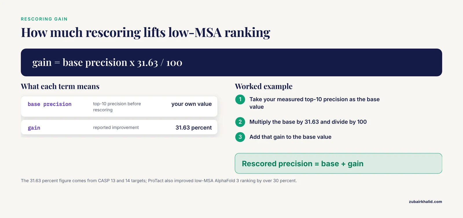
Rescoring tools take predicted complex models and estimate whether each interface is physically and evolutionarily plausible, then re-rank models. PIQLE uses a multi-head graph attention network over inter-residue interaction geometries, combining interaction distances, orientations, and evolutionary features, and it consistently outperformed earlier scorers including DProQA, TRScore, GNN-DOVE, and DOVE across independent test sets [8]. ProTact takes a different route, a geometric graph network with SE(3)-invariance (its outputs do not change under 3D rotation and translation) and physics-informed complementarity constraints, and it improved average top-10 precision by 31.63 percent on CASP 13 and 14 targets and by 31.94 percent on the DIPS-Plus dataset [7]. Most importantly for everyday work, ProTact used as a rescoring layer on AlphaFold 3 outputs improved ranking by over 30 percent in low-MSA contexts, precisely where raw AlphaFold confidence is weakest [7].
This reflects a broader movement. Reviewers describe the field shifting from pattern memorization toward physics-integrated machine learning, through three complementary routes: feeding physics-based sampling results into the model, building strict geometric inductive biases into architectures, and constraining generation with physical energy functions [3].
Pros
- Catches overconfident interfaces that raw confidence scores miss.
- Cheap to run on models you already have.
- Directly targets the low-MSA and antibody failure modes.
Cons
- Another tool with its own training biases to understand.
- Scores models, it does not generate them.
Molecular Dynamics and Physics-Based Refinement
Molecular dynamics (MD) simulates atomic motion over time using physical force fields, turning a static prediction into a stability test. A practical pattern has emerged in recent studies. Predict a complex with AlphaFold 3, then simulate. For the Nipah virus W protein bound to 14-3-3σ, researchers generated 1000 candidate heterotetramer models, filtered them on structural criteria, refined nine survivors, ran 100-nanosecond MD simulations in OpenMM, and advanced three complexes to microsecond-scale trajectories to confirm stability [9]. For the cancer-relevant PODXL-Ezrin interaction, docking, MD, and virtual screening built on predicted structures revealed how the R495W mutation allosterically destabilizes the resting Ezrin conformation [10]. And for the albumin-CD36 fatty acid handoff, docking and AlphaFold 3 produced different interface architectures, with the predicted interface shifting depending on which fatty acid was loaded into albumin [11].
Pros
- Tests whether an interface survives thermal motion.
- Quantifies mutation effects on stability.
- Reveals ligand- and state-dependent interface changes.
Cons
- Force fields approximate reality, and simulation length is always a compromise.
- Cannot rescue a fundamentally wrong topology.
- Expensive in analyst time as much as in compute.
How Accurate Is AlphaFold 3 for Protein-Protein Interactions?
Benchmark numbers help most when you know what they measure. CAPRI criteria grade a predicted complex by how closely its interface and chain placement match a reference structure, and DockQ condenses those ideas into a continuous score from 0 to 1 [5][12]. With that framing, three findings define the current picture.
First, co-folding beats docking on average. On the 235-complex protein-RNA benchmark, the AlphaFold Server achieved an 87.0 percent success rate within its top five predictions, against 44.7 percent for the best docking server, HDOCK [5]. The benchmark concerns protein-RNA rather than protein-protein pairs, so treat the numbers as an indication of the gap between the paradigms rather than as PPI-specific rates. The same benchmark carries a warning for co-folding enthusiasts: nine complexes were solved by docking and missed by AlphaFold [5].
Second, generalization is the weak point. On the LiPP benchmark of 311 lipid-protein complexes, AlphaFold 3 posted the highest success rate at 76.1 percent under a 2 angstrom RMSD criterion, but the rate fell to 47.2 percent for structures unlike anything seen during training [4]. The same study found that AlphaFold 3 and Chai-1 largely preserved intramolecular and intermolecular geometric constraints while DiffDock-L and RoseTTAFold AA frequently violated them [4]. Extrapolated to protein-protein work, the lesson is that accuracy is highest where the training data is densest, and novel fold combinations deserve extra skepticism.
Third, difficulty is concentrated in identifiable classes. Antibody-antigen prediction suffers from severe data scarcity and pattern memorization rather than principled generalization [3]. Low-MSA pairs, where few homologs exist to reveal co-evolving residues, are weak for the same reason, and physics-informed rescoring recovers much of the lost ranking performance there [7]. On the positive side, published end-to-end cases show what success looks like. A high-confidence AlphaFold 3 model of the Tle1-Tla1-VgrG4a ternary complex from Pseudomonas aeruginosa had all three predicted interfaces confirmed in vivo, with key charged residues identified for mutagenesis [6]. An AlphaFold 3 analysis of the monkeypox virus G9 M142I mutation correctly anticipated a weakened subcomplex interaction that ELISA then confirmed experimentally [13]. Independent structural data usually agrees with AlphaFold-family models, with most cross-links in a large Arabidopsis dataset mapping within the expected 35 angstrom constraint on PDB and AlphaFold structures [14], though instructive disagreements occur [15].
An AlphaFold Tutorial for Protein-Protein Interaction Prediction
The following seven steps take you from a hypothesis to a defensible model. They assume two protein partners and use the AlphaFold Server as the primary engine.
- Frame the question and choose your constructs. Decide what the model must answer: does A bind B, which interface, or does a mutation change binding. Pull the canonical sequences for each partner from a curated reference database. Check whether each protein is mostly ordered or contains long intrinsically disordered regions ( stretches that have no stable fold on their own ). The AFDB monomer model is a quick disordered-region probe, since low per-residue confidence flags disorder. Long disordered tails degrade co-folding, so consider modeling folded domains, but remember that truncation can delete the very segment that mediates binding [16][9].
- Prepare the input. Format each chain in FASTA, one header line starting with the greater-than symbol followed by the one-letter sequence. Set stoichiometry explicitly: enter the same sequence twice for a homodimer, three times for a homotrimer. Add cofactors, metals, glycans, or modifications when they are biologically part of the interface, because interface architecture can depend on ligand state [11].
- Submit through the AlphaFold Server. Paste the chains, set copy numbers, attach any ligands or modifications, and submit. Free non-commercial access carries daily job limits, so batch related questions into single jobs where possible. Each job returns a handful of ranked candidate models with confidence annotations.
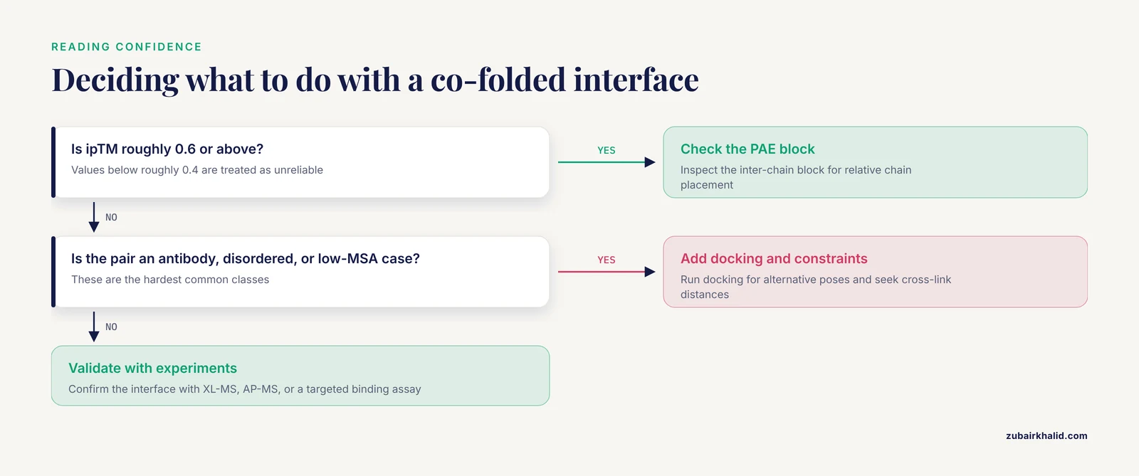
- Read the confidence outputs correctly. Four numbers matter. pLDDT is per-residue confidence on a 0 to 100 scale and describes each fold. PAE (predicted aligned error) is a matrix of expected distance errors in angstroms between every residue pair, and the inter-chain block of that matrix is what tells you whether the two chains are placed confidently relative to each other. pTM estimates global fold similarity, while ipTM (interface TM-score) estimates the correctness of the interface specifically. A widely used working rule treats ipTM values of roughly 0.6 and above as encouraging and values below roughly 0.4 as unreliable, though these thresholds came from earlier multimer benchmarks and are heuristics, not guarantees.
- Inspect the interface against biology. Look at residues in contact, conventionally heavy atoms within about 4 angstroms, and ask whether salt bridges, hydrogen bonds, and hydrophobic contacts land on residues with known function. Map your variants of interest onto the interface, a strategy used systematically in rice, where GWAS mutations were checked against predicted interaction sites and 18 affected complexes were then modeled explicitly [2]. Compare the predicted interface with known motifs, for example the conserved phosphoserine-binding groove of 14-3-3 proteins [9].
- Sample broadly and rescore. Repeat predictions to collect more candidates, cluster them by interface, and rescore rather than trusting the top-ranked model alone. PIQLE provides interface quality estimates that beat earlier scorers on independent benchmarks [8]. ProTact rescoring of AlphaFold 3 outputs improved ranking by over 30 percent in low-MSA settings [7]. For antibody-antigen or engineered-protein pairs, add a docking run as an independent pose generator and look for agreement between methods [5][3].
- Refine and validate. Simulate the leading candidates with MD in a package such as OpenMM to check interface stability over 100 nanoseconds or longer [9]. Then bring in experiments. Cross-linking mass spectrometry (XL-MS) chemically links residues that sit near each other and reports them as distance constraints, typically satisfied within about 35 angstroms for common crosslinkers [14]. Affinity purification mass spectrometry (AP-MS) confirms which partners coexist in a complex [16]. Targeted assays close the loop, including ELISA-style binding measurements [13], co-purification [6], and biomolecular fluorescence complementation (BIFC), where two halves of a fluorescent protein fused to the partners report proximity in living cells [17].
Worked Scenarios from the Recent Literature
A stable bacterial toxin assembly
The Type VI secretion system of Pseudomonas aeruginosa fires the antibacterial toxin Tle1 into rival cells. AlphaFold 3 produced a high-confidence ternary complex in which a VgrG4a trimer carries one Tle1 monomer and one Tla1 adaptor monomer. All three predicted interfaces, Tle1-Tla1, Tla1-VgrG4a, and Tle1-VgrG4a, were then confirmed in vivo, and the charged residues mediating them were identified for follow-up mutagenesis. A separate model of the Tle1-Tli1a immunity pair suggested a neutralization mechanism that leaves the catalytic pocket open, and Tli1a localized to the outer membrane exactly where periplasmic toxin neutralization would be expected [6]. This is the clean success case: folded domains, good homolog coverage, and a model accurate enough to design experiments from.
Mutations that reshape interfaces
The M142I substitution in the monkeypox virus G9 protein illustrates why multi-scale modeling matters. The mutation left the G9 monomer fold intact, shifted the conformation of the G9/A16 subcomplex, and widened the interfacial gap within the predicted G9/A16/A56/K2 quaternary assembly, reducing docking stability. ELISA experiments confirmed the reduced binding affinity that the models predicted [13]. The practical lesson is to model monomer, binary, and higher-order assemblies separately, because mutation effects can appear only at the level of the full complex. Genome-scale versions of the same logic work too. In rice, researchers used AFDB structures partitioned into domains, found 102 stress-associated GWAS mutations likely to affect structure or function, noted 25 sitting near predicted protein-protein interaction sites, modeled complexes for 18 affected proteins, and flagged six mutations predicted to strengthen binding, all with open-access tools [2].
A disordered viral protein anchored by phosphorylation
The Nipah virus W protein is an intrinsically disordered product of the P gene that binds 14-3-3σ, a family of phosphoserine-recognizing adapters. Because disorder defeats straightforward co-folding, the study generated 1000 heterotetramer models with AlphaFold 3 and filtered them on two structural criteria: feasible disulfide pairing between Cys421 residues of the W dimer, and preservation of contacts between phosphorylated Ser449 and Arg56, Arg129, and Tyr130 in the 14-3-3σ binding site. Nine models passed, survived 100-nanosecond MD refinement in OpenMM, and three remained stable through microsecond-scale simulation [9]. When a partner is disordered, large model pools plus biology-based filters beat a single prediction.
When models and experiments disagree
Disagreement is information. A cross-linking mass spectrometry survey of the Escherichia coli interactome, using the MS-cleavable crosslinker DSSO, produced 21,599 cross-linked spectrum matches covering 2,334 residue pairs and 137 protein-protein interactions, 47 of them previously unreported. For the ElaB-YqjD complex, the experimental distance constraints mismatched the AlphaFold 3 prediction [15]. The same survey contextualizes why this matters: only 8 percent of reported E. coli interactions in the IntAct database are annotated as direct physical associations, and XL-MS accounts for just 11 percent of those high-confidence annotations [15]. Separately, docking and AlphaFold 3 gave different interface architectures for the albumin-CD36 fatty acid transfer system, and the predicted interface shifted with the fatty acid loaded into albumin [11]. Independent constraints arbitrate when computational methods diverge.
A niche but useful extension: crystal packing
AlphaFold models also feed adjacent structural problems. MASCL combines AlphaFold predictions with symmetrical docking to simulate how proteins pack in a crystal lattice, scored by PackQ, a stringent DockQ-based metric where values above 0.36 count as successful. On the P41212 and P43212 space groups it predicted packing interfaces for 26.8 percent and 30.1 percent of targets within the top 100 models, rising to 57.9 percent and 39.8 percent within the top 25 when the starting dimeric assembly itself was reasonably accurate [12]. If crystallization is your bottleneck, predicted interfaces are worth mining.
How AlphaFold Fits Next to Experimental PPI Methods
No computational method replaces the experimental interactome toolkit, and the strongest recent work treats them as complementary layers. AP-MS delivers lists of interacting proteins without geometry, as in the KIAA1143 study that identified 73 high-confidence interactors and then turned to structural prediction to understand them [16]. XL-MS delivers residue-level distance constraints that can confirm or contradict models [14][15]. Reviews of plant interactome biology now explicitly position AlphaFold 3 beside proximity labeling, yeast three-hybrid, and mass spectrometry as parts of one toolkit, chosen per question [18]. Comparative modeling studies likewise benchmark several predictors side by side, for example AlphaFold 3, ESMFold, SWISS-MODEL, RoseTTAFold, and TrRosetta when characterizing bacterial outer membrane beta-barrel proteins and their complexes [19]. AlphaFold 3's distinctive contribution is the structural hypothesis: it converts a list of partners into a testable interface.
Common Mistakes and Limitations
- Treating confidence scores as proof. ipTM and related metrics are estimates that can be miscalibrated outside the training distribution, and held-out benchmarks show accuracy dropping sharply on genuinely novel targets [4].
- Getting stoichiometry wrong. If the biology is a trimer and you submit one copy, the model will invent a dimer. Always set chain copies deliberately.
- Ignoring ligands, cofactors, and modification state. Interface architecture can change with the bound ligand, as shown for albumin loaded with different fatty acids [11]. Model the state that matters biologically.
- Modeling disordered segments as if they were folded. Proteins with extensive intrinsic disorder, such as KIAA1143, need construct design and experimental partners rather than naive co-folding [16].
- Accepting the first returned pose. One run gives one dominant answer. Sample repeatedly, cluster by interface, and rescore with dedicated tools.
- Skipping experimental arbitration. Cross-link distances, interactor lists, and binding assays exist precisely because models disagree with reality sometimes, and the ElaB-YqjD mismatch is a concrete example [15].
- Misreading the PAE matrix. Judge the inter-chain block, which reports relative chain placement, not the diagonal, which reports each fold's internal confidence.
- Over-trusting antibody-antigen models. Sparse co-evolution and training-data scarcity make this the hardest class, and hybrid physics-aware methods are still maturing [3].
- Expecting dynamics from a static model. AlphaFold 3 returns conformations, not conformational ensembles, and predicting multiple states and dynamics remains an open challenge for the field [1]. MD simulation is the standard complement [9].
- Reporting models without standard metrics. When a reference structure exists, grade predictions with CAPRI-style criteria or DockQ-family scores so your numbers are comparable to everyone else's [5][12].
Frequently Asked Questions
Is AlphaFold 3 the best protein-protein interaction prediction tool?
It is the strongest default starting point for stable, well-folded protein pairs, but it is not universally the best. Antibody-antigen pairs, disordered partners, and low-MSA systems perform better when you add docking, rescoring, or experimental constraints alongside co-folding.
What is the difference between the AlphaFold Server and a local AlphaFold installation?
The AlphaFold Server is the official web interface, free for non-commercial use with daily job limits and little parameter control. A local installation, either licensed AlphaFold 3 or the open AlphaFold-Multimer, costs GPU hardware and setup effort but permits batch screening, custom sampling settings, and complete data privacy.
Which confidence score should I check first for a predicted protein-protein interface?
Check ipTM first, because it estimates interface correctness specifically, then inspect the inter-chain block of the PAE matrix for the relative placement of the two chains. pTM and pLDDT describe the individual folds and say little about the interface itself.
Can AlphaFold 3 model antibody-antigen complexes reliably?
Not reliably in general. Sparse co-evolutionary signal and limited training data make antibody-antigen the hardest common class, so treat high confidence with suspicion and support predictions with docking, rescoring, and experimental constraints.
Do docking and molecular dynamics still matter if I use AlphaFold 3?
Yes. Docking provides MSA-independent alternative poses and occasionally solves cases co-folding misses, while molecular dynamics tests whether a predicted interface remains stable under thermal motion and quantifies mutation effects.
How do I validate a protein-protein interaction predicted by AlphaFold 3?
Layer independent evidence. Cross-linking mass spectrometry gives residue distance constraints, affinity purification mass spectrometry confirms the partner set, and a targeted assay such as site-directed mutagenesis of predicted interface residues, a binding measurement, or fluorescence complementation tests the interface itself.
Sources
- The transformative impact of AI-enabled AlphaFold 3: evolution, current status, and future prospects in structural biology.
- Employing AlphaFold and TED to explore the structural impacts of stress-associated mutations in rice.
- From memorization to generalization: Why physics will improve machine learning -based prediction of protein complexes.
- The LiPP Benchmark Set for Modeling Lipid-Protein Complexes: Comparison of Co-Folding and Docking Methods.
- Evaluation of protein-RNA Docking Web Servers for Template-Free Docking and Comparison with the AlphaFold Server.
- "Molecular insights into the bactericidal toxin Tle1 of Pseudomonas aeruginosa: Interaction with VgrG, its adaptor, and its immunity protein".
- Accurate protein-protein interactions modeling through physics-informed geometric invariant learning.
- PIQLE: protein-protein interface quality estimation by deep graph learning of multimeric interaction geometries.
- In silico study of 14-3-3σ and Nipah virus W proteins interaction.
- Structural and Dynamic Insights into Podocalyxin-Ezrin Interaction as a Target in Cancer Progression.
- Molecular Mechanisms of Interaction of Human Serum Albumin with the CD36 Receptor: Insights from Molecular Dynamics Simulations.
- Using AlphaFold and Symmetrical Docking to Predict Protein-Protein Interactions for Exploring Potential Crystallization Conditions.
- Structural Insights into the Impact of the M142I Mutation in Monkeypox Virus G9 Protein on Subcomplex Formation Revealed by AlphaFold 3 Modeling.
- Mapping the architecture of protein complexes in Arabidopsis using cross-linking mass spectrometry.
- Protein-protein interactions in Escherichia coli: Using cross-linking/mass spectrometry (XLMS) to reveal our current knowledge gaps in interactomes.
- Mapping the interactome of recombinant human protein KIAA1143.
- Generating models for isoform-specific PKM-KIBRA interactions with BIFC, stabilization and AlphaFold 3.
- Capturing protein-protein interactions in plants: recent advances, challenges, and opportunities.
- Identification and Characterization of Outer Membrane Proteins and Membrane Spanning Protein Complexes in Brucella melitensis.