I-TASSER vs. AlphaFold: Which Tool Should You Use?
By Dr. Zubair Khalid, DVM, MS, PhD ·

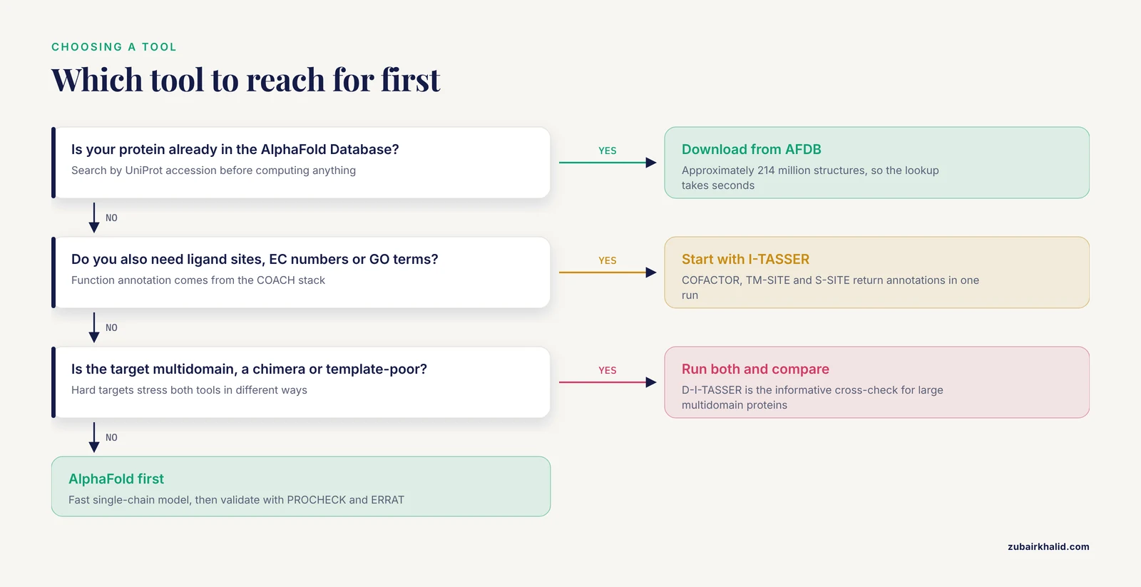
In one sentence, AlphaFold is a deep learning system that predicts a protein's 3D structure almost instantly from its sequence and co-evolutionary signals, while I-TASSER is a hybrid pipeline that threads your sequence onto structural templates, refines the fold through Monte Carlo fragment assembly simulations, and then annotates function from the resulting model. The rule of thumb is simple: reach for AlphaFold first when you need a fast, high-accuracy single-chain structure, especially for a protein already in the AlphaFold Database, and reach for I-TASSER when you also need ligand-binding sites, enzyme commission numbers, gene ontology terms, or a template-grounded interpretation of why the model looks the way it does. For difficult or ambiguous targets, running both and comparing the models is standard practice, and the published head-to-head studies show that neither tool wins every time.
This guide explains the difference between I-TASSER and AlphaFold in practical terms, walks through a short AlphaFold tutorial and the I-TASSER workflow, and gives you concrete criteria for deciding between them on your next project.
Why This Comparison Still Matters
At the 14th Critical Assessment of Structure Prediction (CASP14, the community-wide blind benchmark for structure prediction), John Moult announced that the protein structure prediction problem had been "solved" for single protein chains after five decades of effort. AlphaFold2, developed by DeepMind, achieved a median Global Distance Test (GDT) score of 92.4 across 87 of 92 evaluated domains, a level of accuracy that exceeded all prior expectations and stood out prominently among all competing methods [1]. That result changed the field overnight and earned the DeepMind team recognition that eventually extended to the Nobel Prize [1].
A reasonable conclusion from that headline number would be that traditional pipelines like I-TASSER are obsolete. The reality is more nuanced. The I-TASSER lineage has continued to evolve, most recently into D-I-TASSER, which integrates deep learning potentials with the classical threading and fragment assembly machinery and, in benchmark tests and CASP15 experiments, outperformed both AlphaFold2 and AlphaFold3 on single-domain and multidomain proteins [2]. Independent comparative studies in which researchers model the same protein with AlphaFold, I-TASSER, and other servers routinely find that the best model comes from different tools depending on the target [3][4][5]. Understanding what each method actually computes is the only reliable way to choose between them.
What Each Tool Actually Does
I-TASSER: Threading, Fragment Assembly, and Function Annotation
I-TASSER stands for Iterative Threading ASSEmbly Refinement, and the name describes the pipeline exactly. Given an amino acid sequence, the server first recognizes structural templates from the Protein Data Bank (PDB) using multiple threading alignment approaches. Threading means the sequence is aligned to proteins of known structure even when sequence similarity is faint, which lets I-TASSER detect remote homologs that simple BLAST-style searches would miss. Continuous structural fragments excised from those template alignments are then reassembled into full-length atomic models through iterative replica-exchange Monte Carlo simulations, a stochastic sampling procedure that explores many alternative conformations and, in principle, settles into low free-energy states [6][7][8].
The lowest free-energy conformations are identified by structure clustering, a second round of assembly simulation starting from the cluster centroids removes steric clashes and refines the global topology, and final atomic models are produced by a two-step atomic-level energy minimization. Model confidence is reported as a confidence score based on the significance of the threading alignments and the density of the structure clustering, while residue-level local quality estimates come from ResQ, a support vector regression method built on the variation across modeling simulations and the uncertainty of the homologous alignments [6].
The second half of the pipeline is where I-TASSER diverges sharply from AlphaFold. The predicted models are matched against the BioLiP database of ligand-protein interactions to detect homologous function templates. From those matches, the suite derives ligand-binding site (LBS) predictions, Enzyme Commission (EC) numbers, and Gene Ontology (GO) terms, using three complementary algorithms (COFACTOR, TM-SITE, and S-SITE) whose consensus is computed by COACH with support vector machines [6]. For a newly sequenced enzyme from a metagenome, for example, researchers can go from raw sequence to a 3D model plus a functional annotation in one submission to the Zhanglab webserver [9].
Access options have kept pace with demand. The I-TASSER gateway runs the full pipeline on the XSEDE supercomputer infrastructure to handle the volume of community submissions [8]. For groups that need throughput beyond what any public server allows, GPU-I-TASSER parallelizes the replica-exchange Monte Carlo simulations onto GPU architectures using OpenACC and achieves an average 10-fold speedup with comparable prediction accuracy on a benchmark of 71 protein structures [10]. The multi-domain branch, I-TASSER-MTD, extends the pipeline with a deep-learning-based protocol for large multidomain proteins [11], and the 2025 D-I-TASSER release added domain splitting and assembly for automated modeling of large multidomain structures, folding 81 percent of protein domains and 73 percent of full-chain sequences in the human proteome with results highly complementary to AlphaFold2 [2].
AlphaFold: Deep Learning and the 214-Million-Structure Database
AlphaFold2 replaced the template-first logic of classical pipelines with an end-to-end deep learning architecture. The model reads a multiple sequence alignment (MSA), extracts co-evolutionary signals that reveal which residues contact each other, and applies an attention-based module that reasons about residue pair geometry directly, producing a 3D coordinate set in a single forward pass rather than through physical sampling. The CASP14 result of a 92.4 median GDT across evaluated domains reflects that architectural leap [1].
For most researchers, the practical entry point is not running the model at all but querying the AlphaFold Protein Structure Database (AFDB), maintained by EMBL-EBI and Google DeepMind. The database hosts approximately 214 million predicted structures covering nearly the entire genome across model organisms, and its 2025 update aligned coverage with the UniProt 2025_03 release, added isoform structures with their underlying multiple sequence alignments, and introduced a redesigned entry page with a dedicated domains tab, integrated annotations in an interactive 3D viewer, and data delivery through the website, FTP, Google Cloud, and updated APIs [1][12]. If your protein of interest has a UniProt accession, there is a good chance someone has already computed your structure and you can simply download it.
AlphaFold 3 extended the framework with a substantially updated diffusion-based architecture that predicts the joint structure of complexes containing proteins, nucleic acids, small molecules, ions, and modified residues. Its accuracy gains are substantial where earlier tools struggled: far greater accuracy for protein-ligand interactions than state-of-the-art docking tools, much higher accuracy for protein-nucleic acid interactions than nucleic-acid-specific predictors, and substantially higher antibody-antigen accuracy than AlphaFold-Multimer version 2.3 [13]. For protein-protein complexes, AlphaFold-Multimer remains a workhorse, and published studies use it to discover entirely novel interaction networks, such as the identification of high-confidence interactions and two structurally unique novel complexes among predation-essential hypothetical proteins in Bdellovibrio [14].
Confidence reporting is built into every AlphaFold output. The per-residue pLDDT score expresses local confidence on a 0 to 100 scale, and interface and global confidence metrics such as pTM accompany complex predictions. These scores are essential for interpretation, and later sections cover how to use them and where they mislead.
The Wider Ecosystem
Neither tool exists in isolation. AI techniques have been integrated into established frameworks including SWISS-MODEL, Rosetta, and I-TASSER itself, and the field now evaluates models with standard metrics such as the template modeling (TM) score, the global distance test total score (GDT_TS), and the local distance difference test (lDDT), with CAMEO (Continuous Automated Model EvaluatiOn) providing continuous evaluation that complements the biennial CASP experiment [15]. AlphaFold2 structures now serve as inputs to downstream machine learning pipelines for protein function prediction, for example frameworks that combine ESM-2 protein language model embeddings with graph neural networks over AF2-predicted structures to improve molecular function, cellular component, and biological process annotation across the human proteome [16]. In other words, the choice between I-TASSER or AlphaFold is less about picking a winner and more about picking the right entry point for the question you are asking.
Side-by-Side Comparison
The table below is the core of the I-TASSER vs. AlphaFold decision. Entries reflect the primary literature and official documentation described above.
| Criterion | I-TASSER (and D-I-TASSER) | AlphaFold (AF2, AF3, AFDB) |
|---|---|---|
| Core method | Multiple threading alignments to PDB templates, replica-exchange Monte Carlo fragment assembly, structure clustering, two-step energy minimization [6][7] | End-to-end deep learning on MSA co-evolution plus residue-pair geometry, diffusion-based architecture in AF3 [1][13] |
| Template dependence | Explicit and central, templates constrain the fold and explain the result | Implicit, templates inform training but the model can fold template-poor targets |
| Single-chain accuracy | Strong overall, D-I-TASSER outperformed AlphaFold2 and AlphaFold3 in benchmarks and CASP15 tests [2] | Median GDT of 92.4 at CASP14 for single chains, historically the benchmark standard [1] |
| Multidomain handling | Dedicated protocols in I-TASSER-MTD and D-I-TASSER with domain splitting and assembly [2][11] | Good for many targets, chimera and fusion proteins remain nontrivial [17] |
| Complexes | Not the pipeline's focus | AlphaFold-Multimer and AF3 predict protein complexes, nucleic acids, ligands, ions, and modified residues [13][14] |
| Function annotation | Built in, COFACTOR, TM-SITE, S-SITE, and COACH return ligand-binding sites, EC numbers, and GO terms from BioLiP matches [6] | None built in, structures are inputs to separate annotation tools [16] |
| Local quality output | ResQ residue-level quality estimates plus a global confidence score [6] | Per-residue pLDDT, plus pTM and interface metrics for complexes |
| Database of precomputed models | None, each target is computed | AFDB with approximately 214 million structures, isoform coverage, MSAs, FTP, Google Cloud, and APIs [1][12] |
| Throughput at scale | GPU-I-TASSER gives a 10-fold speedup on GPUs for local large-scale runs [10] | AFDB delivers genome-scale coverage instantly, D-I-TASSER folded 73 percent of full-chain human sequences for comparison [12][2] |
| Typical access | Public webserver on XSEDE infrastructure [8], downloadable source including the GPU version [10] | Webserver and AFDB lookups, downloadable model code, AF3 server for complexes |
| Interpretability | High, threading alignments show which PDB structures shaped the model | Lower, the neural network's reasoning is not exposed as alignments |
| Refinement needs | Refinement is part of the pipeline | Outputs often benefit from external refinement and validation in comparative studies [3][4] |
The Real Differences That Decide Your Choice
Difference 1: Where the Structural Information Comes From
The deepest difference between I-TASSER and AlphaFold is the source of structural signal. I-TASSER asks, which known structures look like fragments of my protein, and how can I reassemble them under physical energy terms. AlphaFold asks, what does the co-evolutionary record in the sequence family imply about residue contacts, and what fold satisfies those constraints. This has practical consequences. If your protein has close structural templates in the PDB, I-TASSER's threading will find them and the final report will show you exactly which templates drove the model, which is valuable for writing a paper or planning mutations [7]. If your protein has no good templates but a deep sequence family, AlphaFold's co-evolutionary inference can succeed where threading struggles. If your protein is a short orphan with neither templates nor deep homologs, both approaches degrade, and that failure mode is discussed under limitations below.
Difference 2: Function Annotation Is a Workflow Saver
If your endpoint is a structure, either tool suffices. If your endpoint is a function, I-TASSER delivers it in the same run. The COACH consensus over COFACTOR, TM-SITE, and S-SITE produces ligand-binding site predictions, EC numbers, and GO terms derived from structure-based matching against known proteins [6]. With AlphaFold, structure-based function annotation is a separate step, and recent research shows it is a productive one, since AF2 structures combined with ESM-2 embeddings and graph neural networks outperform existing methods for predicting molecular function, cellular component, and biological process annotations on the human proteome [16]. The difference is workflow length, not possibility.
Difference 3: Speed, Scale, and Cost of Compute
An AlphaFold prediction is effectively instantaneous at inference time, and for proteome-scale questions the AFDB answers them with zero compute on your side, delivering data through the website, FTP, Google Cloud, and APIs [12]. I-TASSER's Monte Carlo sampling is computationally heavier, which is precisely why the GPU-I-TASSER implementation exists, moving the replica-exchange simulations to GPUs for a 10-fold average speedup while preserving accuracy [10]. If you need thousands of structures tonight, AFDB plus batch AlphaFold-style inference is the natural route. If you need dozens of carefully annotated models with template provenance, I-TASSER server turnaround is usually acceptable.
Difference 4: What Happens with Hard Targets
Multidomain proteins, chimeras, and proteins with sparse evolutionary information stress both tools in different ways. The D-I-TASSER results are the clearest recent evidence that the hybrid approach can lead on hard targets, outperforming both AlphaFold2 and AlphaFold3 on single-domain and multidomain proteins in benchmarks and CASP15 [2]. Conversely, artificial fusion proteins in which two domains are joined by a linker remain difficult for every method, including AlphaFold2, because the spatial freedom allowed by the linker and the mutual orientation of the domains cannot be resolved from sequence alone, and prediction must be done with care [17]. Neither tool rescues you from physics.
Difference 5: Validation Culture
Comparative studies rarely crown a single winner. In one head-to-head of AlphaFold, I-TASSER, and Robetta on the ovarian cancer biomarker HE4, every server's Ramachandran plot placed 100 percent of residues in allowed regions except I-TASSER before refinement, yet after molecular dynamics refinement with GROMACS the Robetta-derived model achieved the highest ERRAT overall quality factor at 97.701, ahead of the third refined model at 94.643 and the AlphaFold model at 93.750 [3]. A separate study of the SARS-CoV-2 NSP6 transmembrane protein generated models with eight servers including SWISS-MODEL, Phyre2, AlphaFold, C-Quark, Galaxyweb, I-TASSER, LOMETS, and Robetta, refined them with six different servers into 48 candidate models, and evaluated everything with ERRAT, Ramachandran plots, Z-scores, PROQ, and TM-align, confirming the protein as roughly 68 to 78 percent alpha-helical in the process [4]. A third comparison on the TBK1-binding protein 1 found Robetta produced the best validated model among Robetta, I-TASSER, and AlphaFold using PROCHECK through the SAVESv6.0 server [5]. The lesson is consistent: tool choice matters less than running a serious validation and refinement pipeline, and cross-checking two or more predictors is cheap insurance.
When to Use I-TASSER
Choose I-TASSER when one or more of the following describe your project.
You need function annotation with the structure. The integrated COFACTOR, TM-SITE, S-SITE, and COACH stack returns ligand-binding sites, EC numbers, and GO terms from the final model in a single submission [6]. Metagenome-derived enzymes are a textbook case, as in the analysis of the recombinant AldII protein, where I-TASSER provided both the 3D model and structure-based functional context for a novel class II aldolase-like protein [9].
You want template provenance and interpretability. Because the method starts from multiple threading alignments, the output tells you which PDB structures underlie the model and how confident each alignment is [7]. For manuscripts, grant reviews, and mutagenesis planning, this traceability is a genuine advantage over a neural network's opaque output.
Your target is multidomain or otherwise hard, and you want a second opinion. I-TASSER-MTD and the newer D-I-TASSER were built specifically for multidomain modeling, and D-I-TASSER outperformed both AlphaFold2 and AlphaFold3 in recent benchmarks and CASP15 tests while producing human proteome-scale predictions complementary to AlphaFold2 [2][11]. If AlphaFold's model looks suspicious for a large multidomain protein, a D-I-TASSER run is the most informative cross-check currently available.
You need local-scale throughput on your own hardware. GPU-I-TASSER brings the pipeline in-house with a 10-fold speedup over the CPU version at comparable accuracy [10]. That combination of control and speed suits core facilities and labs with recurring modeling workloads.
Your protein has recognizable remote homologs but sparse sequence family. Threading can detect structural similarity that MSA-based deep learning may underweight when the sequence family is thin [7].
When to Use AlphaFold
Your protein is already in AFDB. Before computing anything, search the AlphaFold Database by UniProt accession. With approximately 214 million structures aligned to UniProt 2025_03, including isoforms, plus downloadable MSAs and API access, the lookup takes seconds and the structure is fully downloadable for downstream work [1][12].
You need a fast, high-accuracy single-chain model. The CASP14 median GDT of 92.4 remains the reference point for single-chain accuracy [1], and for a standard globular protein with a decent sequence family, an AlphaFold prediction is usually the right first model.
Your question involves complexes, ligands, or nucleic acids. AlphaFold 3's diffusion-based architecture predicts joint structures of proteins with nucleic acids, small molecules, ions, and modified residues, with substantially improved protein-ligand accuracy over docking tools and improved antibody-antigen accuracy over AlphaFold-Multimer v2.3 [13]. For protein-protein interaction discovery, AlphaFold-Multimer has been used to surface novel high-confidence interactions among previously uncharacterized bacterial proteins [14].
You are building a downstream ML or analysis pipeline. AF2 structures are now standard inputs for function prediction frameworks, interaction fingerprint reconstruction, and proteome-scale analyses, and the AFDB APIs make them scriptable at scale [12][18][16].
A Practical AlphaFold Tutorial and the I-TASSER Workflow
Checking AFDB Before You Compute Anything
This three-step AlphaFold tutorial covers the most common use case, retrieving and judging a precomputed model.
- Go to the AlphaFold Protein Structure Database at https://alphafold.ebi.ac.uk and search by protein name or UniProt accession. The 2025 redesign adds a domains tab and integrates annotations directly with the interactive 3D viewer, so you can inspect predicted domain boundaries alongside the model [12].
- Download the structure in PDB or mmCIF format. Files are available through the website, FTP, and Google Cloud, and updated APIs support programmatic access, for example retrieving one structure per UniProt entry at proteome scale [12]. A typical retrieval for UniProt entry Q14508 (the HE4 biomarker used in comparative modeling studies) follows the documented AFDB file naming pattern of the form AF-UNIPROTACCESSION-F1-model with a version suffix, available from the entry page [12][3].
- Read the per-residue pLDDT track before trusting anything. Regions with very high pLDDT are predicted with high local confidence, while low pLDDT regions, which commonly include disordered tails and flexible loops, should be treated as hypotheses rather than coordinates. If you plan docking or simulations, trim or down-weight low-confidence segments and validate the model with tools such as PROCHECK and ERRAT, exactly as the published comparative workflows do [3][4][5].
Running the I-TASSER Server
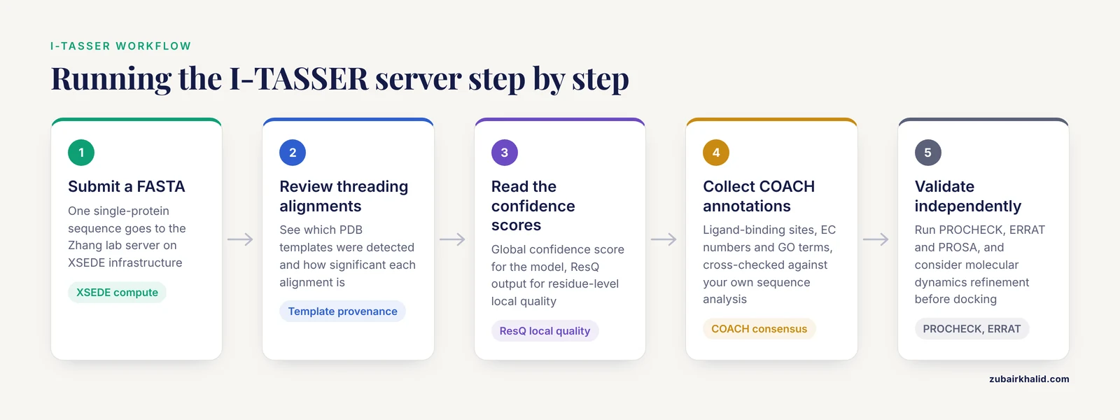
The I-TASSER workflow mirrors this in structure but adds functional output.
- Prepare a single-protein FASTA sequence. Submit it at the I-TASSER server hosted by the Zhang lab, which runs the pipeline on XSEDE compute infrastructure [7][8].
- Review the threading alignments in the results. These show which PDB templates were detected and their significance, and they anchor the interpretation of the final model [7].
- Read the confidence score for global model quality and the ResQ output for residue-level local quality.
- Collect the COACH-derived functional annotations, including ligand-binding sites, EC numbers, and GO terms, and cross-check them against your own sequence analysis [6].
- If the model will be used for docking or refined further, run an independent validation pass with PROCHECK, ERRAT, PROSA, and comparable tools, and consider molecular dynamics refinement as the HE4 and NSP6 studies did [3][4].
For large in-house workloads, clone and build GPU-I-TASSER from the Zhang group repository and run the same pipeline on GPU hardware [10].
A Sensible Combined Workflow
For a novel protein with no experimental structure, a defensible pipeline looks like this: check AFDB first, run AlphaFold if the protein is missing, submit the same sequence to I-TASSER for its template analysis and function annotation, then compare the two models with TM-align or a comparable structural alignment tool and validate both with Ramachandran and ERRAT-style assessments before committing to either [3][4][2][5]. The extra server submission costs hours, not days, and the comparative studies above show the models frequently disagree in exactly the regions where you most need to know.
Worked Scenarios
Scenario A, annotating a metagenomic enzyme. You cloned a novel gene from an environmental sample and the sequence matches no characterized protein. I-TASSER is the better first tool because its threading may connect the sequence to a distant enzyme family and its COACH stack will propose ligand-binding sites and EC numbers from the model [6][9]. Confirm with an AlphaFold model for the fold itself.
Scenario B, modeling a host-virus membrane protein. For the SARS-CoV-2 NSP6 protein, where no crystal structure exists, the published pipeline used eight prediction servers including both AlphaFold and I-TASSER, then six refinement servers, then multi-tool validation [4]. Replicate that logic: for membrane proteins and drug targets, breadth beats loyalty to one predictor.
Scenario C, characterizing a viral protein with no homologs. The feline calicivirus leader of the capsid protein has no similarity beyond its genus, which forced researchers to triangulate among AlphaFold 2, AlphaFold 3, and I-TASSER's QUARK de novo method to argue for a putative transmembrane domain, predictions then supported experimentally by membrane binding and permeation assays [19]. When sequence similarity fails, multiple independent structure predictions plus experiments are the honest path.
Scenario D, mapping a novel interaction network. You have a set of hypothetical proteins implicated in a phenotype and want interaction hypotheses. AlphaFold-Multimer is the appropriate tool, as demonstrated in the Bdellovibrio predation study where Markov clustering of predicted interactions revealed two structurally unique novel complexes [14]. I-TASSER is not designed for this question.
Scenario E, building a proteome-scale function predictor. You need structures as features for machine learning. Pull them from AFDB through its APIs rather than computing anything, then layer your own model on top, following the ESM-2 plus graph network pattern demonstrated on the human proteome [12][16].
Common Mistakes and Limitations
Treating a prediction as an experimental structure. AlphaFold2's CASP14 performance was extraordinary, but the CASP announcement concerned single protein chains, and the same review tracing AlphaFold's journey notes that multi-chain complex prediction, mutation effects, and other facets remain active research areas [1]. A predicted model is a hypothesis with quantified confidence, not a crystal structure.
Trusting pLDDT on short and spurious sequences. AlphaFold2, AlphaFold3, and ESMFold all assign unexpectedly high pLDDT scores to short spurious protein sequences arising from gene prediction errors, and discrimination between spurious and real proteins only improves beyond roughly 100 amino acids. High pLDDT on a short open reading frame is not evidence that the protein exists or folds in vivo [20].
Expecting chimera and fusion protein models to be reliable out of the box. The mutual orientation of artificially fused domains separated by a linker is a genuinely hard prediction problem even for AlphaFold2, and the methodology requires care with linkers, domain boundaries, and validation [17].
Skipping validation and refinement. The published head-to-head studies refined models with GROMACS molecular dynamics and up to six refinement servers, then validated with PROCHECK, ERRAT, PROSA, PROSA Z-scores, and TM-align before drawing conclusions [3][4]. Submitting a raw server output into a docking pipeline without this step is the single most common avoidable error in the literature.
Assuming one tool always wins. The same comparative studies crowned Robetta, not AlphaFold or I-TASSER, as the best model source for HE4 after refinement and for TBK1-binding protein 1 [3][5]. Meanwhile D-I-TASSER beat both AlphaFold2 and AlphaFold3 on multidomain benchmarks [2]. Tool superiority is target-dependent, which is why consensus approaches exist.
Overlooking the ligand-binding caveat. Although AlphaFold 3 sharply improved protein-ligand geometry prediction [13], structural data for protein-drug complexes remains limited across the field, and structure-based drug screening methods have had to reconstruct binding modes from fragment-level data to expand coverage [18]. Treat predicted binding poses as starting points for experimental or computational follow-up, not as docked poses.
Ignoring confounders in function annotation. Structure-based EC and GO transfer is only as good as the detected templates. For proteins with weak template matches, I-TASSER's own confidence scores flag this, and the annotations should be read accordingly [6].
Forgetting that individual research questions need domain expertise and, where relevant, experimental validation. Computational predictions guide experiments, they do not replace them, and specific projects warrant consultation with structural biology collaborators.
Frequently Asked Questions
Is AlphaFold always more accurate than I-TASSER?
No. AlphaFold2 set the single-chain benchmark at CASP14 with a median GDT of 92.4 [1], but the hybrid D-I-TASSER pipeline outperformed both AlphaFold2 and AlphaFold3 on single-domain and multidomain proteins in recent benchmarks and CASP15 tests [2]. Accuracy is target-dependent, and independent head-to-head studies have found other servers producing the best-validated models for specific proteins [3][5].
Do I need to install anything to use either tool?
For routine work, no. The AlphaFold Database and the I-TASSER server are both web-accessible, with AFDB offering website, FTP, Google Cloud, and API delivery [12][7][8]. Installation only becomes relevant for large-scale workloads, where GPU-I-TASSER provides a 10-fold accelerated local version of the I-TASSER pipeline [10].
Can I use these predicted structures for drug docking?
Yes, with caution. AlphaFold 3 delivers substantially improved protein-ligand interaction accuracy over prior docking tools [13], but structural data for protein-drug complexes remains scarce field-wide, so predicted poses should be validated and treated as hypotheses for screening rather than settled answers [18]. Validate any model you dock into with Ramachandran and ERRAT-style checks first [3][4].
Which confidence score should I look at?
Use per-residue pLDDT for AlphaFold models, checking low-confidence regions before any downstream use [20], and use the I-TASSER confidence score plus ResQ residue-level estimates for I-TASSER models [6]. For complexes, interface metrics matter more than global scores. Field-standard global metrics include TM-score, GDT_TS, and lDDT when comparing models against references [15].
Can either tool predict protein-protein interactions?
AlphaFold can. AlphaFold-Multimer predicts multichannel protein complexes and has been used to discover novel interaction networks among previously uncharacterized bacterial proteins [14], and AlphaFold 3 extends complex prediction to nucleic acids, ligands, ions, and modified residues with substantially improved accuracy [13]. I-TASSER is a single-protein structure and function pipeline, not a complex predictor, so use it for the monomer models and function annotation instead [6].
Should I run both tools on the same protein?
Usually yes when the protein is novel or difficult. Comparative studies on HE4, NSP6, and TBK1-binding protein 1 all modeled each target with multiple servers including both AlphaFold and I-TASSER before selecting and refining the best model [3][4][5], and D-I-TASSER's human proteome results were explicitly complementary to AlphaFold2's [2]. The two models disagreeing is itself diagnostic information about which regions of your protein are hard to predict.
Sources
- From CASP13 to the Nobel Prize: DeepMind's AlphaFold Journey in Revolutionizing Protein Structure Prediction and Beyond.
- Deep-learning-based single-domain and multidomain protein structure prediction with D-I-TASSER
- Three-Dimensional Structure of Human Epididymis Protein 4 (HE4): A Protein Modelling of an Ovarian Cancer Biomarker Through In Silico Approach
- Computational Modeling and High-confidence Tertiary Structure Prediction of the SARS-CoV-2 NSP6 Protein: Implications for Viral Pathogenesis and Host Interaction
- Comparative In Silico Analysis and Functional Characterization of TANK-Binding Kinase 1-Binding Protein 1
- The I-TASSER Suite: protein structure and function prediction
- I-TASSER server: new development for protein structure and function predictions
- I-TASSER gateway: A protein structure and function prediction server powered by XSEDE
- 3D STRUCTURE AND FUNCTION ANALYSIS OF RECOMBINANT ALDII PROTEIN FROM Uncultured Acidilobus sp. USING I-TASSER
- GPU-I-TASSER: a GPU accelerated I-TASSER protein structure prediction tool
- I-TASSER-MTD: a deep-learning-based platform for multi-domain protein structure and function prediction
- AlphaFold Protein Structure Database 2025: a redesigned interface and updated structural coverage
- Accurate structure prediction of biomolecular interactions with AlphaFold 3
- Using AlphaFold-Multimer to study novel protein-protein interactions of predation essential hypothetical proteins in Bdellovibrio
- AI-Driven Deep Learning Techniques in Protein Structure Prediction
- Integrating ESM-2 and Graph Neural Networks with AlphaFold-2 Structures for Enhanced Protein Function Prediction
- Fusion of two unrelated protein domains in a chimera protein and its 3D prediction: Justification of the x-ray reference structures as a prediction benchmark.
- Decomposing compounds enables reconstruction of interaction fingerprints for structure-based drug screening.
- The Feline calicivirus Leader of the Capsid (LC) Protein Contains a Putative Transmembrane Domain, Binds to the Cytoplasmic Membrane, and Exogenously Permeates Cells
- Folding the unfoldable 2: using AlphaFold and ESMFold to explore spurious proteins.