From DNA to Protein Sequence: Preparing Input for AlphaFold
By Dr. Zubair Khalid, DVM, MS, PhD ·

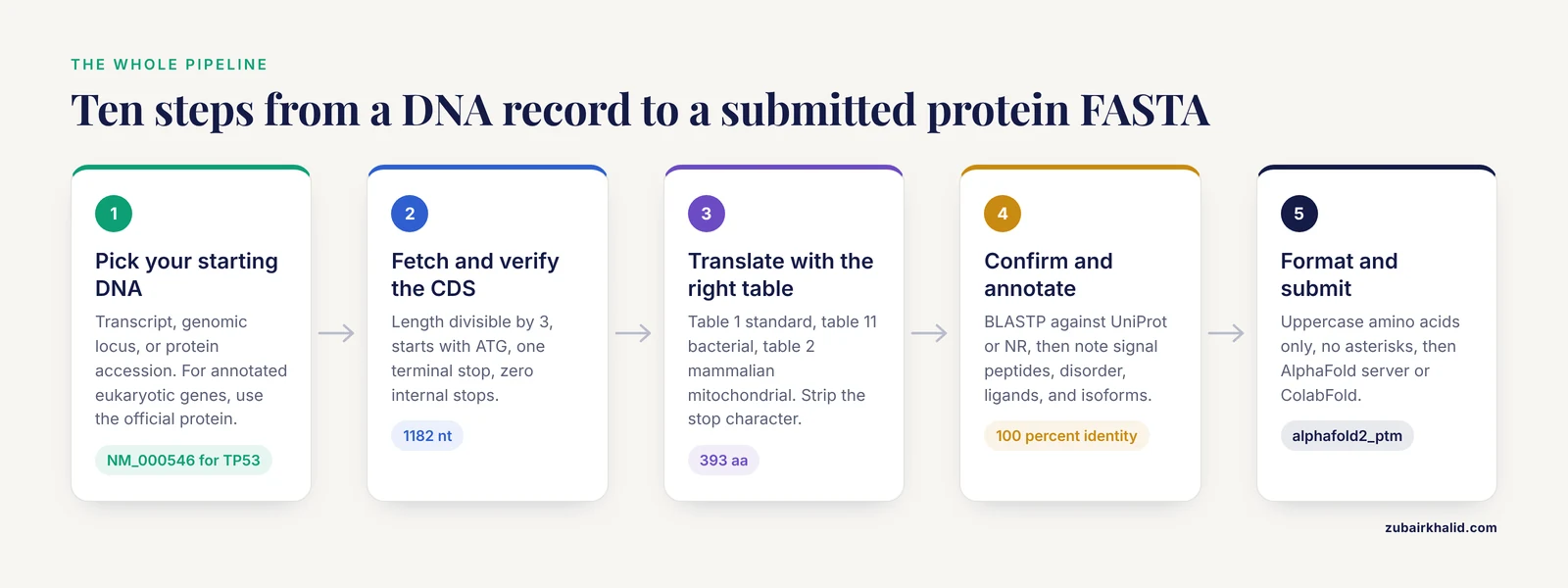
By the end of this article you will be able to take a DNA record for a gene of interest, extract or verify its coding sequence, translate it into a correct amino acid sequence, format that sequence as a protein FASTA file, and submit it to the AlphaFold server or a local ColabFold installation for structure prediction. You will also know how to read the confidence metrics in the output and how to recognize the most frequent failures in this workflow before they waste an hour of compute time.
Here is what you need on hand:
- A computer with Python 3.9 or newer, or access to a terminal on a Linux or macOS workstation
- The Biopython package (
pip install biopython) for the translation steps - Optionally, the NCBI EDirect utilities (
efetch) or a web browser for retrieving sequences - Optionally, ColabFold installed locally if you want to run predictions on your own hardware
- A gene accession number or FASTA file of the DNA you care about
Everything below uses free, standard tools. No proprietary software is required.
What AlphaFold Actually Accepts as Input
AlphaFold does not translate DNA. AlphaFold2 predicts a protein structure from an amino acid sequence plus a multiple sequence alignment (MSA, a table of evolutionarily related protein sequences that tells the network which positions are conserved). AlphaFold 3 extends this to complexes of proteins with other molecules, including nucleic acids and ligands [1]. All versions of AlphaFold2 and AlphaFold 3 take an amino acid sequence as the protein input, and the amino acid sequence, not the underlying gene, is what the neural network sees [2].
This distinction matters more than newcomers expect. If you paste a nucleotide sequence into a protein-only predictor, one of two things happens. The tool rejects the alphabet outright, or, worse, it treats the letters A, C, G, and T as if they were the amino acids alanine, cysteine, glycine, and threonine and returns a confident-looking model of a nonsense polymer. Either way you get no structure of your real protein. The translation step, with the correct genetic code and the correct reading frame, is the actual work in going from DNA to protein for AlphaFold.
There is one important exception to keep straight. AlphaFold 3, available through the public AlphaFold server, does accept nucleotide sequences as separate entities in a complex. Studies have used it to model transcription factor binding to DNA elements [3], protein complexes bound to DNA in viral transcription machinery [4], and protein complexes with single-stranded DNA [1]. But even in those cases, the protein components must still be entered as amino acid sequences. The DNA is a partner in the complex, never a substitute for translating your gene. Work on transcription factor variants showed that AlphaFold 3 can model TF-DNA complexes and that differences in the interface-predicted template modeling (ipTM, a confidence score for the predicted interface) tracked experimental binding outcomes, which shows how useful a correct protein-plus-DNA input can be when both partners are prepared properly [5].
Getting a Clean Coding Sequence
A coding sequence (CDS) is the stretch of DNA that encodes a protein, from the start codon through the stop codon, with introns removed. For bacteria and other prokaryotes, genes are usually contiguous, so a CDS is a simple open reading frame (ORF, a stretch of codons uninterrupted by stop codons). For eukaryotes, the CDS is spliced together from exons and cannot be read straight out of the genome. Choosing the right starting DNA is the first fork in the road.
Step 1: Decide what DNA you actually have
There are three common starting points, and they lead to different amounts of work:
- A RefSeq mRNA or transcript accession, such as
NM_000546for human TP53. This contains the spliced CDS with untranslated regions (UTRs) on both ends. You will need to locate the CDS within it. - A genomic locus, such as a region of
NC_000913.3for E. coli or a chromosome scaffold for a eukaryote. For prokaryotes you can find the ORF directly. For eukaryotes you must splice using an annotation file, or better, skip translation entirely and pull the annotated protein. - A protein accession, such as an
NP_RefSeq or a UniProt entry. You already have the amino acid sequence and can skip to the formatting step.
A practical rule for eukaryotic genes: if an official protein accession exists, use it. Translating a spliced CDS yourself is a valuable skill and a good consistency check, but for a well-annotated human, mouse, or model organism gene, the RefSeq protein record has already been curated and reviewed.
Step 2: Retrieve the sequence
With EDirect installed, you can fetch the translated CDS of a RefSeq transcript directly from NCBI in one command. The -format fasta_cds_aa output gives you the protein translation of every CDS annotated on the record:
efetch -db nuccore -id NM_000546.6 -format fasta_cds_aa > tp53_aa.fastaExpected output looks like this:
>NP_000537.3 tumor protein p53 [Homo sapiens]
MEEPQSDPSVEPPLSQETFSDLWKLLPENNVLSPLPSQAMDDLMLSPDDIEQWFTEDPGP
DEAPRMPEAAPPVAPAPAAPTPAAPAPAPSWPLSSSVPSQKTYQGSYGFRLGFLHSGTAK
...
If you instead want the nucleotide CDS so you can translate it yourself and verify the result, use:
efetch -db nuccore -id NM_000546.6 -format fasta_cds > tp53_cds.fastaOn the web, the NCBI Gene and Nucleotide database pages expose the same translations under the CDS features of a GenBank flatfile, and the NCBI ORFfinder tool will find and translate ORFs in any sequence you paste in. These routes are equivalent for a well-annotated record.
For genomes without official protein annotations, structure prediction has become part of the annotation pipeline itself. A workflow for the highly divergent microsporidian Vairimorpha necatrix genome started from predicted protein-coding DNA sequences, ran structure predictions with ColabFold, and then used Foldseek searches against the PDB and AlphaFold databases to assign functions, with manual curation improving accuracy [6]. That pattern, translate the predicted CDS, predict the structure, search the structure, is now standard for divergent genomes where sequence similarity alone fails.
Translating DNA to Protein
Step 3: Verify the coding sequence before translating
Four checks catch most problems in seconds. All of them are properties of any correct CDS:
- The length is divisible by 3, because codons are triplets
- It starts with a start codon, usually ATG, though bacteria use GUG and UUG as functional starts under the standard code as well
- It ends with a stop codon: TAA, TAG, or TGA
- It contains no stop codons before the final position
A sequence failing any of these is in the wrong frame, is not a CDS, or contains an error. Do not translate and submit anyway. Fix the input first.
Step 4: Translate with the correct genetic code
The genetic code is the mapping from the 64 codons to amino acids and stops. Table 1 of the standard code covers most eukaryotic nuclear and many viral genes. Table 11 is the bacterial and plant plastid code, which differs from the standard code mainly in alternative start codons. Mammalian mitochondrial code is table 2. Using the wrong table is a real and silent failure mode, because most codons are identical across tables and the damage shows up at starts and stops only. Biopython lets you specify the table explicitly:
from Bio.Seq import Seq

# Read the CDS you fetched in step 2
with open("tp53_cds.fasta") as fh:
    lines = fh.read().split("\n")
header = lines
dna = "".join(lines[1:]).upper()

print("Length:", len(dna), "nt")
print("Divisible by 3:", len(dna) % 3 == 0)
print("Starts with ATG:", dna.startswith("ATG"))
print("Ends with stop:", dna[-3:] in ("TAA", "TAG", "TGA"))

# Table 1 is the standard code, use table 11 for bacterial genes
protein = str(Seq(dna).translate(table=1, to_stop=False))

internal_stops = protein[:-1].count("*")
print("Internal stops:", internal_stops)

protein = protein.rstrip("*")

with open("tp53_aa.fasta", "w") as out:
    out.write(">TP53_human translated from NM_000546.6\n")
    for i in range(0, len(protein), 60):
        out.write(protein[i:i+60] + "\n")

print("Protein length:", len(protein), "aa")
Expected output:
Length: 1182 nt
Divisible by 3: True
Starts with ATG: True
Ends with stop: True
Internal stops: 0
Protein length: 393 aa
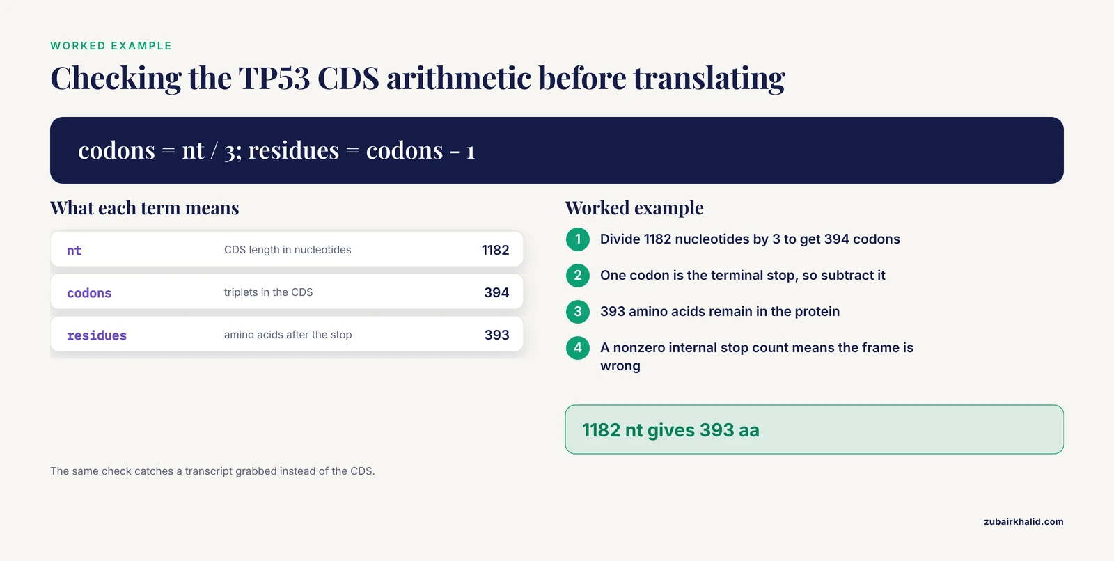
The arithmetic is the built-in sanity check: 1182 nucleotides divided by 3 gives 394 codons, one of which is the stop, leaving 393 amino acids. If your file is one or two nucleotides longer than the CDS because you grabbed a full transcript instead of the CDS, every codon after the offset is wrong and the internal stop count will be nonzero.
Step 5: When to skip manual translation
For eukaryotic genes with introns, fetching the annotated protein directly is faster and safer than reconstructing the CDS. Two reliable sources are the NCBI Protein database (accessions beginning NP_ for curated records or XP_ for predicted ones) and UniProt, the expertly curated protein knowledgebase. Paste the resulting FASTA straight into the formatting step. Keep the accession in the FASTA header so you can trace the model back to its source record later.
An alternative worth knowing for divergent organisms: if your predicted CDS does not match any database protein, translate it anyway and let structure carry the annotation. Structural homology searches against the AlphaFold2 database have uncovered domain families invisible to sequence methods, including previously unrecognized BEN DNA-binding domains in C. elegans and a new subtype corresponding to the domain of unknown function DUF4806 [7]. A model of a novel protein can be the evidence that identifies it.
Checking the Protein Before Prediction
Step 6: Confirm the translation against a database
Run a BLASTP search of your translated protein against the non-redundant protein database or UniProt. You are looking for two things. First, a near-identical hit (often 100 percent coverage and identity for a known gene) confirms the frame and code were right. Second, if your top hit is a different protein or no strong hit exists, you have either translated the wrong region or found something genuinely novel, and you should resolve which before predicting.
If you have any doubt about the boundaries of a folded domain, compare your sequence against related proteins. In one published example, the boundaries of the exonuclease and polymerase domains of the white spot syndrome virus DNA polymerase were defined precisely by aligning the AlphaFold model against the equivalent viral enzymes, even though that polymerase carries over 1000 additional amino acids compared with the herpes simplex virus 1 enzyme [8]. Knowing where the conserved core ends and the species-specific additions begin tells you which parts of the prediction to trust.
Step 7: Annotate what AlphaFold cannot see
AlphaFold knows nothing about your protein beyond its sequence and its homologs. Before submitting, make a short annotation of features that affect interpretation:
- Signal peptides and targeting sequences. These N-terminal segments are cleaved after import. Predict the mature N-terminus and consider modeling the mature chain separately, because the transit peptide itself is often disordered.
- Low-complexity and disordered regions. Predicted models routinely contain unstructured regions, and these complicate functional interpretation of the model [9]. Long acidic or repetitive tails frequently come out as extended spaghetti with low confidence. That is usually a correct reflection of biology, not a failure of prediction.
- Cofactors, metals, and modified residues. AlphaFold does not know which ligand is bound unless you provide it. For example, a study of the bifunctional M. tuberculosis AdaA-AlkA enzyme used AlphaFold prediction validated against the E. coli Ada and AlkA crystal structures as domain-specific references when no experimental structure of their target existed, then identified an interdomain cavity and screened approved drugs into it [10]. The prediction was reliable for the protein fold, and the ligand was a separate analysis.
- Known processing and isoforms. If the gene has splice isoforms, predict the isoform you actually study, not the canonical one by default.
Nucleic acid binding is worth flagging specifically, because it changes what you can do downstream. Tools such as BindUP-Alpha predict DNA- and RNA-binding proteins from either experimental structures or predicted models by analyzing electrostatic patches on the protein surface, and they work on AlphaFold models directly [9]. The xBind webserver similarly accepts a single-chain protein sequence in FASTA format and can run an AlphaFold prediction on the fly before predicting protein-DNA and protein-RNA binding sites at the residue level [11]. If your translated protein is likely to bind DNA, plan for a complex prediction rather than a monomer.
Formatting FASTA and Submitting
Step 8: Format the protein FASTA correctly
AlphaFold servers and ColabFold are forgiving about whitespace but strict about the alphabet. Follow these rules:
- One sequence per prediction request unless you are deliberately modeling a complex
- Uppercase standard amino acid letters only. Remove any
*stop characters,Xfor ambiguous residues is tolerated but lowers local confidence, and unusual characters will cause rejection - A descriptive header starting with
>helps you match outputs to inputs - Line lengths of 60 to 80 characters are conventional but not required
A clean file looks like this:
>TP53_human_NM_000546.6
MEEPQSDPSVEPPLSQETFSDLWKLLPENNVLSPLPSQAMDDLMLSPDDIEQWFTEDPGP
DEAPRMPEAAPPVAPAPAAPTPAAPAPAPSWPLSSSVPSQKTYQGSYGFRLGFLHSGTAK
SVTCTYSPALNKMFCQLAKTCPVQLWVDSTPPPGTRVRAMAIYKQSQHMTEVVRRCPH
For a complex, give each chain its own entry in the same file or a separate FASTA, and label chains clearly. The AlphaFold server accepts proteins, nucleic acids, ions, and small molecules as separate entities in one prediction, which is what allows modeling of protein-DNA interfaces [3], multimeric transcription assemblies [4], and enzyme-oligonucleotide complexes [1].
Step 9: Submit to the AlphaFold server or run ColabFold
The AlphaFold server (alphafoldserver.com) is the simplest route. You paste protein sequences in FASTA format, optionally add DNA, RNA, ligand, or ion entities, and the server returns structures with per-residue confidence and pairwise alignment error data. It requires no installation and no GPU. For most people preparing a single protein or a small complex, it is the right first choice, and published studies now use it as the primary prediction engine for aptamer structures [12], DNA-recognition models [3], and viral protein complexes [13].
For batch jobs, custom MSAs, or full control, install ColabFold locally:
pip install "colabfold[alphafold-minus-jax]"Then run a prediction:
colabfold_batch --model-type alphafold2_ptm --num-recycle 3 tp53_aa.fasta af2_out/Expected console output includes MSA generation against your chosen databases, followed by model construction with per-model confidence printed as ranking is performed. The output directory contains:
tp53_aa_unrelaxed_rank_001_alphafold2_ptm_model_1_v3.pdb, the top-ranked modeltp53_aa_scores_rank_001_alphafold2_ptm_model_1_v3.json, containing pLDDT and PAE datatp53_aa.a3m, the multiple sequence alignment actually used- Relaxed PDB files if the
-amberflag was supplied
The -model-type flag matters. Use alphafold2_ptm for a single chain, and alphafold2_multimer_v3 when your FASTA contains multiple protein chains. The -msa-mode option controls where the alignment comes from, which is the single biggest lever on accuracy for proteins with deep or shallow homology. Template usage is controlled with -templates when you want the model informed by experimentally solved relatives.
Step 10: Read the confidence metrics
Three numbers tell you whether to believe the model:
- pLDDT (predicted local distance difference test) is a per-residue score from 0 to 100. The conventional reading is that scores above 90 indicate very high confidence where backbone and most side chains are well placed, 70 to 90 indicates a confident backbone with less certain side chains, 50 to 70 indicates low confidence, and below 50 should be treated as disordered rather than structured.
- PAE (predicted aligned error) reports, for every pair of residues, the expected error in their relative positions. Low PAE between two domains means the model has evidence for how they pack against each other. High PAE between domains means the relative arrangement is a guess.
- ipTM applies to interfaces in complexes. A study benchmarking AlphaFold 3 against SNP-SELEX binding data for six transcription factors found that differences in AF3's ipTM score aligned with experimental allele preferences and generally outperformed energy-based metrics from FoldX, and that combining both metrics was more reliable than either alone [5]. When you model a protein-DNA or protein-protein complex, ipTM is the number that tells you whether the interface prediction is evidence or speculation.
Confidence behaves differently in different regimes, and the published record gives useful calibration points. AlphaFold reproduced the NMR-determined structure of the small anti-CRISPR protein AcrIE5 correctly, a case of a compact single domain with homologs [14]. For leucine zipper coiled-coils, an evaluation against over 2000 experimentally tested human zippers mapped out where AlphaFold2 and AlphaFold 3 models of dimerization are and are not reliable, a reminder that oligomeric state prediction has its own error profile [2]. For protein-DNA complexes, AF3 predicted the enzyme fold of type IA topoisomerases with high confidence, especially the N-terminal domain, but confidence dropped for complexes with longer oligonucleotides beyond roughly 25 bases [1]. Read your metrics in that light: small, homolog-rich, single domains tend to be excellent, long nucleic acid partners and novel folds tend to be weaker.
Key Options and Commands at a Glance
| Task | Tool and command or option | What it does |
|---|---|---|
| , - | , - | , - |
| Fetch translated CDS from a transcript | efetch -db nuccore -id ACC -format fasta_cds_aa | Returns the protein translations of all CDS features on the record |
| Fetch the nucleotide CDS | efetch -db nuccore -id ACC -format fasta_cds | Returns the spliced coding DNA for your own translation |
| Verify CDS properties | Length divisible by 3, starts with ATG, one terminal stop, zero internal stops | A single failed check usually means wrong frame or wrong region |
| Translate with Biopython | Seq(dna).translate(table=1) | Table 1 is the standard code, table 11 is bacterial, table 2 is mammalian mitochondrial |
| Strip stop characters | protein.rstrip("*") | Removes the terminal stop, never submit * to a predictor |
| Confirm the protein | BLASTP against UniProt or NR | A near-identical hit validates frame and code |
| Find ORFs interactively | NCBI ORFfinder | Finds and translates all ORFs in a pasted sequence |
| Single-chain prediction | colabfold_batch, model-type alphafold2_ptm file.fasta out/ | AF2-style monomer prediction with automatic MSA |
| Complex prediction | colabfold_batch, model-type alphafold2_multimer_v3 file.fasta out/ | Models multiple protein chains and their interfaces |
| Protein-DNA complexes | AlphaFold server with DNA entities | AF3 accepts nucleic acids alongside proteins, expect lower confidence with longer oligos [1] |
| Interface confidence | Read ipTM from AF3 output | ΔipTM tracked experimental TF-DNA binding preferences in a benchmark [5] |
Tips for Better Models
These tips come up repeatedly in practice and in the published literature.
Feed the model the mature chain when a signal peptide exists. The precursor sequence with a cleavable N-terminal peptide predicts the peptide as a floppy appendage, which lowers the apparent quality of the N-terminal domain you actually care about. Trimming to the mature N-terminus, if the cleavage site is known from annotation or a signal peptide predictor, produces a cleaner model.
Do not panic about disordered tails. Predicted structures frequently include unstructured and disordered regions, and their presence is a known feature of AlphaFold output that complicates but does not invalidate functional analysis [9]. A long tail with pLDDT under 50 is usually a true disorder call. Trim it for visualization, keep it for completeness, and never build functional claims on its exact conformation.
Use deep homology when you can, and know when you cannot. The MSA is the source of most of AlphaFold's power. For a protein with thousands of homologs, monomer folds come out near-experimental accuracy in favorable cases, as demonstrated when AlphaFold reproduced the NMR structure of AcrIE5 [14]. For proteins from divergent or streamlined genomes, structural search of the prediction against PDB and AlphaFold databases can rescue annotation when sequence searches fail [6][7].
Validate against any experimental anchor you have. When a domain of your protein has a relative with a crystal or NMR structure, overlay your model on that structure. The AdaA-AlkA study used E. coli Ada (1U8B) and AlkA (1DIZ) as domain-specific structural references to validate its AlphaFold model of the mycobacterial enzyme [10]. The same habit applies to any prediction: one experimental anchor converts a pretty picture into an argument.
Treat nucleic acid complexes as exploratory. AlphaFold 3 has produced useful models of TF-DNA recognition [3], herpesvirus late transcription factor complexes bound near promoter DNA [4], and DNA-RNA-protein assemblies in RNA design workflows, where AF3 confidence metrics helped distinguish functional from nonfunctional designed sequences [15]. But the same body of work documents weakening confidence with longer oligonucleotides [1]. Keep DNA partners short, check ipTM at the interface, and where a variant or mutation matters, combine structural reasoning with an experimental assay such as EMSA, as the rice DREB1 study did when mutation analysis verified the predicted roles of R40 and E66 in CRT element binding [3].
Remember that complexes may be context-dependent. In the AcrIE5 study, the protein did not bind individual Cascade subunits with high affinity but instead recognized a composite interface of the assembled type I-E Cascade [14]. If biology assembles your protein only in a larger context, a monomer model answers the wrong question, and the complex prediction is the relevant one.
Use generative and design workflows carefully. When new sequences are generated computationally, AlphaFold and ESMFold serve as structural validators that the designed sequences fold as intended [16]. The same validation logic applies in reverse to your input: if a designed or mutated sequence produces a model whose fold collapses relative to the wild type, that is a signal worth investigating, not noise to ignore.
Common Mistakes and Limitations
Submitting DNA where protein is expected. The single most common error in going from DNA to protein for AlphaFold is pasting nucleotides into a protein-only field. AlphaFold2-based tools predict from amino acid sequences, and the nucleotide sequence is never a valid protein input [2]. Double-check the alphabet of your FASTA before submitting. A quick way is seqkit fx2tab -n -l -H tp53_aa.fasta to print the sequence alphabet and length, or simply scan the first line of residues for T, which appears in DNA but not in real protein sequences except in rare engineered residues.
Reading the wrong frame or the wrong strand. Six possible reading frames exist per double-stranded DNA segment, three per strand. A frame shift of even one nucleotide produces a completely different protein with internal stops. The internal stop check in step 3 catches this reliably, which is why it should never be skipped.
Using the wrong genetic code. Bacterial genes frequently initiate at GUG or UUG codons, which translate as valine or leucine under the standard table but as N-formylmethionine or methionine at a true start under the bacterial table. Mitochondrial codes reassign stop codons. If your protein starts with V or L where every homolog starts with M, you used the wrong table or picked up the codon after the real start.
Forgetting introns. Translating unspliced eukaryomic genomic DNA produces a sequence riddled with internal stops and frameshifts. This is the failure that makes people conclude their gene "does not work" in AlphaFold. Splice first, using the annotated CDS or an official protein accession.
Leaving stop symbols and junk characters in the FASTA. A trailing * is harmless to most viewers but is not an amino acid, and some pipelines choke on it. Strip it. Also strip whitespace, digits from header lines copied into the sequence, and any lowercase masking that survived a soft-masked genome export if your tool is case-sensitive.
Modeling the wrong isoform or the precursor instead of the mature chain. Many human genes encode multiple isoforms with different domain compositions. Predicting isoform 1 when your experiments use isoform 3 produces a model that looks right and answers the wrong question. Similarly, secreted proteins and organelle-imported proteins carry N-terminal targeting peptides that are cleaved in vivo. Decide explicitly whether you are modeling the precursor or the mature chain and record the choice.
Overreading confidence in complex and nucleic-acid predictions. AF3's protein components in complexes can be excellent while the nucleic acid partner or its interface is weak, and this degrades further with longer oligonucleotides [1]. A high-resolution protein with a low-resolution DNA pose is a normal outcome, not a bug. Use ipTM and the PAE block between protein and DNA residues to judge how much of the interface story to keep [5]. The same caution applies to multimeric interfaces, where evaluations such as the leucine zipper study show that dimerization and pairing specificity have their own reliability envelope [2].
Expecting AlphaFold to know chemistry it was not told about. Post-translational modifications, bound metals, disulfide patterns in unusual topologies, and ligand identities are not inferred from sequence alone. Where they matter for function, model them explicitly as entities in AF3 or analyze them separately, as in the docking and molecular dynamics studies that followed AF3 structure generation for aptamer-protein systems [12][17].
Ignoring the possibility that the protein is disordered. If BLASTP finds no homologs, pLDDT stays low everywhere, and PAE is uniformly high, the honest conclusion may be that this sequence is mostly intrinsically disordered. AlphaFold output does not give you a structure in that case because there is no single structure to give. Sequence-based and surface-property predictors of nucleic acid binding can still be informative for such proteins [9].
Assuming one tool answers every question. AlphaFold predicts structure, not function, dynamics, or binding energetics. Studies that got real biological insight paired AlphaFold predictions with docking, molecular dynamics, and experimental assays [12][17], or with NMR ground truth [14]. Plan your validation before you run the prediction, not after.
Frequently Asked Questions
Can AlphaFold predict structure directly from DNA?
No for the protein itself. AlphaFold2 predicts protein structures from amino acid sequences, and AlphaFold 3, while it can accept DNA and RNA as partner entities in a complex, still requires protein components as amino acid sequences [2][1]. Translating the coding sequence is always your job.
What is the fastest way to get the protein sequence from a gene?
For an annotated gene, fetch the official protein accession from NCBI or UniProt directly. If you must translate yourself, efetch -db nuccore -id ACC -format fasta_cds_aa returns the translated CDS for annotated records in one command.
My translated protein has internal stop codons. What went wrong?
An internal stop almost always means the wrong reading frame, unspliced genomic DNA, or a truncated CDS missing the true start. Return to the annotation, confirm the CDS coordinates, and retranslate. A correct CDS has exactly one stop codon, at the end.
Which genetic code table should I use?
Use the standard table (table 1) for eukaryotic nuclear genes and most viruses, the bacterial table (table 11) for bacterial genes, and the appropriate mitochondrial table for mitochondrial genomes. The wrong table usually changes only start and stop handling, but that is enough to corrupt the N-terminus or add a false internal stop.
Should I use the AlphaFold server or ColabFold?
Use the AlphaFold server for one-off predictions and for complexes with DNA, RNA, ligands, or ions, since it requires no installation and accepts nucleic acid entities. Use ColabFold when you need batch predictions, custom MSAs, control over templates and recycling, or local hardware. ColabFold runs AF2-style models and multimer predictions from the command line.
What does pLDDT below 50 mean?
It conventionally indicates a region the model does not consider structured, and in practice these regions are often genuinely intrinsically disordered. Treat low-pLDDT segments as flexible or disordered rather than as an unreliable version of a real fold, and avoid building structural arguments on them.
Can I include DNA in my prediction?
Yes, with AlphaFold 3 through the AlphaFold server. Studies have modeled transcription factors bound to their DNA motifs [3], large transcription complexes positioned on promoter DNA [4], and topoisomerases with single-stranded DNA [1]. Keep oligonucleotides short, check interface confidence through ipTM and PAE, and remember confidence drops for longer nucleic acid partners [1].
Why is my model's tail floppy or missing structure?
Long low-complexity and terminal regions are frequently disordered, and AlphaFold reflects that disorder as low pLDDT and extended conformations rather than failing outright [9]. This is expected behavior. Check whether the tail is a known disordered region, trim it for clearer visualization of the folded core, and interpret the folded core on its own metrics.
Sources
- Substrate binding of human and bacterial type IA topoisomerase: An experimentation with AlphaFold 3.0.
- Assessing the validity of leucine zipper constructs predicted by AlphaFold.
- AlphaFold 3-Assisted Deciphering of the DNA Recognition by DREB1 Transcription Factors in Rice.
- Structure Prediction of Complexes Controlling Beta- and Gamma-Herpesvirus Late Transcription Using AlphaFold 3.
- A structure-guided approach to noncoding variant evaluation for transcription factor binding using AlphaFold 3.
- Functional annotation of a divergent genome using sequence and structure-based similarity.
- Unanticipated broad phylogeny of BEN DNA-binding domains revealed by structural homology searches.
- AlphaFold modeling of the white spot syndrome virus polymerase.
- BindUP-Alpha: A Webserver for Predicting DNA-and RNA-binding Proteins based on Experimental and Computational Structural Models☆.
- Structural characterizations of the Mycobacterium tuberculosis AdaA-AlkA (Rv1317c) bifunctional enzyme and identification of enprofylline as a candidate synergistic DNA-repair targeting agent through integrated computational and experimental approaches.
- xBind: an integrated webserver for large language model-enabled cross-molecular protein binding site prediction.
- Structure-guided optimization of the A58 DNA aptamer targeting the SARS-CoV-2 nucleocapsid protein through integrated computational analysis.
- Genome characterization and receptor-binding protein identification of Klebsiella phage vB_VIPKPNMC05, a member of a novel viral family Pituviridae.
- Structural and mechanistic investigation of the anti-CRISPR protein AcrIE5 using NMR spectroscopy and AlphaFold modeling.
- Design of function-regulating RNA via deep learning and AlphaFold 3.
- Controllable protein design via autoregressive direct coupling analysis conditioned on principal components.
- Structure-Guided Optimization of a LipL32-Targeting DNA Aptamer through Molecular Dynamics and Free-Energy Analysis.